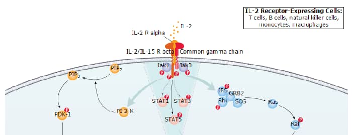
Interleukin 2 was initially identified as a T cell growth factor that is produced by T cells following activation by mitogens or antigens. Since then, it has been shown that in addition to its T cell growth factor activity, IL-2 can also stimulate the growth and differentiation of B cells, natural killer (NK) cells, lymphokine activated killer (LAK) cells, monocytes/macrophages and oligodendrocytes. Mature porcine and human IL-2 share approximately 72% amino acid sequence identity.The biological activity of IL-2 is mediated by the binding of IL-2 to cell surface receptor complexes. The functional high-affinity receptor of IL-2 is composed of three distinct polypeptide chains, the IL-2 receptor alpha, beta and gamma subunits. The intermediate-affinity IL-2 receptor complex, which lacks the alpha subunit, but contains both the beta and gamma subunits, is also capable of transducing the IL-2 signal. In T cells, the beta and gamma subunits are shared with the IL-15 receptor complex. The gamma chain of the IL-2 receptor complex is also a subunit of IL-4, IL-7, and IL-9 receptor complexes.
Porcine IL-2 Antibody (100312) [FITC]
R&D Systems | Catalog # IC6521F

Scientific images may be resized, cropped, or adjusted for brightness or contrast for presentation purposes. These updates are limited to presentation and do not affect the underlying scientific interpretation of the data. Where available, additional source data may be provided upon request.
Key Product Details
Species Reactivity
Applications
Label
Antibody Source
Product Specifications
Immunogen
Ala21-Thr154
Accession # P26891
Specificity
Clonality
Host
Isotype
Scientific Data Images for Porcine IL-2 Antibody (100312) [FITC]
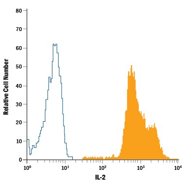
Detection of IL‑2 in Porcine PBMCs by Flow Cytometry.
Porcine peripheral blood mononuclear cells (PBMCs) were stained with Mouse Anti-Porcine IL-2 Fluorescein-conjugated Monoclonal Antibody (Catalog # IC6521F, filled histogram) or isotype control antibody (Catalog # IC0041F, open histogram). To facilitate intracellular staining, cells were fixed with Flow Cytometry Fixation Buffer (Catalog # FC004) and permeabilized with Flow Cytometry Permeabilization/Wash Buffer I (Catalog # FC005). View our protocol for Staining Intracellular Molecules.Applications for Porcine IL-2 Antibody (100312) [FITC]
Intracellular Staining by Flow Cytometry
Sample: Porcine peripheral blood mononuclear cells (PBMCs) fixed with Flow Cytometry Fixation Buffer (Catalog # FC004) and permeabilized with Flow Cytometry Permeabilization/Wash Buffer I (Catalog # FC005)
Spectra Viewer
Plan Your Experiments
Use our spectra viewer to interactively plan your experiments, assessing multiplexing options. View the excitation and emission spectra for our fluorescent dye range and other commonly used dyes.
Spectra Viewer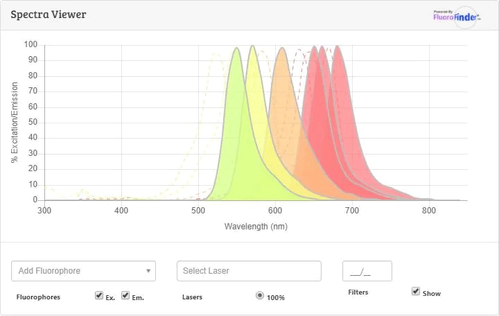
Flow Cytometry Panel Builder
Bio-Techne Knows Flow Cytometry
Save time and reduce costly mistakes by quickly finding compatible reagents using the Panel Builder Tool.
Advanced Features
- Spectra Viewer - Custom analysis of spectra from multiple fluorochromes
- Spillover Popups - Visualize the spectra of individual fluorochromes
- Antigen Density Selector - Match fluorochrome brightness with antigen density

Formulation, Preparation, and Storage
Purification
Formulation
Shipping
Stability & Storage
- 12 months from date of receipt, 2 to 8 °C as supplied.
Background: IL-2
References
- Taniguchi, T. and Y. Minami (1993) Cell 73:5.
- Waldmann, T. et al. (1998) Int. Rev. Immunol. 16:205.
- Nelson, B.H. and D.M. Willeford (1998) Adv. Immunol. 70:1.
Long Name
Alternate Names
Entrez Gene IDs
Gene Symbol
UniProt
Additional IL-2 Products
Product Documents for Porcine IL-2 Antibody (100312) [FITC]
Certificate of Analysis
To download a Certificate of Analysis, please enter a lot or batch number in the search box below.
Note: Certificate of Analysis not available for kit components.
Product Specific Notices for Porcine IL-2 Antibody (100312) [FITC]
For research use only
Related Research Areas
Customer Reviews for Porcine IL-2 Antibody (100312) [FITC]
There are currently no reviews for this product. Be the first to review Porcine IL-2 Antibody (100312) [FITC] and earn rewards!
Have you used Porcine IL-2 Antibody (100312) [FITC]?
Submit a review and receive an Amazon gift card!
$25/€18/£15/$25CAN/¥2500 for a review with an image
$10/€7/£6/$10CAN/¥1110 for a review without an image
Submit a review
Protocols
Find general support by application which include: protocols, troubleshooting, illustrated assays, videos and webinars.
- 7-Amino Actinomycin D (7-AAD) Cell Viability Flow Cytometry Protocol
- Extracellular Membrane Flow Cytometry Protocol
- Flow Cytometry Protocol for Cell Surface Markers
- Flow Cytometry Protocol for Staining Membrane Associated Proteins
- Flow Cytometry Staining Protocols
- Flow Cytometry Troubleshooting Guide
- Intracellular Flow Cytometry Protocol Using Alcohol (Methanol)
- Intracellular Flow Cytometry Protocol Using Detergents
- Intracellular Nuclear Staining Flow Cytometry Protocol Using Detergents
- Intracellular Staining Flow Cytometry Protocol Using Alcohol Permeabilization
- Intracellular Staining Flow Cytometry Protocol Using Detergents to Permeabilize Cells
- Propidium Iodide Cell Viability Flow Cytometry Protocol
- Protocol for Liperfluo
- Protocol for the Characterization of Human Th22 Cells
- Protocol for the Characterization of Human Th9 Cells
- Protocol: Annexin V and PI Staining by Flow Cytometry
- Protocol: Annexin V and PI Staining for Apoptosis by Flow Cytometry
- Troubleshooting Guide: Fluorokine Flow Cytometry Kits
- View all Protocols, Troubleshooting, Illustrated assays and Webinars
Associated Pathways