![Western Blot: RhoA Antibody [NB100-91273]](https://resources.rndsystems.com/images/products/RhoA-Antibody-Western-Blot-NB100-91273-img0034.jpg "Western Blot: RhoA Antibody [NB100-91273]")
Loading...
Key Product Details
Species Reactivity
Validated:
Human, Mouse, Rat, Primate
Cited:
Human, Mouse, Rat
Applications
Validated:
Immunohistochemistry, Immunohistochemistry-Paraffin, Western Blot, Immunocytochemistry/ Immunofluorescence
Cited:
Immunohistochemistry-Paraffin, Western Blot
Label
Unconjugated
Antibody Source
Polyclonal Rabbit IgG
Loading...
Product Specifications
Immunogen
A synthetic peptide from amino acid region 100-150 of human RhoA conjugated to blue carrier protein was used as the antigen. The peptide is homologous in many species including rat, mouse, chicken, dog, bovine, frog, zebrafish and orangutan.
Reactivity Notes
Marmoset
Clonality
Polyclonal
Host
Rabbit
Isotype
IgG
Theoretical MW
22 kDa.
Disclaimer note: The observed molecular weight of the protein may vary from the listed predicted molecular weight due to post translational modifications, post translation cleavages, relative charges, and other experimental factors.
Disclaimer note: The observed molecular weight of the protein may vary from the listed predicted molecular weight due to post translational modifications, post translation cleavages, relative charges, and other experimental factors.
Scientific Data Images for RhoA Antibody
Western Blot: RhoA Antibody [NB100-91273]
Western Blot: RhoA Antibody [NB100-91273] - Rat spinal cord lysate using Rabbit antibody to Transforming protein RhoA: whole serum at 1:2000 dilution. One single band is detected.![Immunocytochemistry/ Immunofluorescence: RhoA Antibody [NB100-91273]](https://resources.rndsystems.com/images/products/RhoA-Antibody-Immunocytochemistry-Immunofluorescence-NB100-91273-img0024.jpg "Immunocytochemistry/ Immunofluorescence: RhoA Antibody [NB100-91273]")
Immunocytochemistry/ Immunofluorescence: RhoA Antibody [NB100-91273]
Immunocytochemistry/Immunofluorescence: RhoA Antibody [NB100-91273] - Rat trigeminal at 1:500 dilution using Rabbit antibody to Transforming protein RhoA: whole serum (NB100-91273, in red), Sheep antibody to extracellular, N-terminal part of Sortilin: whole serum (NB100-98771, in green), DAPI counter stained appearing in blue.![Immunohistochemistry: RhoA Antibody [NB100-91273]](https://resources.rndsystems.com/images/products/RhoA-Antibody-Immunohistochemistry-NB100-91273-img0040.jpg "Immunohistochemistry: RhoA Antibody [NB100-91273]")
Immunohistochemistry: RhoA Antibody [NB100-91273]
Immunohistochemistry: RhoA Antibody [NB100-91273] - IHC on rat brain (paraffin sections) using Rabbit antibody to RhoA at a concentration of 15 ug/ml, incubated overnight and developed with DAB Ni.![Immunocytochemistry/ Immunofluorescence: RhoA Antibody [NB100-91273]](https://resources.rndsystems.com/images/products/RhoA-Antibody-Immunocytochemistry-Immunofluorescence-NB100-91273-img0008.jpg "Immunocytochemistry/ Immunofluorescence: RhoA Antibody [NB100-91273]")
Immunocytochemistry/ Immunofluorescence: RhoA Antibody [NB100-91273]
Immunocytochemistry/Immunofluorescence: RhoA Antibody [NB100-91273] - Rat trigeminal at 1:500 dilution using Rabbit antibody to Transforming protein RhoA: whole serum (NB100-91273, in red), Sheep antibody to extracellular, N-terminal part of Sortilin: whole serum (NB100-98771, in green), DAPI counter stained appearing in blue.![Immunocytochemistry/ Immunofluorescence: RhoA Antibody [NB100-91273]](https://resources.rndsystems.com/images/products/RhoA-Antibody-Immunocytochemistry-Immunofluorescence-NB100-91273-img0022.jpg "Immunocytochemistry/ Immunofluorescence: RhoA Antibody [NB100-91273]")
Immunocytochemistry/ Immunofluorescence: RhoA Antibody [NB100-91273]
Immunocytochemistry/Immunofluorescence: RhoA Antibody [NB100-91273] - Rat trigeminal at 1:500 dilution using Rabbit antibody to Transforming protein RhoA: whole serum (NB100-91273, in red), Sheep antibody to extracellular, N-terminal part of Sortilin: whole serum (NB100-98771, in green), DAPI counter stained appearing in blue.![Immunocytochemistry/ Immunofluorescence: RhoA Antibody [NB100-91273]](https://resources.rndsystems.com/images/products/RhoA-Antibody-Immunocytochemistry-Immunofluorescence-NB100-91273-img0023.jpg "Immunocytochemistry/ Immunofluorescence: RhoA Antibody [NB100-91273]")
Immunocytochemistry/ Immunofluorescence: RhoA Antibody [NB100-91273]
Immunocytochemistry/Immunofluorescence: RhoA Antibody [NB100-91273] - Rat trigeminal at 1:500 dilution using Rabbit antibody to Transforming protein RhoA: whole serum (NB100-91273, in red), Sheep antibody to extracellular, N-terminal part of Sortilin: whole serum (NB100-98771, in green), DAPI counter stained appearing in blue.![Immunohistochemistry-Paraffin: RhoA Antibody [NB100-91273]](https://resources.rndsystems.com/images/products/RhoA-Antibody-Immunohistochemistry-Paraffin-NB100-91273-img0029.jpg "Immunohistochemistry-Paraffin: RhoA Antibody [NB100-91273]")
Immunohistochemistry-Paraffin: RhoA Antibody [NB100-91273]
Immunohistochemistry-Paraffin: RhoA Antibody [NB100-91273] - Rat brain (paraffin section) using Rabbit antibody to Transforming protein RhoA (Arha, Arha2): whole serum at 1 : 300 dilution incubated overnight at 4C and developed with ABC/DAB/Ni.![Immunohistochemistry-Paraffin: RhoA Antibody [NB100-91273]](https://resources.rndsystems.com/images/products/RhoA-Antibody-Immunohistochemistry-Paraffin-NB100-91273-img0031.jpg "Immunohistochemistry-Paraffin: RhoA Antibody [NB100-91273]")
Immunohistochemistry-Paraffin: RhoA Antibody [NB100-91273]
Immunohistochemistry-Paraffin: RhoA Antibody [NB100-91273] - Rat brain (paraffin section) using Rabbit antibody to Transforming protein RhoA (Arha, Arha2): whole serum at 1 : 300 dilution incubated overnight at 4C and developed with ABC/DAB/Ni.![Immunohistochemistry-Paraffin: RhoA Antibody [NB100-91273]](https://resources.rndsystems.com/images/products/RhoA-Antibody-Immunohistochemistry-Paraffin-NB100-91273-img0032.jpg "Immunohistochemistry-Paraffin: RhoA Antibody [NB100-91273]")
Immunohistochemistry-Paraffin: RhoA Antibody [NB100-91273]
Immunohistochemistry-Paraffin: RhoA Antibody [NB100-91273] - Rat brain (paraffin section) using Rabbit antibody to Transforming protein RhoA (Arha, Arha2): whole serum at 1 : 300 dilution incubated overnight at 4C and developed with ABC/DAB/Ni.![Immunohistochemistry-Paraffin: RhoA Antibody [NB100-91273]](https://resources.rndsystems.com/images/products/RhoA-Antibody-Immunohistochemistry-Paraffin-NB100-91273-img0016.jpg "Immunohistochemistry-Paraffin: RhoA Antibody [NB100-91273]")
Immunohistochemistry-Paraffin: RhoA Antibody [NB100-91273]
Immunohistochemistry-Paraffin: RhoA Antibody [NB100-91273] - IHC on rat brain (paraffin section) using Rabbit antibody to Transforming protein RhoA (Arha, Arha2): whole serum at 1 : 300 dilution incubated overnight at 4C and developed with ABC/DAB/Ni.![Immunohistochemistry-Paraffin: RhoA Antibody [NB100-91273]](https://resources.rndsystems.com/images/products/RhoA-Antibody-Immunohistochemistry-Paraffin-NB100-91273-img0035.jpg "Immunohistochemistry-Paraffin: RhoA Antibody [NB100-91273]")
Immunohistochemistry-Paraffin: RhoA Antibody [NB100-91273]
Immunohistochemistry-Paraffin: RhoA Antibody [NB100-91273] - IHC on mouse brain (cryo section) using Rabbit antibody to RhoA at a concentration of 15 ug/ml, incubated overnight and developed with DAB Ni.
Western Blot: RhoA Antibody [NB100-91273] -
Melatonin injection restores renal cortical fibrosis in a CKD mouse model via increased expression of miR-4516. (A) Hematoxylin and eosin (H&E) staining was performed on kidney sections from a CKD mouse model following melatonin injection, or melatonin inhibition with miR-4516 inhibitor (scale bar = 1000 μm). (B,C) Expression of miR-4516 and ITGA9 was detected in the kidney cortex in each group by qPCR (n = 3). (D–F) Western blot analysis for ITGA9, Rac1, RhoA, CDC42, collagen type 1, and fibronectin expression using samples from the kidney cortex of each mouse model group (n = 3). Protein levels were determined by densitometry relative to alpha -tubulin. The values represent mean +/- SEM. * p < 0.05, ** p < 0.01 vs. healthy kidney, ##p < 0.01 vs. phosphate buffered saline (PBS), $$p < 0.01 vs. melatonin. Image collected and cropped by CiteAb from the following open publication (https://pubmed.ncbi.nlm.nih.gov/32727098), licensed under a CC-BY license. Not internally tested by Novus Biologicals.
Western Blot: RhoA Antibody [NB100-91273] -
Cytoskeleton reorganization and ITGA9-Rho GTPase signaling pathways are activated due to decreased miR-4516 expression following P-cresol exposure. (A,B) Expression of miR-4516 and ITGA9 was detected in human proximal tubular epithelial (TH1) cells with P-cresol (0.1, 0.25, and 0.5 mM) or indoxyl sulfate (0.2, 0.4, and 0.8 mM) exposure for 72 h (n = 3). The values represent mean +/- SEM. * p < 0.05, ** p < 0.01 vs. control. (C,D) Western blot analysis for ITGA9, Rac1, RhoA, and CDC42 in TH1 cells after exposure to various doses of P-cresol (0, 0.1, 0.25, and 0.5 mM) for 72 h (n = 3). Protein expression was determined by densitometry relative to beta -actin. The values represent mean +/- SEM. ** p < 0.01 vs. control. Image collected and cropped by CiteAb from the following open publication (https://pubmed.ncbi.nlm.nih.gov/32727098), licensed under a CC-BY license. Not internally tested by Novus Biologicals.Applications for RhoA Antibody
Application
Recommended Usage
Immunocytochemistry/ Immunofluorescence
1:500
Immunohistochemistry
1:1000
Immunohistochemistry-Paraffin
1:1000
Western Blot
1:1000
Reviewed Applications
Read 1 review rated 3 using NB100-91273 in the following applications:
Formulation, Preparation, and Storage
Purification
Unpurified
Reconstitution
Reconstitute in 0.1 ml of sterile water. Centrifuge to remove any insoluble material. Glycerol may be added (1:1) for additional stability. Please note the sample size is provided in reconstituted format.
Formulation
Lyophilized from whole antisera
Preservative
No Preservative
Concentration
This product is unpurified. The exact concentration of antibody is not quantifiable.
Shipping
The product is shipped with polar packs. Upon receipt, store it immediately at the temperature recommended below.
Stability & Storage
Store at 4C short term. Aliquot and store at -20C long term. Avoid freeze-thaw cycles.
Calculators
Background: RhoA
Alternate Names
h12, oncogene RHO H12, ras homolog gene family, member A, Rho cDNA clone 12, Rho12, RhoA, RHOH12ARH12ARHAAplysia ras-related homolog 12, small GTP binding protein RhoA, transforming protein RhoA
Gene Symbol
RHOA
UniProt
Additional RhoA Products
Product Documents for RhoA Antibody
Certificate of Analysis
To download a Certificate of Analysis, please enter a lot or batch number in the search box below.
Product Specific Notices for RhoA Antibody
This product is for research use only and is not approved for use in humans or in clinical diagnosis. Primary Antibodies are guaranteed for 1 year from date of receipt.
Citations for RhoA Antibody
 Powered by Bioz
Customer Reviews for RhoA Antibody (1)
3 out of 5
1 Customer Rating
Have you used RhoA Antibody?
Submit a review and receive an Amazon gift card!
$25/€18/£15/$25CAN/¥2500 Yen for a review with an image
$10/€7/£6/$10CAN/¥1110 Yen for a review without an image
Submit a review
Customer Images
Showing
1
-
1 的
1 review
Showing All
Filter By:
-
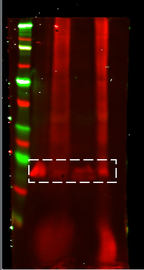
Application: Western BlotSample Tested: mouse embryonic fibroblast cell line, Sample Amount: 40 ugSpecies: MouseVerified Customer | Posted 05/09/2017Testing antibody from Novus for RhoA (seen in white dashed box). Perhaps too much protein (40ug) was loaded. Band location was as expected (~25kDa).MEFs cultured in 10% FBS in DMEM for 48 hours

There are no reviews that match your criteria.
Protocols
Find general support by application which include: protocols, troubleshooting, illustrated assays, videos and webinars.
- Antigen Retrieval Protocol (PIER)
- Antigen Retrieval for Frozen Sections Protocol
- Appropriate Fixation of IHC/ICC Samples
- Cellular Response to Hypoxia Protocols
- Chromogenic IHC Staining of Formalin-Fixed Paraffin-Embedded (FFPE) Tissue Protocol
- Chromogenic Immunohistochemistry Staining of Frozen Tissue
- ClariTSA™ Fluorophore Kits
- Detection & Visualization of Antibody Binding
- Fluorescent IHC Staining of Frozen Tissue Protocol
- Graphic Protocol for Heat-induced Epitope Retrieval
- Graphic Protocol for the Preparation and Fluorescent IHC Staining of Frozen Tissue Sections
- Graphic Protocol for the Preparation and Fluorescent IHC Staining of Paraffin-embedded Tissue Sections
- Graphic Protocol for the Preparation of Gelatin-coated Slides for Histological Tissue Sections
- ICC Cell Smear Protocol for Suspension Cells
- ICC Immunocytochemistry Protocol Videos
- ICC for Adherent Cells
- IHC Sample Preparation (Frozen sections vs Paraffin)
- Immunocytochemistry (ICC) Protocol
- Immunocytochemistry Troubleshooting
- Immunofluorescence of Organoids Embedded in Cultrex Basement Membrane Extract
- Immunofluorescent IHC Staining of Formalin-Fixed Paraffin-Embedded (FFPE) Tissue Protocol
- Immunohistochemistry (IHC) and Immunocytochemistry (ICC) Protocols
- Immunohistochemistry Frozen Troubleshooting
- Immunohistochemistry Paraffin Troubleshooting
- Preparing Samples for IHC/ICC Experiments
- Preventing Non-Specific Staining (Non-Specific Binding)
- Primary Antibody Selection & Optimization
- Protocol for Heat-Induced Epitope Retrieval (HIER)
- Protocol for Making a 4% Formaldehyde Solution in PBS
- Protocol for VisUCyte™ HRP Polymer Detection Reagent
- Protocol for the Fluorescent ICC Staining of Cell Smears - Graphic
- Protocol for the Fluorescent ICC Staining of Cultured Cells on Coverslips - Graphic
- Protocol for the Preparation & Fixation of Cells on Coverslips
- Protocol for the Preparation and Chromogenic IHC Staining of Frozen Tissue Sections
- Protocol for the Preparation and Chromogenic IHC Staining of Frozen Tissue Sections - Graphic
- Protocol for the Preparation and Chromogenic IHC Staining of Paraffin-embedded Tissue Sections
- Protocol for the Preparation and Chromogenic IHC Staining of Paraffin-embedded Tissue Sections - Graphic
- Protocol for the Preparation and Fluorescent ICC Staining of Cells on Coverslips
- Protocol for the Preparation and Fluorescent ICC Staining of Non-adherent Cells
- Protocol for the Preparation and Fluorescent ICC Staining of Stem Cells on Coverslips
- Protocol for the Preparation and Fluorescent IHC Staining of Frozen Tissue Sections
- Protocol for the Preparation and Fluorescent IHC Staining of Paraffin-embedded Tissue Sections
- Protocol for the Preparation of Gelatin-coated Slides for Histological Tissue Sections
- Protocol for the Preparation of a Cell Smear for Non-adherent Cell ICC - Graphic
- R&D Systems Quality Control Western Blot Protocol
- TUNEL and Active Caspase-3 Detection by IHC/ICC Protocol
- The Importance of IHC/ICC Controls
- Troubleshooting Guide: Immunohistochemistry
- Troubleshooting Guide: Western Blot Figures
- Western Blot Conditions
- Western Blot Protocol
- Western Blot Protocol for Cell Lysates
- Western Blot Troubleshooting
- Western Blot Troubleshooting Guide
- View all Protocols, Troubleshooting, Illustrated assays and Webinars
Loading...