
SHARPIN Antibody - BSA Free
Novus Biologicals | Catalog # NBP2-04116

![Immunoprecipitation: SHARPIN Antibody [NBP2-04116]](https://resources.rndsystems.com/images/products/SHARPIN-Antibody-Immunoprecipitation-NBP2-04116-img0001.jpg "Immunoprecipitation: SHARPIN Antibody [NBP2-04116]")
Scientific images may be resized, cropped, or adjusted for brightness or contrast for presentation purposes. These updates are limited to presentation and do not affect the underlying scientific interpretation of the data. Where available, additional source data may be provided upon request.
Key Product Details
Species Reactivity
Validated:
Cited:
Predicted:
Applications
Validated:
Cited:
Label
Antibody Source
Format
Product Specifications
Immunogen
Reactivity Notes
Clonality
Host
Isotype
Scientific Data Images for SHARPIN Antibody - BSA Free
Immunoprecipitation: SHARPIN Antibody [NBP2-04116]
Immunoprecipitation: SHARPIN Antibody [NBP2-04116] - Samples: Whole cell lysate from HeLa (15 and 50 ug for WB; 1 mg for IP, 20% of IP loaded), 293T (T; 50 ug) and Jurkat (J; 50 ug) cells. Antibodies: Affinity purified rabbit anti-SHARPIN antibody NBP2-04116 used for WB at 0.1 ug/ml (A) and 1 ug/ml (B) and used for IP at 6 ug/mg lysate. SHARPIN was also immunoprecipitated by rabbit anti-SHARPIN antibody, which recognizes an upstream epitope. Detection: Chemiluminescence with exposure times of 3 minutes (A) and 30 seconds (B).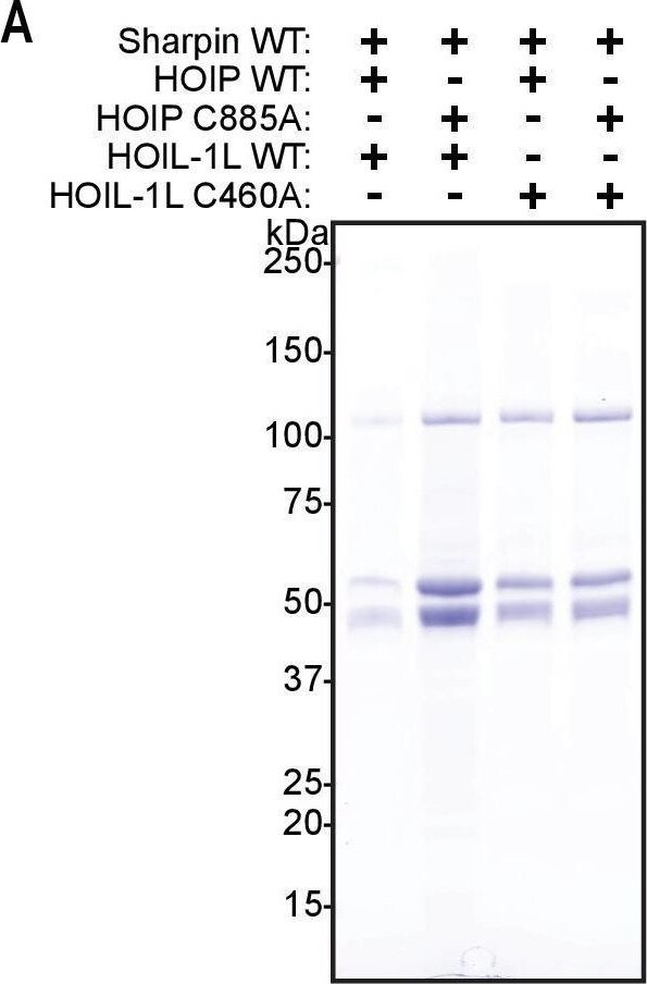
Western Blot: SHARPIN Antibody - BSA Free [NBP2-04116] -
Purification of linear ubiquitin chain assembly complex (LUBAC) containing catalytically inert HOIL-1-interacting protein (HOIP) and heme-oxidized IRP2 ubiquitin ligase 1 (HOIL-1L) proteins.(A) SDS-PAGE analysis of different purified LUBAC. (B) Immunoblot analysis of different purified LUBAC. Image collected and cropped by CiteAb from the following open publication (https://pubmed.ncbi.nlm.nih.gov/34142657), licensed under a CC-BY license. Not internally tested by Novus Biologicals.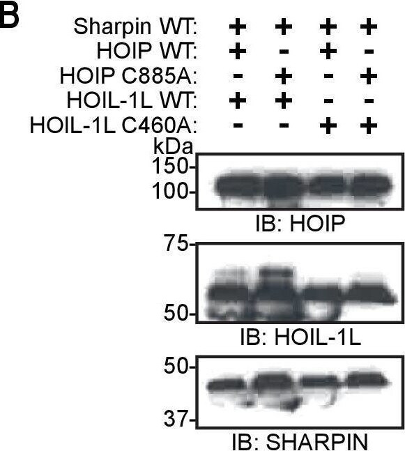
Western Blot: SHARPIN Antibody - BSA Free [NBP2-04116] -
Purification of linear ubiquitin chain assembly complex (LUBAC) containing catalytically inert HOIL-1-interacting protein (HOIP) and heme-oxidized IRP2 ubiquitin ligase 1 (HOIL-1L) proteins.(A) SDS-PAGE analysis of different purified LUBAC. (B) Immunoblot analysis of different purified LUBAC. Image collected and cropped by CiteAb from the following open publication (https://pubmed.ncbi.nlm.nih.gov/34142657), licensed under a CC-BY license. Not internally tested by Novus Biologicals.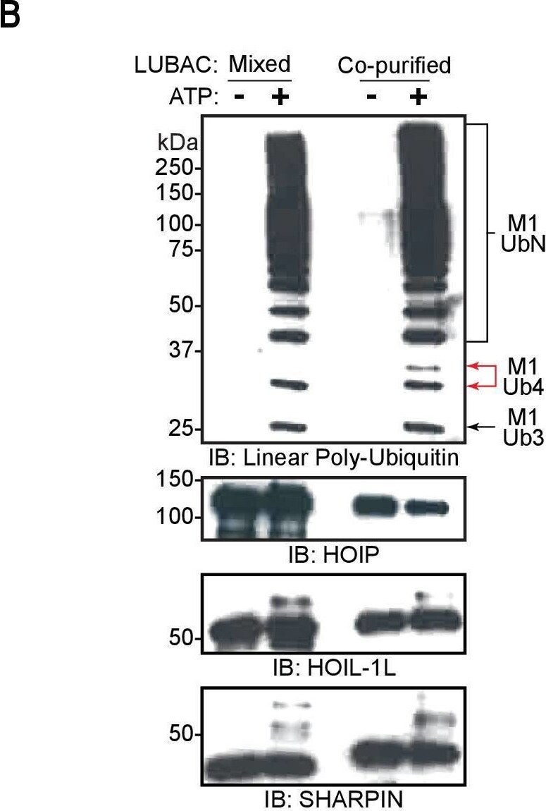
Western Blot: SHARPIN Antibody - BSA Free [NBP2-04116] -
Linear ubiquitin chain assembly complex (LUBAC) assembles heterotypic poly-ubiquitin chains containing M1 and non-Lys linkages in vitro.(A) Time course of co-purified LUBAC in vitro ubiquitin chain assembly reaction. (B) Comparison of in vitro chain assembly between HOIL-1-interacting protein (HOIP), heme-oxidized IRP2 ubiquitin ligase 1 (HOIL-1L), and Shank-associated RH domain-interacting protein (SHARPIN) mixed at 1:1:1 molar ratio versus co-purified LUBAC. (C) LUBAC in vitro chain assembly using different ubiquitin K to R mutants. (D) LUBAC in vitro chain assembly using K0 ubiquitin. (E) Ubiquitin chain restriction (UbiCRest) analysis of poly-ubiquitin chains assembled by LUBAC in vitro. All experiments were performed in triplicate representative results are shown.Anti-linear ubiquitin antibody validation.(A) Detection of mono-ubiquitin and di-ubiquitin of different linkages by anti-linear ubiquitin antibody. (B) Detection of longer Lys48 and Lys63-linked ubiquitin chains by anti-linear ubiquitin antibody.Independently purified HOIL-1-interacting protein (HOIP), heme-oxidized IRP2 ubiquitin ligase 1 (HOIL-1L), and Shank-associated RH domain-interacting protein (SHARPIN) mixed at an equimolar ratio cannot reconstitute the trimeric linear ubiquitin chain assembly complex (LUBAC).Independent mass photometry (MP) measurements and weighted average made from HOIP, HOIL-1L, and SHARPIN mixed to a final concentration of 4 pM of each protein. Image collected and cropped by CiteAb from the following open publication (https://pubmed.ncbi.nlm.nih.gov/34142657), licensed under a CC-BY license. Not internally tested by Novus Biologicals.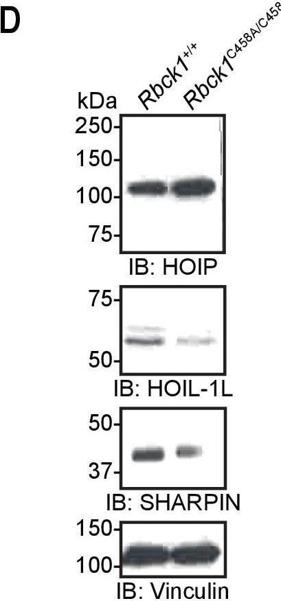
Western Blot: SHARPIN Antibody - BSA Free [NBP2-04116] -
Generation of Hoil-1lC458A/C458A mice.(A) Sequences of genomic DNA around C458 codon, gRNAs, and donor oligonucleotide used to target heme-oxidized IRP2 ubiquitin ligase 1 (HOIL-1L) C458A mutation. (B) Sanger sequencing confirming correct mutations at target sites. (C) Genotyping results of Hoil-1l+/+, Hoil-1l+/C458A, and Hoil-1lC458A/C458A mice. Hpy188III digest of a PCR fragment confirming correct targeting where a silent mutation is inserted. (D) Immunoblot analysis of linear ubiquitin chain assembly complex (LUBAC) component expression in mouse embryonic fibroblasts (MEFs) derived from Hoil-1l+/+ and Hoil-1lC458A/C458A mice. Image collected and cropped by CiteAb from the following open publication (https://pubmed.ncbi.nlm.nih.gov/34142657), licensed under a CC-BY license. Not internally tested by Novus Biologicals.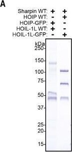
Western Blot: SHARPIN Antibody - BSA Free [NBP2-04116] -
GFP-tagged LUBAC purification.(A) Purification of LUBAC containing GFP-tagged HOIP or HOIL-1L. (B) Mass photometry measurement of GFP-tagged LUBAC showing 27 kDa shift in the monomeric (250kDa) and dimeric (500kDa) mass of the complex. Image collected and cropped by CiteAb from the following open publication (https://pubmed.ncbi.nlm.nih.gov/34142657), licensed under a CC-BY license. Not internally tested by Novus Biologicals.Applications for SHARPIN Antibody - BSA Free
Immunocytochemistry/ Immunofluorescence
Immunohistochemistry
Immunoprecipitation
Western Blot
Formulation, Preparation, and Storage
Purification
Formulation
Format
Preservative
Concentration
Shipping
Stability & Storage
Background: SHARPIN
Long Name
Alternate Names
Gene Symbol
UniProt
Additional SHARPIN Products
Product Documents for SHARPIN Antibody - BSA Free
Certificate of Analysis
To download a Certificate of Analysis, please enter a lot or batch number in the search box below.
Product Specific Notices for SHARPIN Antibody - BSA Free
This product is for research use only and is not approved for use in humans or in clinical diagnosis. Primary Antibodies are guaranteed for 1 year from date of receipt.
Related Research Areas
Citations for SHARPIN Antibody - BSA Free
 Powered by Bioz
Customer Reviews for SHARPIN Antibody - BSA Free
There are currently no reviews for this product. Be the first to review SHARPIN Antibody - BSA Free and earn rewards!
Have you used SHARPIN Antibody - BSA Free?
Submit a review and receive an Amazon gift card!
$25/€18/£15/$25CAN/¥2500 for a review with an image
$10/€7/£6/$10CAN/¥1110 for a review without an image
Submit a review
Protocols
Find general support by application which include: protocols, troubleshooting, illustrated assays, videos and webinars.
- Antigen Retrieval Protocol (PIER)
- Antigen Retrieval for Frozen Sections Protocol
- Appropriate Fixation of IHC/ICC Samples
- Cellular Response to Hypoxia Protocols
- Chromogenic IHC Staining of Formalin-Fixed Paraffin-Embedded (FFPE) Tissue Protocol
- Chromogenic Immunohistochemistry Staining of Frozen Tissue
- ClariTSA™ Fluorophore Kits
- Detection & Visualization of Antibody Binding
- Fluorescent IHC Staining of Frozen Tissue Protocol
- Graphic Protocol for Heat-induced Epitope Retrieval
- Graphic Protocol for the Preparation and Fluorescent IHC Staining of Frozen Tissue Sections
- Graphic Protocol for the Preparation and Fluorescent IHC Staining of Paraffin-embedded Tissue Sections
- Graphic Protocol for the Preparation of Gelatin-coated Slides for Histological Tissue Sections
- ICC Cell Smear Protocol for Suspension Cells
- ICC Immunocytochemistry Protocol Videos
- ICC for Adherent Cells
- IHC Sample Preparation (Frozen sections vs Paraffin)
- Immunocytochemistry (ICC) Protocol
- Immunocytochemistry Troubleshooting
- Immunofluorescence of Organoids Embedded in Cultrex Basement Membrane Extract
- Immunofluorescent IHC Staining of Formalin-Fixed Paraffin-Embedded (FFPE) Tissue Protocol
- Immunohistochemistry (IHC) and Immunocytochemistry (ICC) Protocols
- Immunohistochemistry Frozen Troubleshooting
- Immunohistochemistry Paraffin Troubleshooting
- Immunoprecipitation Protocol
- Preparing Samples for IHC/ICC Experiments
- Preventing Non-Specific Staining (Non-Specific Binding)
- Primary Antibody Selection & Optimization
- Protocol for Heat-Induced Epitope Retrieval (HIER)
- Protocol for Making a 4% Formaldehyde Solution in PBS
- Protocol for VisUCyte™ HRP Polymer Detection Reagent
- Protocol for the Fluorescent ICC Staining of Cell Smears - Graphic
- Protocol for the Fluorescent ICC Staining of Cultured Cells on Coverslips - Graphic
- Protocol for the Preparation & Fixation of Cells on Coverslips
- Protocol for the Preparation and Chromogenic IHC Staining of Frozen Tissue Sections
- Protocol for the Preparation and Chromogenic IHC Staining of Frozen Tissue Sections - Graphic
- Protocol for the Preparation and Chromogenic IHC Staining of Paraffin-embedded Tissue Sections
- Protocol for the Preparation and Chromogenic IHC Staining of Paraffin-embedded Tissue Sections - Graphic
- Protocol for the Preparation and Fluorescent ICC Staining of Cells on Coverslips
- Protocol for the Preparation and Fluorescent ICC Staining of Non-adherent Cells
- Protocol for the Preparation and Fluorescent ICC Staining of Stem Cells on Coverslips
- Protocol for the Preparation and Fluorescent IHC Staining of Frozen Tissue Sections
- Protocol for the Preparation and Fluorescent IHC Staining of Paraffin-embedded Tissue Sections
- Protocol for the Preparation of Gelatin-coated Slides for Histological Tissue Sections
- Protocol for the Preparation of a Cell Smear for Non-adherent Cell ICC - Graphic
- R&D Systems Quality Control Western Blot Protocol
- TUNEL and Active Caspase-3 Detection by IHC/ICC Protocol
- The Importance of IHC/ICC Controls
- Troubleshooting Guide: Immunohistochemistry
- Troubleshooting Guide: Western Blot Figures
- Western Blot Conditions
- Western Blot Protocol
- Western Blot Protocol for Cell Lysates
- Western Blot Troubleshooting
- Western Blot Troubleshooting Guide
- View all Protocols, Troubleshooting, Illustrated assays and Webinars