Human Skin Tissue MicroArray (Cancer)
Novus Biologicals | Catalog # NBP2-30229
Key Product Details
Species
Applications
Product Summary for Human Skin Tissue MicroArray (Cancer)
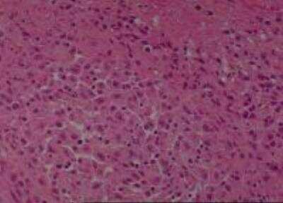
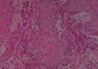
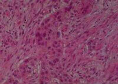
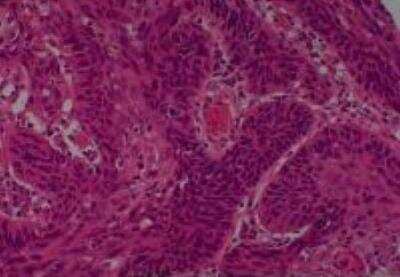
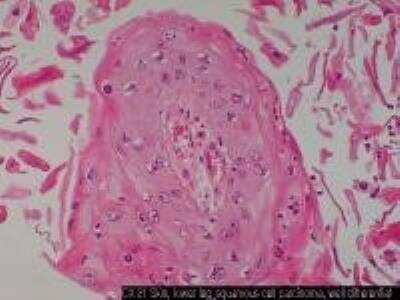
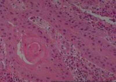
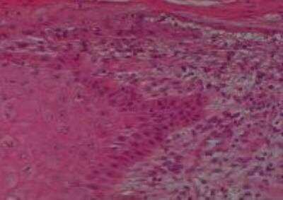
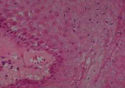
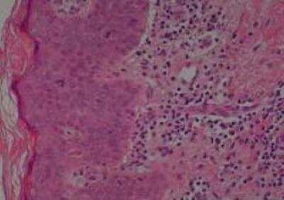
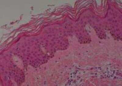
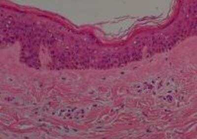
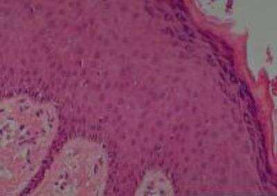
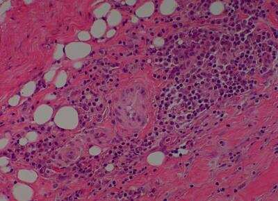
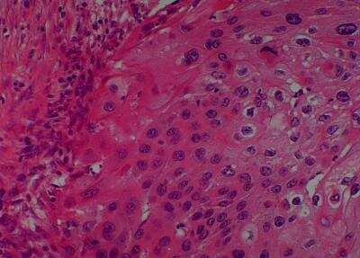
Skin cancer-normal
No. of samples: 49
No. of patients: 47
Core diameter: 2.0 mm
Format: Formalin Fixed
Section thickness: 4 micrometer
Please see manual for tissue information and location.
All tissues are fixed in 10% neutral buffered formalin for 12 to 24 hours, dehydrated with gradient ethanol, cleared with xylene, and embedded in paraffin. Then each slide is tested for immunohistochemistry on multiple antibodies including the p53 protein. Quality control is ensured as every tissue block is collected and arranged by certified pathologists, who used an identical process for their own research.
About 100,000 cells are included in each sample with 2 mm core diameter. Some slides may have less than 60 cores due to sample loss when obtaining multiple sections. Maximum number of missing cores in a slide is less than 10%.
Product Specifications
Application Notes
Type
Tissue Condition
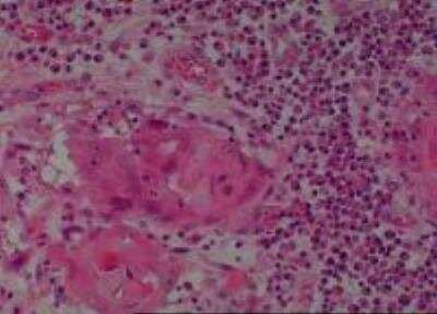
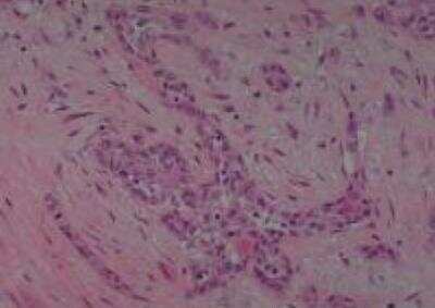
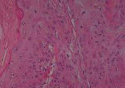
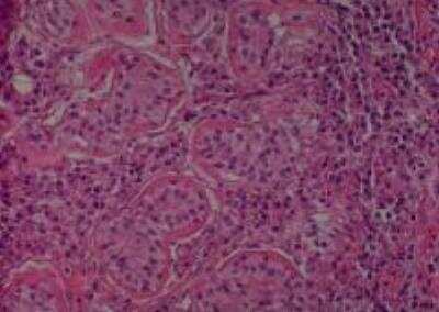
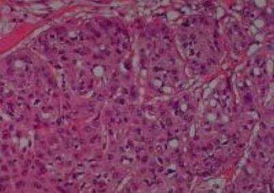
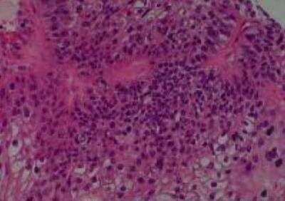
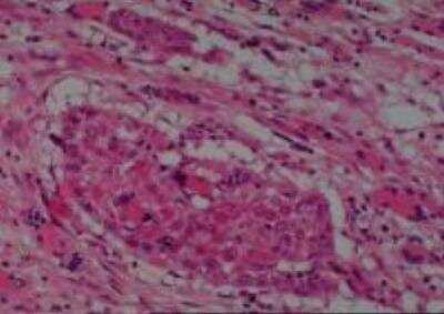
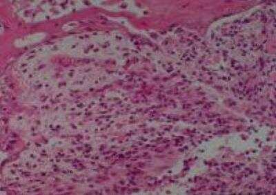
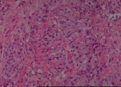
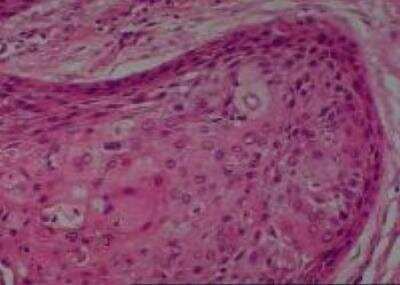
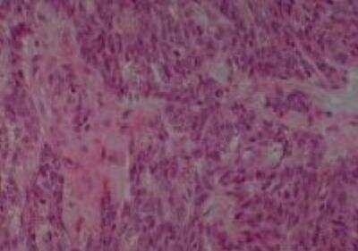
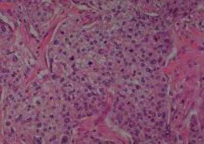
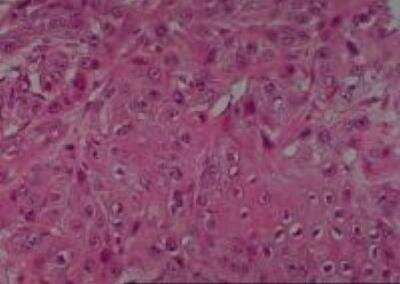
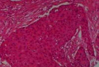
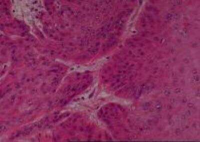
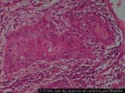
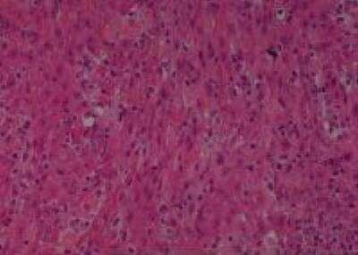
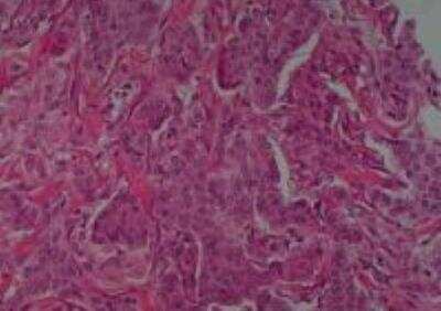
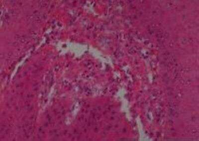
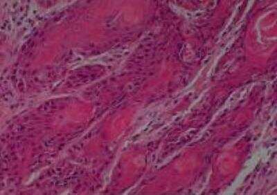
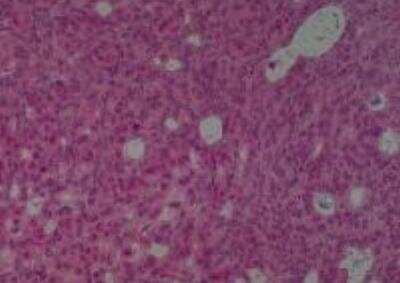
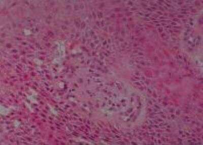
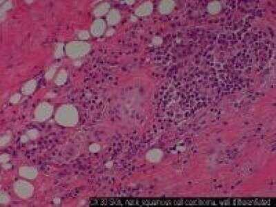
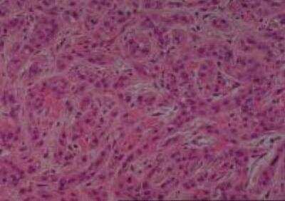
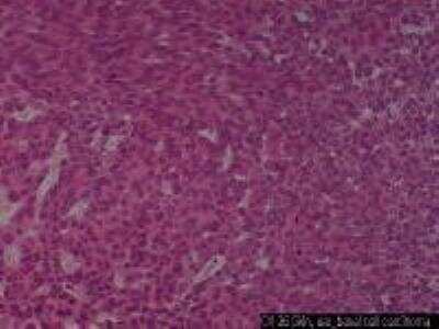
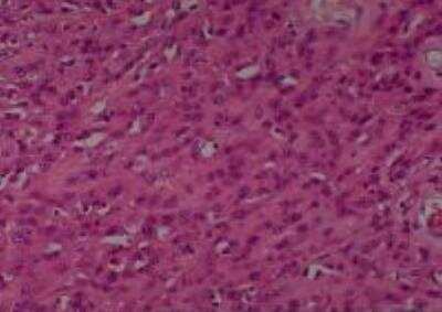
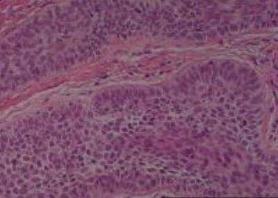
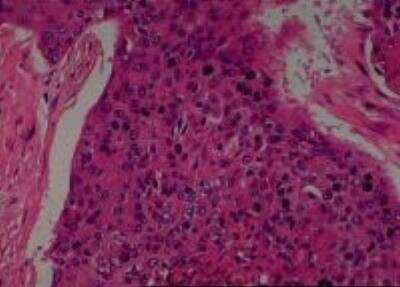
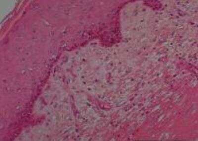
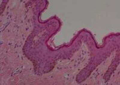
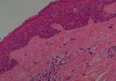
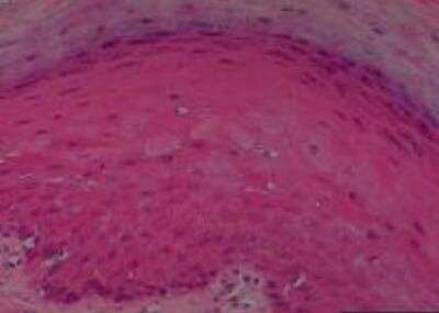
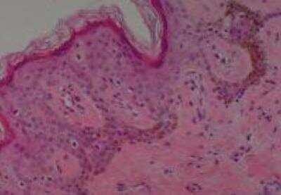
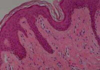
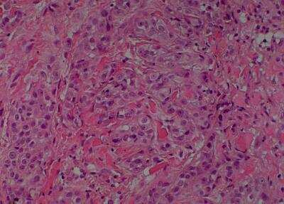
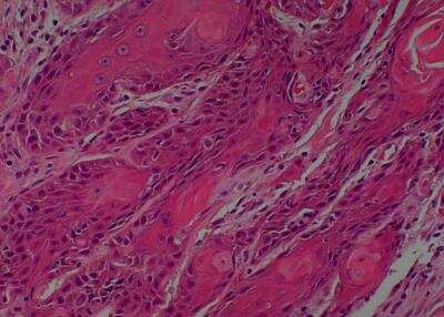
Scientific Data Images for Human Skin Tissue MicroArray (Cancer)
Formulation, Preparation, and Storage
Concentration
Shipping
Storage
Product Documents for Human Skin Tissue MicroArray (Cancer)
Certificate of Analysis
To download a Certificate of Analysis, please enter a lot or batch number in the search box below.
Product Specific Notices for Human Skin Tissue MicroArray (Cancer)
This product is for research use only and is not approved for use in humans or in clinical diagnosis. Tissue Micro Arrays are guaranteed for 1 year from date of receipt.
Citations for Human Skin Tissue MicroArray (Cancer)
 Powered by Bioz
Customer Reviews for Human Skin Tissue MicroArray (Cancer)
There are currently no reviews for this product. Be the first to review Human Skin Tissue MicroArray (Cancer) and earn rewards!
Have you used Human Skin Tissue MicroArray (Cancer)?
Submit a review and receive an Amazon gift card!
$25/€18/£15/$25CAN/¥2500 Yen for a review with an image
$10/€7/£6/$10CAN/¥1110 Yen for a review without an image
Submit a review
Protocols
Find general support by application which include: protocols, troubleshooting, illustrated assays, videos and webinars.
- Antigen Retrieval Protocol (PIER)
- Antigen Retrieval for Frozen Sections Protocol
- Appropriate Fixation of IHC/ICC Samples
- Cellular Response to Hypoxia Protocols
- Chromogenic IHC Staining of Formalin-Fixed Paraffin-Embedded (FFPE) Tissue Protocol
- Chromogenic Immunohistochemistry Staining of Frozen Tissue
- ClariTSA™ Fluorophore Kits
- Detection & Visualization of Antibody Binding
- Fluorescent IHC Staining of Frozen Tissue Protocol
- Graphic Protocol for Heat-induced Epitope Retrieval
- Graphic Protocol for the Preparation and Fluorescent IHC Staining of Frozen Tissue Sections
- Graphic Protocol for the Preparation and Fluorescent IHC Staining of Paraffin-embedded Tissue Sections
- Graphic Protocol for the Preparation of Gelatin-coated Slides for Histological Tissue Sections
- IHC Sample Preparation (Frozen sections vs Paraffin)
- Immunofluorescent IHC Staining of Formalin-Fixed Paraffin-Embedded (FFPE) Tissue Protocol
- Immunohistochemistry (IHC) and Immunocytochemistry (ICC) Protocols
- Immunohistochemistry Frozen Troubleshooting
- Immunohistochemistry Paraffin Troubleshooting
- Preparing Samples for IHC/ICC Experiments
- Preventing Non-Specific Staining (Non-Specific Binding)
- Primary Antibody Selection & Optimization
- Protocol for Heat-Induced Epitope Retrieval (HIER)
- Protocol for Making a 4% Formaldehyde Solution in PBS
- Protocol for VisUCyte™ HRP Polymer Detection Reagent
- Protocol for the Preparation & Fixation of Cells on Coverslips
- Protocol for the Preparation and Chromogenic IHC Staining of Frozen Tissue Sections
- Protocol for the Preparation and Chromogenic IHC Staining of Frozen Tissue Sections - Graphic
- Protocol for the Preparation and Chromogenic IHC Staining of Paraffin-embedded Tissue Sections
- Protocol for the Preparation and Chromogenic IHC Staining of Paraffin-embedded Tissue Sections - Graphic
- Protocol for the Preparation and Fluorescent IHC Staining of Frozen Tissue Sections
- Protocol for the Preparation and Fluorescent IHC Staining of Paraffin-embedded Tissue Sections
- Protocol for the Preparation of Gelatin-coated Slides for Histological Tissue Sections
- TUNEL and Active Caspase-3 Detection by IHC/ICC Protocol
- The Importance of IHC/ICC Controls
- Troubleshooting Guide: Immunohistochemistry
- View all Protocols, Troubleshooting, Illustrated assays and Webinars
FAQs for Human Skin Tissue MicroArray (Cancer)
-
Q: For adult normal frozen skin tissue slides can I choose the donor age/sex?
A: Unfortunately, that is not an option for our commercially available tissue slides.
-
Q: On the web page of NBP2-30229, I can find the patients' information including age, gender, tumor location, diagnosis, pTNM and tissue type. Would you please indicate the meaning of code of tissue type? Also, do you have more detail information about the patients on the tissue microarry?
A: The tissue codes on our information for tissue microarray NBP2-30229 are translated as follows: 1. Normal tissue from a non-cancer patient 2. Normal tissue form a cancer patient, but the cancer involves unrelated organs 3. Normal tissue adjacent to the cancer 4. Benign tumor 5. Tumor of borderline malignancy or uncertain malignant potential 6. Cancer When purchased, the datasheet for the microarray will contain a password and website where you can obtain supplementary data for the donors of the tissues.
-
Q: For adult normal frozen skin tissue slides can I choose the donor age/sex?
A: Unfortunately, that is not an option for our commercially available tissue slides.
-
Q: On the web page of NBP2-30229, I can find the patients' information including age, gender, tumor location, diagnosis, pTNM and tissue type. Would you please indicate the meaning of code of tissue type? Also, do you have more detail information about the patients on the tissue microarry?
A: The tissue codes on our information for tissue microarray NBP2-30229 are translated as follows: 1. Normal tissue from a non-cancer patient 2. Normal tissue form a cancer patient, but the cancer involves unrelated organs 3. Normal tissue adjacent to the cancer 4. Benign tumor 5. Tumor of borderline malignancy or uncertain malignant potential 6. Cancer When purchased, the datasheet for the microarray will contain a password and website where you can obtain supplementary data for the donors of the tissues.