ST2/IL-33R Antibody - BSA Free
Novus Biologicals | Catalog # NBP1-85251
![Western Blot: ST2/IL-33R Antibody [NBP1-85251]](https://resources.rndsystems.com/images/products/ST2-IL-33-R-Antibody-Western-Blot-NBP1-85251-img0008.jpg "Western Blot: ST2/IL-33R Antibody [NBP1-85251]")
Key Product Details
Species Reactivity
Validated:
Cited:
Applications
Validated:
Cited:
Label
Antibody Source
Format
Product Specifications
Immunogen
Clonality
Host
Isotype
Scientific Data Images for ST2/IL-33R Antibody - BSA Free
Western Blot: ST2/IL-33R Antibody [NBP1-85251]
Western Blot: ST2/IL-33 R Antibody [NBP1-85251] - Analysis in human cell line HEL.![Immunohistochemistry-Paraffin: ST2/IL-33R Antibody [NBP1-85251]](https://resources.rndsystems.com/images/products/ST2-IL-33-R-Antibody-Immunohistochemistry-Paraffin-NBP1-85251-img0007.jpg "Immunohistochemistry-Paraffin: ST2/IL-33R Antibody [NBP1-85251]")
Immunohistochemistry-Paraffin: ST2/IL-33R Antibody [NBP1-85251]
Immunohistochemistry-Paraffin: ST2/IL-33 R Antibody [NBP1-85251] - Staining of human placenta shows moderate membranous positivity in trophoblastic cells.![Immunohistochemistry-Paraffin: ST2/IL-33R Antibody [NBP1-85251]](https://resources.rndsystems.com/images/products/ST2-IL-33-R-Antibody-Immunohistochemistry-Paraffin-NBP1-85251-img0004.jpg "Immunohistochemistry-Paraffin: ST2/IL-33R Antibody [NBP1-85251]")
Immunohistochemistry-Paraffin: ST2/IL-33R Antibody [NBP1-85251]
Immunohistochemistry-Paraffin: ST2/IL-33 R Antibody [NBP1-85251] - Staining of human stomach shows moderate membranous positivity in glandular cells.![Immunohistochemistry-Paraffin: ST2/IL-33R Antibody [NBP1-85251]](https://resources.rndsystems.com/images/products/ST2-IL-33-R-Antibody-Immunohistochemistry-Paraffin-NBP1-85251-img0005.jpg "Immunohistochemistry-Paraffin: ST2/IL-33R Antibody [NBP1-85251]")
Immunohistochemistry-Paraffin: ST2/IL-33R Antibody [NBP1-85251]
Immunohistochemistry-Paraffin: ST2/IL-33 R Antibody [NBP1-85251] - Staining of human kidney shows moderate membranous positivity in cells in glomeruli.![Immunohistochemistry-Paraffin: ST2/IL-33R Antibody [NBP1-85251]](https://resources.rndsystems.com/images/products/ST2-IL-33-R-Antibody-Immunohistochemistry-Paraffin-NBP1-85251-img0006.jpg "Immunohistochemistry-Paraffin: ST2/IL-33R Antibody [NBP1-85251]")
Immunohistochemistry-Paraffin: ST2/IL-33R Antibody [NBP1-85251]
Immunohistochemistry-Paraffin: ST2/IL-33 R Antibody [NBP1-85251] - Staining of human lung shows moderate membranous positivity in pneumocytes.Applications for ST2/IL-33R Antibody - BSA Free
Immunohistochemistry
Immunohistochemistry-Paraffin
Western Blot
Reviewed Applications
Read 1 review rated 5 using NBP1-85251 in the following applications:
Formulation, Preparation, and Storage
Purification
Formulation
Format
Preservative
Concentration
Shipping
Stability & Storage
Background: ST2/IL-33R
Long Name
Alternate Names
Gene Symbol
Additional ST2/IL-33R Products
Product Documents for ST2/IL-33R Antibody - BSA Free
Certificate of Analysis
To download a Certificate of Analysis, please enter a lot or batch number in the search box below.
Product Specific Notices for ST2/IL-33R Antibody - BSA Free
This product is for research use only and is not approved for use in humans or in clinical diagnosis. Primary Antibodies are guaranteed for 1 year from date of receipt.
Related Research Areas
Citations for ST2/IL-33R Antibody - BSA Free
 Powered by Bioz
Customer Reviews for ST2/IL-33R Antibody - BSA Free (1)
Have you used ST2/IL-33R Antibody - BSA Free?
Submit a review and receive an Amazon gift card!
$25/€18/£15/$25CAN/¥2500 Yen for a review with an image
$10/€7/£6/$10CAN/¥1110 Yen for a review without an image
Submit a review
Customer Images
-
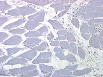
Application: Immunohistochemistry-ParaffinSample Tested: FFPESpecies: HumanVerified Customer | Posted 01/07/2015Antibody did not stain skeletal muscle, as described in the literature

There are no reviews that match your criteria.
Protocols
Find general support by application which include: protocols, troubleshooting, illustrated assays, videos and webinars.
- Antigen Retrieval Protocol (PIER)
- Antigen Retrieval for Frozen Sections Protocol
- Appropriate Fixation of IHC/ICC Samples
- Cellular Response to Hypoxia Protocols
- Chromogenic IHC Staining of Formalin-Fixed Paraffin-Embedded (FFPE) Tissue Protocol
- Chromogenic Immunohistochemistry Staining of Frozen Tissue
- ClariTSA™ Fluorophore Kits
- Detection & Visualization of Antibody Binding
- Fluorescent IHC Staining of Frozen Tissue Protocol
- Graphic Protocol for Heat-induced Epitope Retrieval
- Graphic Protocol for the Preparation and Fluorescent IHC Staining of Frozen Tissue Sections
- Graphic Protocol for the Preparation and Fluorescent IHC Staining of Paraffin-embedded Tissue Sections
- Graphic Protocol for the Preparation of Gelatin-coated Slides for Histological Tissue Sections
- IHC Sample Preparation (Frozen sections vs Paraffin)
- Immunofluorescent IHC Staining of Formalin-Fixed Paraffin-Embedded (FFPE) Tissue Protocol
- Immunohistochemistry (IHC) and Immunocytochemistry (ICC) Protocols
- Immunohistochemistry Frozen Troubleshooting
- Immunohistochemistry Paraffin Troubleshooting
- Preparing Samples for IHC/ICC Experiments
- Preventing Non-Specific Staining (Non-Specific Binding)
- Primary Antibody Selection & Optimization
- Protocol for Heat-Induced Epitope Retrieval (HIER)
- Protocol for Making a 4% Formaldehyde Solution in PBS
- Protocol for VisUCyte™ HRP Polymer Detection Reagent
- Protocol for the Preparation & Fixation of Cells on Coverslips
- Protocol for the Preparation and Chromogenic IHC Staining of Frozen Tissue Sections
- Protocol for the Preparation and Chromogenic IHC Staining of Frozen Tissue Sections - Graphic
- Protocol for the Preparation and Chromogenic IHC Staining of Paraffin-embedded Tissue Sections
- Protocol for the Preparation and Chromogenic IHC Staining of Paraffin-embedded Tissue Sections - Graphic
- Protocol for the Preparation and Fluorescent IHC Staining of Frozen Tissue Sections
- Protocol for the Preparation and Fluorescent IHC Staining of Paraffin-embedded Tissue Sections
- Protocol for the Preparation of Gelatin-coated Slides for Histological Tissue Sections
- R&D Systems Quality Control Western Blot Protocol
- TUNEL and Active Caspase-3 Detection by IHC/ICC Protocol
- The Importance of IHC/ICC Controls
- Troubleshooting Guide: Immunohistochemistry
- Troubleshooting Guide: Western Blot Figures
- Western Blot Conditions
- Western Blot Protocol
- Western Blot Protocol for Cell Lysates
- Western Blot Troubleshooting
- Western Blot Troubleshooting Guide
- View all Protocols, Troubleshooting, Illustrated assays and Webinars
FAQs for ST2/IL-33R Antibody - BSA Free
-
Q: Does this antibody recognize a specific isoform for ST2, particularly ST2 L or not?
A: The immunogen sequence for IL1RL1 Antibody (NBP1-85251) is stated on its datasheet. I have compared this for you with the ST2 isoforms as stated on UniProt. I can tell you that the immunogen matches perfectly with ST2L, and not with ST2S or ST2V. This is because the immunogen sequence corresponds to a region close to the C terminus of ST2L, within the sections missing in ST2S and ST2V (according to UniProt).
Associated Pathways


