10X Citrate Buffer pH 6.0
Novus Biologicals | Catalog # NB900-62075
Loading...
Key Product Details
Species
Human
Applications
Immunohistochemistry
Product Summary for 10X Citrate Buffer pH 6.0
10X citrate buffer at pH 6.0 is 1M.
Loading...
Product Specifications
Specificity
10X Citrate Buffer pH 6.0 for Heat Induced Epitope Recovery
Application Notes
The antigen retrieval protocol is recommended for use in tissues that have been fixed in formalin only. Ensure that the fixed sections are adequately embedded in paraffin. Cut tissue sections to 4-5 microns.
Preparation of Working Solutions
1. The 10X concentrated format should be diluted tenfold with distilled or deionized water.
2. Mix one part of concentrated Antigen Retrieval Solution with nine parts of deionized or distilled water.
3. Shake the bottle vigorously to completely mix the components of the concentrate (the solution may separate into phases over time).
4. Store with cap tightly secured.
Protocol Recommendations
1. Deparaffinize and rehydrate tissue sections.
2. Place slides into 1X retrieval solution in a slide container (e.g. Coplin jar, Tissue-Tek, staining dish or metal slide canister).
3. Retrieve sections under pressure
4. After take-off reagent jar containing slides from pressure cooker, allow the slides to cool for 20 minutes to reach room temperature.
5. Wash slides in deionized water and then with wash buffer. Proceed with immunostaining recommendations in the antibody datasheet.
6. Gently rinse by gradually adding DI water to the solution, then remove slides and rinse with DI water.
Reactivity Notes
Use in Human reported in scientific literature (PMID:33422385)
Formulation, Preparation, and Storage
Formulation
Dilute 1 part buffer in 9 parts distilled water.
Preservative
0.05% Sodium Azide
Concentration
Please see the protocols for proper use of this product. If no protocol is available, contact technical services for assistance.
Shipping
The product is shipped with polar packs. Upon receipt, store it immediately at the temperature recommended below.
Storage
Store at room temperature.
Background: 10X Citrate Buffer pH 6.0
Additional 10X Citrate Buffer pH 6.0 Products
Product Documents for 10X Citrate Buffer pH 6.0
Certificate of Analysis
To download a Certificate of Analysis, please enter a lot or batch number in the search box below.
Product Specific Notices for 10X Citrate Buffer pH 6.0
This product is for research use only and is not approved for use in humans or in clinical diagnosis. Support products are guaranteed for 6 months from date of receipt.
Citations for 10X Citrate Buffer pH 6.0
 Powered by Bioz
Customer Reviews for 10X Citrate Buffer pH 6.0 (1)
5 out of 5
1 Customer Rating
Have you used 10X Citrate Buffer pH 6.0?
Submit a review and receive an Amazon gift card!
$25/€18/£15/$25CAN/¥2500 Yen for a review with an image
$10/€7/£6/$10CAN/¥1110 Yen for a review without an image
Submit a review
Customer Images
Showing
1
-
1 of
1 review
Showing All
Filter By:
-
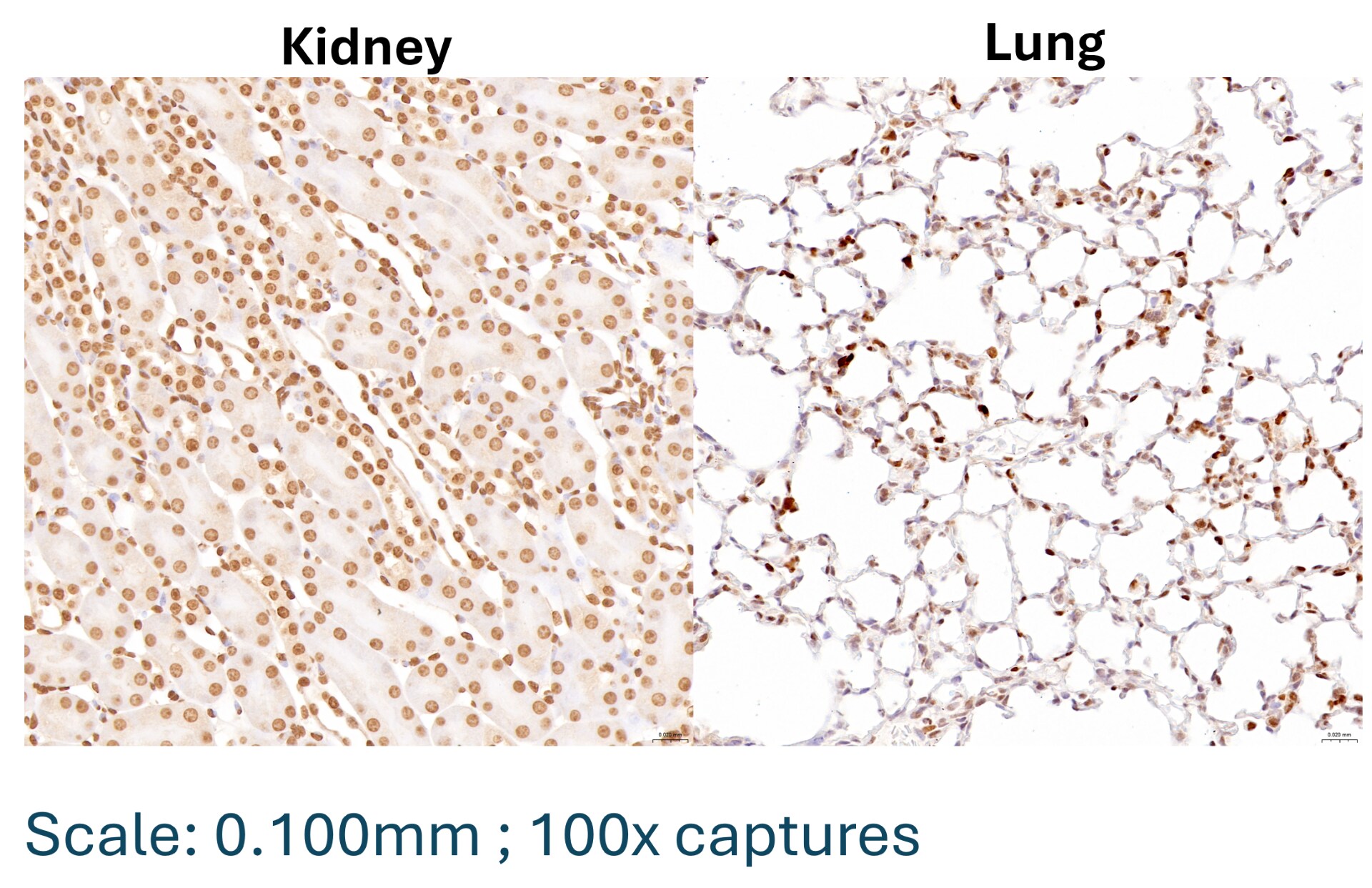
Verified Customer | Posted 05/08/2026Mouse kidney and lung formalin fixed paraffin embedded tissue sections IHC-DAB with heat-induced antigen retrieval conducted using 1x Citrate Buffer pH 6 in the pressure cooker for 15 min.1x Citrate buffer pH 6.0 works excellent for heat-induced antigen retrieval of mouse formalin fixed paraffin embedded tissues using a pressure cooker for 15 min to perform IHC-DAB.

There are no reviews that match your criteria.
Protocols
Find general support by application which include: protocols, troubleshooting, illustrated assays, videos and webinars.
- Antigen Retrieval Protocol (PIER)
- Antigen Retrieval for Frozen Sections Protocol
- Appropriate Fixation of IHC/ICC Samples
- Cellular Response to Hypoxia Protocols
- Chromogenic IHC Staining of Formalin-Fixed Paraffin-Embedded (FFPE) Tissue Protocol
- Chromogenic Immunohistochemistry Staining of Frozen Tissue
- ClariTSA™ Fluorophore Kits
- Detection & Visualization of Antibody Binding
- Fluorescent IHC Staining of Frozen Tissue Protocol
- Graphic Protocol for Heat-induced Epitope Retrieval
- Graphic Protocol for the Preparation and Fluorescent IHC Staining of Frozen Tissue Sections
- Graphic Protocol for the Preparation and Fluorescent IHC Staining of Paraffin-embedded Tissue Sections
- Graphic Protocol for the Preparation of Gelatin-coated Slides for Histological Tissue Sections
- IHC Sample Preparation (Frozen sections vs Paraffin)
- Immunofluorescent IHC Staining of Formalin-Fixed Paraffin-Embedded (FFPE) Tissue Protocol
- Immunohistochemistry (IHC) and Immunocytochemistry (ICC) Protocols
- Immunohistochemistry Frozen Troubleshooting
- Immunohistochemistry Paraffin Troubleshooting
- Preparing Samples for IHC/ICC Experiments
- Preventing Non-Specific Staining (Non-Specific Binding)
- Primary Antibody Selection & Optimization
- Protocol for Heat-Induced Epitope Retrieval (HIER)
- Protocol for Making a 4% Formaldehyde Solution in PBS
- Protocol for VisUCyte™ HRP Polymer Detection Reagent
- Protocol for the Preparation & Fixation of Cells on Coverslips
- Protocol for the Preparation and Chromogenic IHC Staining of Frozen Tissue Sections
- Protocol for the Preparation and Chromogenic IHC Staining of Frozen Tissue Sections - Graphic
- Protocol for the Preparation and Chromogenic IHC Staining of Paraffin-embedded Tissue Sections
- Protocol for the Preparation and Chromogenic IHC Staining of Paraffin-embedded Tissue Sections - Graphic
- Protocol for the Preparation and Fluorescent IHC Staining of Frozen Tissue Sections
- Protocol for the Preparation and Fluorescent IHC Staining of Paraffin-embedded Tissue Sections
- Protocol for the Preparation of Gelatin-coated Slides for Histological Tissue Sections
- TUNEL and Active Caspase-3 Detection by IHC/ICC Protocol
- The Importance of IHC/ICC Controls
- Troubleshooting Guide: Immunohistochemistry
- View all Protocols, Troubleshooting, Illustrated assays and Webinars
Loading...