5-MethylCytosine Antibody (33D3) - BSA Free
Novus Biologicals | Catalog # NBP2-54609
![Immunocytochemistry/ Immunofluorescence: 5-MethylCytosine Antibody (33D3) [NBP2-54609]](https://resources.rndsystems.com/images/products/5-MethylCytosine-Antibody-33D3-Immunocytochemistry-Immunofluorescence-NBP2-54609-img0022.jpg "Immunocytochemistry/ Immunofluorescence: 5-MethylCytosine Antibody (33D3) [NBP2-54609]")
Loading...
Key Product Details
Species Reactivity
Human, Mouse, Rat, Bovine
Applications
Immunofluorescence, Immunocytochemistry/ Immunofluorescence, Methylated DNA Immunoprecipitation, Dot Blot, Surface Plasmon Resonance
Label
Unconjugated
Antibody Source
Monoclonal Mouse IgG1 Clone # 33D3
Format
BSA Free
Loading...
Product Specifications
Immunogen
5-methylcytosine
Clonality
Monoclonal
Host
Mouse
Isotype
IgG1
Scientific Data Images for 5-MethylCytosine Antibody (33D3) - BSA Free
Immunocytochemistry/ Immunofluorescence: 5-MethylCytosine Antibody (33D3) [NBP2-54609]
Immunocytochemistry/Immunofluorescence: 5-MethylCytosine Antibody (33D3) [NBP2-54609] - HeLa cells were stained with the antibody against 5-mC and with DAPI. Cells were fixed with 4% formaldehyde for 10 minutes and blocked with PBS/TX-100 containing 1% BSA. The cells were immunofluorescently labelled with the 5-mC antibody (middle) diluted 1:500 in blocking solution followed by an anti-mouse antibody conjugated to Alexa Fluor 594. The left panel shows staining of the nuclei with DAPI. A merge of the two stainings is shown on the right.![Surface Plasmon Resonance: 5-MethylCytosine Antibody (33D3) [NBP2-54609]](https://resources.rndsystems.com/images/products/5-MethylCytosine-Antibody-33D3-Ovalbumine-Surface-Plasmon-Resonance-NBP2-54609-img0015.jpg "Surface Plasmon Resonance: 5-MethylCytosine Antibody (33D3) [NBP2-54609]")
Surface Plasmon Resonance: 5-MethylCytosine Antibody (33D3) [NBP2-54609]
Surface Plasmon Resonance: 5-MethylCytosine Antibody (33D3) [NBP2-54609] - A synthesized biotin-labeled 5-mC conjugate was immobilized on a sensorchip. Briefly, two flowcells were prepared by sequential injections of EDC/NHS, streptavidin, and ethanolamine. One of these flowcells served as negative control, while biotinylated 5-mC conjugate was injected in the other one, to get an immobilization level of 55 response units (RU). All SPR experiments were performed, using HBS-N buffer (10 mM HEPES,150 mM NaCl, pH 7.4), at a flow rate of 5 uL/min. Interaction assays involved injections of 2 different dilutions of the 5-mC monoclonal antibody over the biotinylated 5-mC conjugate and negative control surfaces, followed by a 3 minute washing step with HBS-N buffer. At the end of each cycle, the streptavidin surface was regenerated by injection of 0.1M citric acid (pH 3). The value of the dissociation constant (kd) obtained by global fitting and 1:1 Langmuir model is 65 nM.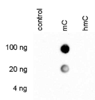
Dot Blot: 5-MethylCytosine Antibody (33D3) [NBP2-54609] - To demonstrate the specificity of the antibody against 5-mC, a Dot blot analysis was performed using hmC, mC and C controls. 100 to 4 ng (equivalent of 5 to 0.2 pmol of C-bases) of the controls were spotted on a membrane. The antibody was used at a dilution of 1:300. Figure shows a high specificity of the antibody for the methylated control.
![Methylated DNA Immunoprecipitation: 5-MethylCytosine Antibody (33D3) [NBP2-54609]](https://resources.rndsystems.com/images/products/5-MethylCytosine-Antibody-33D3-Methylated-DNA-Immunoprecipitation-NBP2-54609-img0023.jpg "Methylated DNA Immunoprecipitation: 5-MethylCytosine Antibody (33D3) [NBP2-54609]")
Methylated DNA Immunoprecipitation: 5-MethylCytosine Antibody (33D3) [NBP2-54609]
Methylated DNA Immunoprecipitation: 5-MethylCytosine Antibody (33D3) [NBP2-54609] - Analysis was performed on 1 ug fragmented human genomic DNA using 0.2 ug of the monoclonal antibody against 5-mC. The fragmented DNA was spiked with controls (methylated DNA (meDNA) as a positive and unmethylated DNA (unDNA) as a negative control) prior to performing the IP. QPCR was performed with primer sets specific for the methylated and unmethylated DNA controls, and for a known methylated (TSH2B) and unmethylated (GAPDH) genomic region. The figure shows the recovery expressed as a percent of input (the relative amount of immunoprecipitated DNA compared to input DNA after qPCR analysis).Applications for 5-MethylCytosine Antibody (33D3) - BSA Free
Application
Recommended Usage
Dot Blot
1:100
Immunocytochemistry/ Immunofluorescence
1:500
Immunofluorescence
1:500
Methylated DNA Immunoprecipitation
0.5 - 1.0 ug/IP
Formulation, Preparation, and Storage
Purification
Protein A purified
Formulation
PBS
Format
BSA Free
Preservative
0.05% Sodium Azide
Concentration
Please see the vial label for concentration. If unlisted please contact technical services.
Shipping
The product is shipped with dry ice or equivalent. Upon receipt, store it immediately at the temperature recommended below.
Stability & Storage
Store at -80C. Avoid freeze-thaw cycles.
Background: 5-MethylCytosine
Alternate Names
5-mC, 5-Methyl Cytosine, Cytosine (5-Methyl)
Additional 5-MethylCytosine Products
Product Documents for 5-MethylCytosine Antibody (33D3) - BSA Free
Certificate of Analysis
To download a Certificate of Analysis, please enter a lot or batch number in the search box below.
Product Specific Notices for 5-MethylCytosine Antibody (33D3) - BSA Free
This product is for research use only and is not approved for use in humans or in clinical diagnosis. Primary Antibodies are guaranteed for 1 year from date of receipt.
Customer Reviews for 5-MethylCytosine Antibody (33D3) - BSA Free
There are currently no reviews for this product. Be the first to review 5-MethylCytosine Antibody (33D3) - BSA Free and earn rewards!
Have you used 5-MethylCytosine Antibody (33D3) - BSA Free?
Submit a review and receive an Amazon gift card!
$25/€18/£15/$25CAN/¥2500 Yen for a review with an image
$10/€7/£6/$10CAN/¥1110 Yen for a review without an image
Submit a review
Protocols
Find general support by application which include: protocols, troubleshooting, illustrated assays, videos and webinars.
- Appropriate Fixation of IHC/ICC Samples
- Cellular Response to Hypoxia Protocols
- ClariTSA™ Fluorophore Kits
- Detection & Visualization of Antibody Binding
- ICC Cell Smear Protocol for Suspension Cells
- ICC Immunocytochemistry Protocol Videos
- ICC for Adherent Cells
- Immunocytochemistry (ICC) Protocol
- Immunocytochemistry Troubleshooting
- Immunofluorescence of Organoids Embedded in Cultrex Basement Membrane Extract
- Immunohistochemistry (IHC) and Immunocytochemistry (ICC) Protocols
- Immunoprecipitation Protocol
- Preparing Samples for IHC/ICC Experiments
- Preventing Non-Specific Staining (Non-Specific Binding)
- Primary Antibody Selection & Optimization
- Protocol for VisUCyte™ HRP Polymer Detection Reagent
- Protocol for the Fluorescent ICC Staining of Cell Smears - Graphic
- Protocol for the Fluorescent ICC Staining of Cultured Cells on Coverslips - Graphic
- Protocol for the Preparation and Fluorescent ICC Staining of Cells on Coverslips
- Protocol for the Preparation and Fluorescent ICC Staining of Non-adherent Cells
- Protocol for the Preparation and Fluorescent ICC Staining of Stem Cells on Coverslips
- Protocol for the Preparation of a Cell Smear for Non-adherent Cell ICC - Graphic
- TUNEL and Active Caspase-3 Detection by IHC/ICC Protocol
- The Importance of IHC/ICC Controls
- View all Protocols, Troubleshooting, Illustrated assays and Webinars
Loading...