ACBD7 Antibody - BSA Free
Novus Biologicals | Catalog # NBP1-56527
![Western Blot: ACBD7 Antibody [NBP1-56527]](https://resources.rndsystems.com/images/products/ACBD7-Antibody-Western-Blot-NBP1-56527-img0002.jpg "Western Blot: ACBD7 Antibody [NBP1-56527]")
Loading...
Key Product Details
Species Reactivity
Validated:
Human, Mouse
Cited:
Mouse
Applications
Validated:
Immunohistochemistry, Immunohistochemistry-Paraffin, Western Blot, Immunocytochemistry/ Immunofluorescence
Cited:
Western Blot, IF/IHC
Label
Unconjugated
Antibody Source
Polyclonal Rabbit IgG
Format
BSA Free
Loading...
Product Specifications
Immunogen
Synthetic peptides corresponding to ACBD7(acyl-Coenzyme A binding domain containing 7) The peptide sequence was selected from the N terminal of ACBD7 (NP_001034933). Peptide sequence MALQADFDRAAEDVRKLKARPDDGELKELYGLYKQAIVGDINIACPGMLD. The peptide sequence for this immunogen was taken from within the described region.
Reactivity Notes
Mouse reactivity reported in scientific literature (PMID: 26880548)
Clonality
Polyclonal
Host
Rabbit
Isotype
IgG
Theoretical MW
10 kDa.
Disclaimer note: The observed molecular weight of the protein may vary from the listed predicted molecular weight due to post translational modifications, post translation cleavages, relative charges, and other experimental factors.
Disclaimer note: The observed molecular weight of the protein may vary from the listed predicted molecular weight due to post translational modifications, post translation cleavages, relative charges, and other experimental factors.
Description
The addition of 50% glycerol is optional for those storing this antibody at -20C and not aliquoting smaller units. However, please note that glycerol may interrupt some downstream antibody applications and should be added with caution.
Scientific Data Images for ACBD7 Antibody - BSA Free
Western Blot: ACBD7 Antibody [NBP1-56527]
Western Blot: ACBD7 Antibody [NBP1-56527] - Human Brain lysate, concentration 0.2-1 ug/ml.![Immunocytochemistry/ Immunofluorescence: ACBD7 Antibody [NBP1-56527]](https://resources.rndsystems.com/images/products/ACBD7-Antibody-Immunofluorescence-NBP1-56527-img0005.jpg "Immunocytochemistry/ Immunofluorescence: ACBD7 Antibody [NBP1-56527]")
Immunocytochemistry/ Immunofluorescence: ACBD7 Antibody [NBP1-56527]
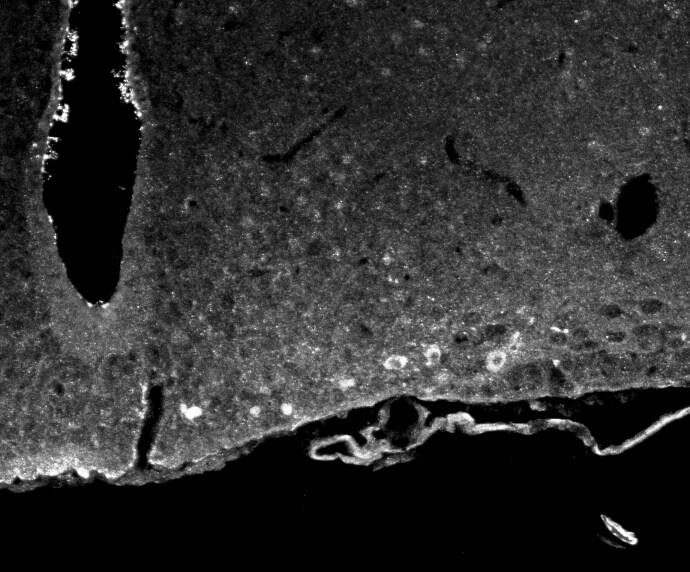
Immunocytochemistry/Immunofluorescence: ACBD7 Antibody [NBP1-56527] - Mouse arcuate nucleus stained with ACBD7 antibody. Image from verified customer review.![Immunohistochemistry-Paraffin: ACBD7 Antibody [NBP1-56527]](https://resources.rndsystems.com/images/products/ACBD7-Antibody-Immunohistochemistry-Paraffin-NBP1-56527-img0003.jpg "Immunohistochemistry-Paraffin: ACBD7 Antibody [NBP1-56527]")
Immunohistochemistry-Paraffin: ACBD7 Antibody [NBP1-56527]
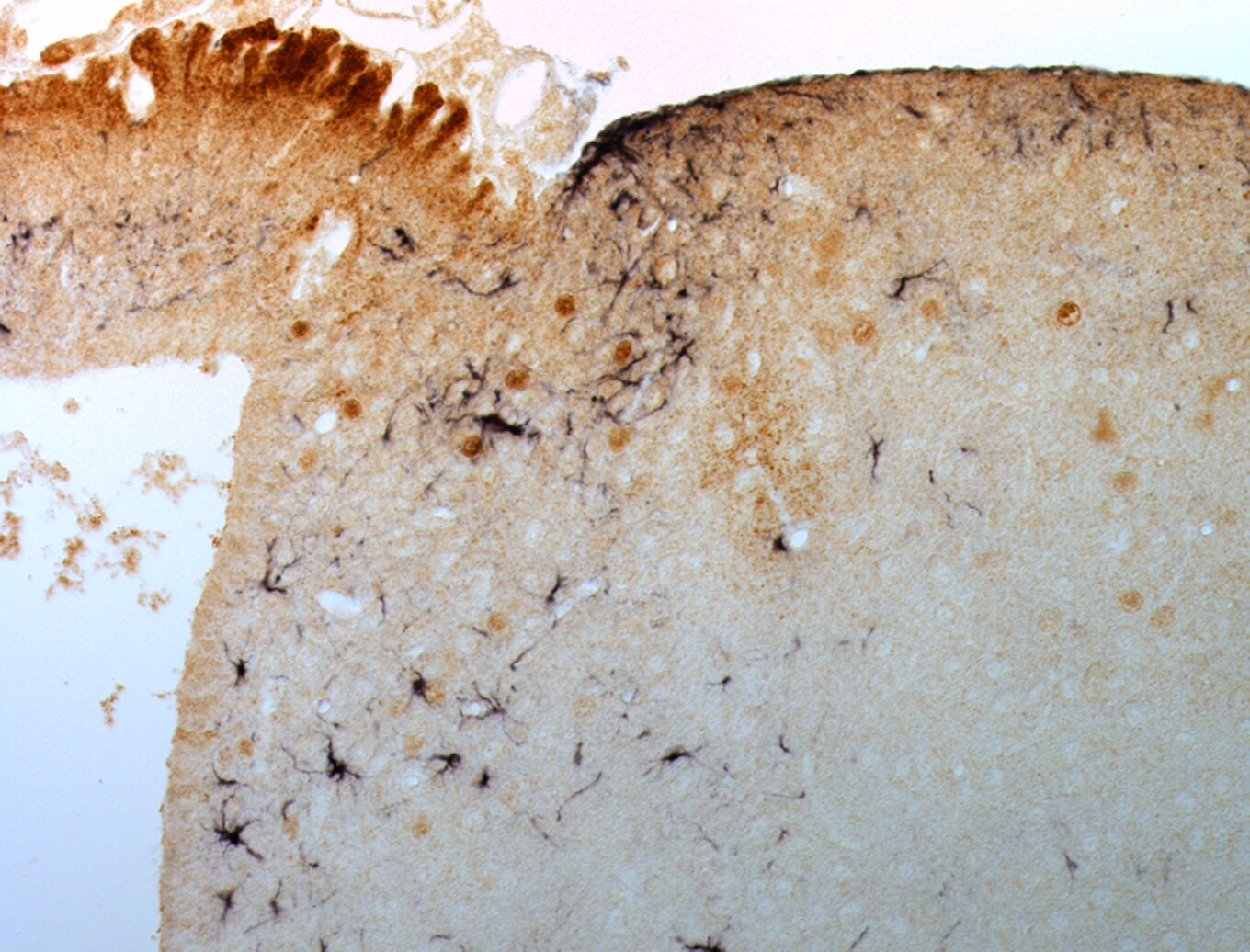
Immunohistochemistry-Paraffin: ACBD7 Antibody [NBP1-56527] - Mouse arcuate nucleus stained with ACBD7 (brown) and GFAP (black) antibodies. Image from verified customer review.![Immunocytochemistry/ Immunofluorescence: ACBD7 Antibody [NBP1-56527]](https://resources.rndsystems.com/images/products/ACBD7-Antibody-Immunocytochemistry-Immunofluorescence-NBP1-56527-img0004.jpg "Immunocytochemistry/ Immunofluorescence: ACBD7 Antibody [NBP1-56527]")
Immunocytochemistry/ Immunofluorescence: ACBD7 Antibody [NBP1-56527]
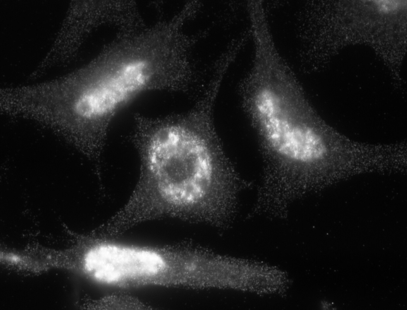
Immunocytochemistry/Immunofluorescence: ACBD7 Antibody [NBP1-56527] - Analysis of ACBD7 in 3T3-L1 cells using ACBD7 antibody. Image from verified customer review.Applications for ACBD7 Antibody - BSA Free
Application
Recommended Usage
Immunocytochemistry/ Immunofluorescence
1:10-1:500
Immunohistochemistry-Paraffin
1:10-1:500
Western Blot
1.0 ug/ml
Application Notes
Use in Immunohistochemistry reported in scientific literature (PMID 26880548).
Reviewed Applications
Read 3 reviews rated 4.7 using NBP1-56527 in the following applications:
Formulation, Preparation, and Storage
Purification
Affinity purified
Formulation
PBS, 2% Sucrose
Format
BSA Free
Preservative
0.09% Sodium Azide
Concentration
0.5 mg/ml
Shipping
The product is shipped with polar packs. Upon receipt, store it immediately at the temperature recommended below.
Stability & Storage
Store at 4C short term. Aliquot and store at -20C long term. Avoid freeze-thaw cycles.
Background: ACBD7
Alternate Names
acyl-CoA binding domain containing 7, acyl-CoA-binding domain-containing protein 7, acyl-Coenzyme A binding domain containing 7, bA455B2.2, FLJ38219, FLJ52263, MGC33893
Entrez Gene IDs
414149 (Human)
Gene Symbol
ACBD7
UniProt
Additional ACBD7 Products
Product Documents for ACBD7 Antibody - BSA Free
Certificate of Analysis
To download a Certificate of Analysis, please enter a lot or batch number in the search box below.
Product Specific Notices for ACBD7 Antibody - BSA Free
This product is for research use only and is not approved for use in humans or in clinical diagnosis. Primary Antibodies are guaranteed for 1 year from date of receipt.
Citations for ACBD7 Antibody - BSA Free
 Powered by Bioz
Customer Reviews for ACBD7 Antibody - BSA Free (3)
4.7 out of 5
3 Customer Ratings
Have you used ACBD7 Antibody - BSA Free?
Submit a review and receive an Amazon gift card!
$25/€18/£15/$25CAN/¥2500 Yen for a review with an image
$10/€7/£6/$10CAN/¥1110 Yen for a review without an image
Submit a review
Customer Images
Showing
1
-
3 of
3 reviews
Showing All
Filter By:
-
Application: ImmunofluorescenceSample Tested:Species: MouseVerified Customer | Posted 03/19/2016Immunofluorescence ACBD7 in the mice arcuate nucleus

-
Application: ImmunocytochemistrySample Tested:Species: MouseVerified Customer | Posted 03/19/2016ACBD7 immunocytochemistry on 3T3-L1
-
Application: ImmunocytochemistrySample Tested:Species: MouseVerified Customer | Posted 03/19/2016DAB IHC-P showing ACBD7 (brown) and GFAP (black) labeling in the mice arcuate nucleus
There are no reviews that match your criteria.
Protocols
Find general support by application which include: protocols, troubleshooting, illustrated assays, videos and webinars.
- Antigen Retrieval Protocol (PIER)
- Antigen Retrieval for Frozen Sections Protocol
- Appropriate Fixation of IHC/ICC Samples
- Cellular Response to Hypoxia Protocols
- Chromogenic IHC Staining of Formalin-Fixed Paraffin-Embedded (FFPE) Tissue Protocol
- Chromogenic Immunohistochemistry Staining of Frozen Tissue
- ClariTSA™ Fluorophore Kits
- Detection & Visualization of Antibody Binding
- Fluorescent IHC Staining of Frozen Tissue Protocol
- Graphic Protocol for Heat-induced Epitope Retrieval
- Graphic Protocol for the Preparation and Fluorescent IHC Staining of Frozen Tissue Sections
- Graphic Protocol for the Preparation and Fluorescent IHC Staining of Paraffin-embedded Tissue Sections
- Graphic Protocol for the Preparation of Gelatin-coated Slides for Histological Tissue Sections
- ICC Cell Smear Protocol for Suspension Cells
- ICC Immunocytochemistry Protocol Videos
- ICC for Adherent Cells
- IHC Sample Preparation (Frozen sections vs Paraffin)
- Immunocytochemistry (ICC) Protocol
- Immunocytochemistry Troubleshooting
- Immunofluorescence of Organoids Embedded in Cultrex Basement Membrane Extract
- Immunofluorescent IHC Staining of Formalin-Fixed Paraffin-Embedded (FFPE) Tissue Protocol
- Immunohistochemistry (IHC) and Immunocytochemistry (ICC) Protocols
- Immunohistochemistry Frozen Troubleshooting
- Immunohistochemistry Paraffin Troubleshooting
- Preparing Samples for IHC/ICC Experiments
- Preventing Non-Specific Staining (Non-Specific Binding)
- Primary Antibody Selection & Optimization
- Protocol for Heat-Induced Epitope Retrieval (HIER)
- Protocol for Making a 4% Formaldehyde Solution in PBS
- Protocol for VisUCyte™ HRP Polymer Detection Reagent
- Protocol for the Fluorescent ICC Staining of Cell Smears - Graphic
- Protocol for the Fluorescent ICC Staining of Cultured Cells on Coverslips - Graphic
- Protocol for the Preparation & Fixation of Cells on Coverslips
- Protocol for the Preparation and Chromogenic IHC Staining of Frozen Tissue Sections
- Protocol for the Preparation and Chromogenic IHC Staining of Frozen Tissue Sections - Graphic
- Protocol for the Preparation and Chromogenic IHC Staining of Paraffin-embedded Tissue Sections
- Protocol for the Preparation and Chromogenic IHC Staining of Paraffin-embedded Tissue Sections - Graphic
- Protocol for the Preparation and Fluorescent ICC Staining of Cells on Coverslips
- Protocol for the Preparation and Fluorescent ICC Staining of Non-adherent Cells
- Protocol for the Preparation and Fluorescent ICC Staining of Stem Cells on Coverslips
- Protocol for the Preparation and Fluorescent IHC Staining of Frozen Tissue Sections
- Protocol for the Preparation and Fluorescent IHC Staining of Paraffin-embedded Tissue Sections
- Protocol for the Preparation of Gelatin-coated Slides for Histological Tissue Sections
- Protocol for the Preparation of a Cell Smear for Non-adherent Cell ICC - Graphic
- R&D Systems Quality Control Western Blot Protocol
- TUNEL and Active Caspase-3 Detection by IHC/ICC Protocol
- The Importance of IHC/ICC Controls
- Troubleshooting Guide: Immunohistochemistry
- Troubleshooting Guide: Western Blot Figures
- Western Blot Conditions
- Western Blot Protocol
- Western Blot Protocol for Cell Lysates
- Western Blot Troubleshooting
- Western Blot Troubleshooting Guide
- View all Protocols, Troubleshooting, Illustrated assays and Webinars
Loading...