ARP10 Antibody - BSA Free
Novus Biologicals | Catalog # NBP1-91682
![Western Blot: ARP10 Antibody [NBP1-91682]](https://resources.rndsystems.com/images/products/ARP10-Antibody-Western-Blot-NBP1-91682-img0005.jpg "Western Blot: ARP10 Antibody [NBP1-91682]")
Loading...
Key Product Details
Species Reactivity
Validated:
Human
Cited:
Human
Applications
Validated:
Immunohistochemistry, Immunohistochemistry-Paraffin, Western Blot, Immunocytochemistry/ Immunofluorescence
Cited:
Western Blot, Immunocytochemistry/ Immunofluorescence, SDS-Page
Label
Unconjugated
Antibody Source
Polyclonal Rabbit IgG
Format
BSA Free
Loading...
Product Specifications
Immunogen
This antibody was developed against Recombinant Protein corresponding to amino acids: CSSCAWELVDFIKAHDHLNLGIFASRLYYHWCKPQQKGLRLLCGSQVPVEVMGFPEFADCWENFVDHEKPLSFNPYKMLEELDKNSRAIKRRLERIKQS
Clonality
Polyclonal
Host
Rabbit
Isotype
IgG
Theoretical MW
24 kDa.
Disclaimer note: The observed molecular weight of the protein may vary from the listed predicted molecular weight due to post translational modifications, post translation cleavages, relative charges, and other experimental factors.
Disclaimer note: The observed molecular weight of the protein may vary from the listed predicted molecular weight due to post translational modifications, post translation cleavages, relative charges, and other experimental factors.
Scientific Data Images for ARP10 Antibody - BSA Free
Western Blot: ARP10 Antibody [NBP1-91682]
ARP10-Antibody-Western-Blot-NBP1-91682-img0005.jpg![Western Blot: ARP10 Antibody [NBP1-91682]](https://resources.rndsystems.com/images/products/ARP10-Antibody-Western-Blot-NBP1-91682-img0004.jpg "Western Blot: ARP10 Antibody [NBP1-91682]")
Western Blot: ARP10 Antibody [NBP1-91682]
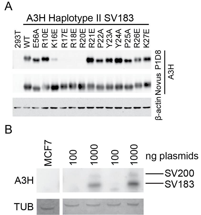
Western Blot: ARP10 Antibody [NBP1-91682] - A) APOBEC3H (A3H) haplotype II SV183 and various mutants expressed in 293T cells. B) The Novus anti-ARP10 antibody detected two splice variants of A3H, SV183 and SV200 in MCF-7 cell lysates. Transient transfection, overnight incubation with primary. WB image submitted by a verified customer review.
Immunohistochemistry-Paraffin: ARP10 Antibody [NBP1-91682] -
Immunohistochemistry-Paraffin: ARP10 Antibody [NBP1-91682] - Staining of human liver shows no positivity in hepatocytes as expected.
Western Blot: ARP10 Antibody [NBP1-91682] -
Western Blot: ARP10 Antibody [NBP1-91682] - Generation & validation of HIV-1 Vif separation-of-function molecular/viral probes.A) A schematic of the Vif protein encoded by each HIV-1 molecular clone showing amino acid differences responsible for the hyper- & hypo-Vif functionality relative to lab-Vif (HIV-1 IIIB/NL4-3) against stable A3H haplotype II. B) Immunoblots showing the expression levels of the indicated A3 proteins stably expressed in SupT11 cells. In this experiment untagged A3H is detected with the mouse monoclonal antibody P3A3-A10. C) HIV-1 spreading infection kinetics for the indicated viruses on A3-expressing SupT11 cells lines described in panel B. The hyper-, lab-, & hypo-Vif isolates spread with similar kinetics on cells expressing a control vector, A3D, A3F, or A3G, but showed clear phenotypic differences on cells expressing low, intermediate (int), & high levels of stable A3H haplotype II. Delta-Vif virus replication was evident in control vector expressing SupT11 cells, delayed in A3D expressing cells, & suppressed under all other conditions (some symbols eclipsed). Image collected & cropped by CiteAb from the following publication (https://dx.plos.org/10.1371/journal.pgen.1004761), licensed under a CC-BY license. Not internally tested by Novus Biologicals.![ARP10 Antibody - BSA Free Immunohistochemistry: ARP10 Antibody - BSA Free [NBP1-91682]](https://resources.rndsystems.com/images/products/nbp1-91682_rabbit-polyclonal-arp10-antibody-29520251646023.jpg "Immunohistochemistry: ARP10 Antibody - BSA Free [NBP1-91682]")
Immunohistochemistry: ARP10 Antibody - BSA Free [NBP1-91682]
Staining of human liver shows no positivity in hepatocytes as expected.![ARP10 Antibody - BSA Free Immunohistochemistry: ARP10 Antibody - BSA Free [NBP1-91682]](https://resources.rndsystems.com/images/products/nbp1-91682_rabbit-polyclonal-arp10-antibody-305202510132921.jpg "Immunohistochemistry: ARP10 Antibody - BSA Free [NBP1-91682]")
Immunohistochemistry: ARP10 Antibody - BSA Free [NBP1-91682]
Staining of human skin shows no positivity in squamous epithelial cells as expected.![ARP10 Antibody - BSA Free Western Blot: ARP10 Antibody - BSA Free [NBP1-91682]](https://resources.rndsystems.com/images/products/nbp1-91682_rabbit-polyclonal-arp10-antibody-295202516173712.jpg "Western Blot: ARP10 Antibody - BSA Free [NBP1-91682]")
Western Blot: ARP10 Antibody - BSA Free [NBP1-91682]
Analysis in human cell line HDLM-2 and human cell line RT-4.![ARP10 Antibody - BSA Free Immunohistochemistry: ARP10 Antibody - BSA Free [NBP1-91682]](https://resources.rndsystems.com/images/products/nbp1-91682_rabbit-polyclonal-arp10-antibody-29520251625723.jpg "Immunohistochemistry: ARP10 Antibody - BSA Free [NBP1-91682]")
Immunohistochemistry: ARP10 Antibody - BSA Free [NBP1-91682]
Staining of human pancreas shows strong cytpoplasmic granular positivity in a subset of exocrine glandular cells.![ARP10 Antibody - BSA Free Immunohistochemistry: ARP10 Antibody - BSA Free [NBP1-91682]](https://resources.rndsystems.com/images/products/nbp1-91682_rabbit-polyclonal-arp10-antibody-295202516153424.jpg "Immunohistochemistry: ARP10 Antibody - BSA Free [NBP1-91682]")
Immunohistochemistry: ARP10 Antibody - BSA Free [NBP1-91682]
Staining of human fallopian tube shows moderate cytoplasmic positivity in glandular cells.Applications for ARP10 Antibody - BSA Free
Application
Recommended Usage
Immunocytochemistry/ Immunofluorescence
Reported in scientific literature (PMID 27650891)
Immunohistochemistry
1:200 - 1:500
Immunohistochemistry-Paraffin
1:200 - 1:500
Western Blot
Reported in scientific literature (PMID: 25411794)/verified customer review.
Application Notes
IHC-Paraffin, HIER pH 6 retrieval method is recommended.
Reviewed Applications
Read 1 review rated 5 using NBP1-91682 in the following applications:
Formulation, Preparation, and Storage
Purification
Affinity purified
Formulation
PBS (pH 7.2) and 40% Glycerol
Format
BSA Free
Preservative
0.02% Sodium Azide
Concentration
Concentrations vary lot to lot. See vial label for concentration. If unlisted please contact technical services.
Shipping
The product is shipped with polar packs. Upon receipt, store it immediately at the temperature recommended below.
Stability & Storage
Store at 4C short term. Aliquot and store at -20C long term. Avoid freeze-thaw cycles.
Background: ARP10
Alternate Names
APOBEC-related protein 10, apolipoprotein B mRNA editing enzyme, catalytic polypeptide-like 3H, ARP-10, ARP10Apolipoprotein B mRNA-editing enzyme catalytic polypeptide-like 3H, DNA dC->dU-editing enzyme APOBEC-3H, EC 3.5.4.-
Entrez Gene IDs
164668 (Human)
Gene Symbol
APOBEC3H
UniProt
Additional ARP10 Products
Product Documents for ARP10 Antibody - BSA Free
Certificate of Analysis
To download a Certificate of Analysis, please enter a lot or batch number in the search box below.
Product Specific Notices for ARP10 Antibody - BSA Free
This product is for research use only and is not approved for use in humans or in clinical diagnosis. Primary Antibodies are guaranteed for 1 year from date of receipt.
Citations for ARP10 Antibody - BSA Free
 Powered by Bioz
Customer Reviews for ARP10 Antibody - BSA Free (1)
5 out of 5
1 Customer Rating
Have you used ARP10 Antibody - BSA Free?
Submit a review and receive an Amazon gift card!
$25/€18/£15/$25CAN/¥2500 Yen for a review with an image
$10/€7/£6/$10CAN/¥1110 Yen for a review without an image
Submit a review
Customer Images
Showing
1
-
1 of
1 review
Showing All
Filter By:
-
Application: Western BlotSample Tested: 293T and MCF-7 cell lysatesSpecies: HumanVerified Customer | Posted 11/17/2017A) APOBEC3H (A3H) haplotype II SV183 and various mutants expressed in 293T cells. B) The Novus anti-ARP10 antibody detected two splice variants of A3H, SV183 and SV200 in MCF-7 cell lysates. Transient transfection, overnight incubation with primary.

There are no reviews that match your criteria.
Protocols
Find general support by application which include: protocols, troubleshooting, illustrated assays, videos and webinars.
- Antigen Retrieval Protocol (PIER)
- Antigen Retrieval for Frozen Sections Protocol
- Appropriate Fixation of IHC/ICC Samples
- Cellular Response to Hypoxia Protocols
- Chromogenic IHC Staining of Formalin-Fixed Paraffin-Embedded (FFPE) Tissue Protocol
- Chromogenic Immunohistochemistry Staining of Frozen Tissue
- ClariTSA™ Fluorophore Kits
- Detection & Visualization of Antibody Binding
- Fluorescent IHC Staining of Frozen Tissue Protocol
- Graphic Protocol for Heat-induced Epitope Retrieval
- Graphic Protocol for the Preparation and Fluorescent IHC Staining of Frozen Tissue Sections
- Graphic Protocol for the Preparation and Fluorescent IHC Staining of Paraffin-embedded Tissue Sections
- Graphic Protocol for the Preparation of Gelatin-coated Slides for Histological Tissue Sections
- ICC Cell Smear Protocol for Suspension Cells
- ICC Immunocytochemistry Protocol Videos
- ICC for Adherent Cells
- IHC Sample Preparation (Frozen sections vs Paraffin)
- Immunocytochemistry (ICC) Protocol
- Immunocytochemistry Troubleshooting
- Immunofluorescence of Organoids Embedded in Cultrex Basement Membrane Extract
- Immunofluorescent IHC Staining of Formalin-Fixed Paraffin-Embedded (FFPE) Tissue Protocol
- Immunohistochemistry (IHC) and Immunocytochemistry (ICC) Protocols
- Immunohistochemistry Frozen Troubleshooting
- Immunohistochemistry Paraffin Troubleshooting
- Preparing Samples for IHC/ICC Experiments
- Preventing Non-Specific Staining (Non-Specific Binding)
- Primary Antibody Selection & Optimization
- Protocol for Heat-Induced Epitope Retrieval (HIER)
- Protocol for Making a 4% Formaldehyde Solution in PBS
- Protocol for VisUCyte™ HRP Polymer Detection Reagent
- Protocol for the Fluorescent ICC Staining of Cell Smears - Graphic
- Protocol for the Fluorescent ICC Staining of Cultured Cells on Coverslips - Graphic
- Protocol for the Preparation & Fixation of Cells on Coverslips
- Protocol for the Preparation and Chromogenic IHC Staining of Frozen Tissue Sections
- Protocol for the Preparation and Chromogenic IHC Staining of Frozen Tissue Sections - Graphic
- Protocol for the Preparation and Chromogenic IHC Staining of Paraffin-embedded Tissue Sections
- Protocol for the Preparation and Chromogenic IHC Staining of Paraffin-embedded Tissue Sections - Graphic
- Protocol for the Preparation and Fluorescent ICC Staining of Cells on Coverslips
- Protocol for the Preparation and Fluorescent ICC Staining of Non-adherent Cells
- Protocol for the Preparation and Fluorescent ICC Staining of Stem Cells on Coverslips
- Protocol for the Preparation and Fluorescent IHC Staining of Frozen Tissue Sections
- Protocol for the Preparation and Fluorescent IHC Staining of Paraffin-embedded Tissue Sections
- Protocol for the Preparation of Gelatin-coated Slides for Histological Tissue Sections
- Protocol for the Preparation of a Cell Smear for Non-adherent Cell ICC - Graphic
- R&D Systems Quality Control Western Blot Protocol
- TUNEL and Active Caspase-3 Detection by IHC/ICC Protocol
- The Importance of IHC/ICC Controls
- Troubleshooting Guide: Immunohistochemistry
- Troubleshooting Guide: Western Blot Figures
- Western Blot Conditions
- Western Blot Protocol
- Western Blot Protocol for Cell Lysates
- Western Blot Troubleshooting
- Western Blot Troubleshooting Guide
- View all Protocols, Troubleshooting, Illustrated assays and Webinars
Loading...