B7-H6 is a transmembrane immune regulatory protein that binds to NKp30 expressed on NK cells. Ligation of NKp30 by B7-H6 induces NK cell activation and target cell cytolysis. B7-H6 is expressed on a wide range of hematopoietic, carcinoma, and melanoma tumor cells. The expression of NKp30 ligands on tumor cells correlates with tumor cell sensitivity to NKp30-dependent cell lysis.
B7-H6 Antibody (17B1.3) - Azide and BSA Free
Novus Biologicals | Catalog # NBP3-09049
Recombinant Monoclonal Antibody
![Flow Cytometry: B7-H6 Antibody (17B1.3) - Azide and BSA Free [NBP3-09049]](https://resources.rndsystems.com/images/products/B7-H6-Antibody-17B1-3-Flow-Cytometry-NBP3-09049-img0001.jpg "Flow Cytometry: B7-H6 Antibody (17B1.3) - Azide and BSA Free [NBP3-09049]")
Loading...
Key Product Details
Species Reactivity
Human
Applications
Flow Cytometry, Immunocytochemistry/ Immunofluorescence
Label
Unconjugated
Antibody Source
Recombinant Monoclonal Mouse IgG1 kappa Clone # 17B1.3
Format
Azide and BSA Free
Loading...
Product Specifications
Immunogen
Immunised mice with extracellular domain of human B7-H6 and selected on the basis of specific reactivity by P815.B7-H6, but not with P815.B7-H1.
Specificity
mAb 17B1.3 specifically recognises B7-H6. 17B1.3 reacted with human tumour cell lines constitutively expressing B7-H6 such as HeLa cells. Fab 17B1.3 does not interfere with the binding of B7-H6 to NKp30, but nevertheless blocks NK cell activation.
Clonality
Monoclonal
Host
Mouse
Isotype
IgG1 kappa
Scientific Data Images for B7-H6 Antibody (17B1.3) - Azide and BSA Free
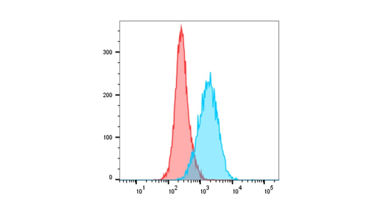
Flow Cytometry: B7-H6 Antibody (17B1.3) - Azide and BSA Free [NBP3-09049]
Flow Cytometry: B7-H6 Antibody (17B1.3) [NBP3-09049] - HEK293T cells were harvested using PBS/1 mM EDTA and stained with B7-H6 antibody and/or secondary antibody. Image from verified customer review.Applications for B7-H6 Antibody (17B1.3) - Azide and BSA Free
Application
Recommended Usage
Flow Cytometry
Optimal dilutions of this antibody should be experimentally determined.
Immunocytochemistry/ Immunofluorescence
Optimal dilutions of this antibody should be experimentally determined.
Application Notes
B7-H6 is not detected in normal human tissues at steady state but is expressed on tumour cells, so tumour cells can be detected by NK cells. This article describes the study of the mechanisms that govern the induction of B7-H6 on primary cells as well as its consequences in vitro and in vivo.
Reviewed Applications
Read 1 review rated 5 using NBP3-09049 in the following applications:
Flow Cytometry Panel Builder
Bio-Techne Knows Flow Cytometry
Save time and reduce costly mistakes by quickly finding compatible reagents using the Panel Builder Tool.
Advanced Features
- Spectra Viewer - Custom analysis of spectra from multiple fluorochromes
- Spillover Popups - Visualize the spectra of individual fluorochromes
- Antigen Density Selector - Match fluorochrome brightness with antigen density

Formulation, Preparation, and Storage
Purification
Protein A purified
Formulation
PBS
Format
Azide and BSA Free
Preservative
0.02% Proclin 300
Concentration
1 mg/ml
Shipping
The product is shipped with polar packs. Upon receipt, store it immediately at the temperature recommended below.
Stability & Storage
Store at 4C short term. Aliquot and store at -20C long term. Avoid freeze-thaw cycles.
Background: B7-H6
Long Name
B7 Homolog 6
Alternate Names
B7H6, DKFZp686I21167, DKFZp686O24166, NCR3LG1
Gene Symbol
NCR3LG1
Additional B7-H6 Products
Product Documents for B7-H6 Antibody (17B1.3) - Azide and BSA Free
Certificate of Analysis
To download a Certificate of Analysis, please enter a lot or batch number in the search box below.
Product Specific Notices for B7-H6 Antibody (17B1.3) - Azide and BSA Free
This product is for research use only and is not approved for use in humans or in clinical diagnosis. Primary Antibodies are guaranteed for 1 year from date of receipt.
Related Research Areas
Customer Reviews for B7-H6 Antibody (17B1.3) - Azide and BSA Free (1)
5 out of 5
1 Customer Rating
Have you used B7-H6 Antibody (17B1.3) - Azide and BSA Free?
Submit a review and receive an Amazon gift card!
$25/€18/£15/$25CAN/¥2500 Yen for a review with an image
$10/€7/£6/$10CAN/¥1110 Yen for a review without an image
Submit a review
Customer Images
Showing
1
-
1 of
1 review
Showing All
Filter By:
-
Application: Flow CytometrySample Tested: HEK293T cellsSpecies: HumanVerified Customer | Posted 10/06/2022Blue: 17B1.3 and Alexa-Fluor 488 conjugated secondary antibody. Red: Only secondary antibody.HEK293T cells were harvested using PBS/1 mM EDTA and stained with 17B1.3 and/or secondary antibody.

There are no reviews that match your criteria.
Protocols
Find general support by application which include: protocols, troubleshooting, illustrated assays, videos and webinars.
- 7-Amino Actinomycin D (7-AAD) Cell Viability Flow Cytometry Protocol
- Appropriate Fixation of IHC/ICC Samples
- Cellular Response to Hypoxia Protocols
- ClariTSA™ Fluorophore Kits
- Detection & Visualization of Antibody Binding
- Extracellular Membrane Flow Cytometry Protocol
- Flow Cytometry Protocol for Cell Surface Markers
- Flow Cytometry Protocol for Staining Membrane Associated Proteins
- Flow Cytometry Staining Protocols
- Flow Cytometry Troubleshooting Guide
- ICC Cell Smear Protocol for Suspension Cells
- ICC Immunocytochemistry Protocol Videos
- ICC for Adherent Cells
- Immunocytochemistry (ICC) Protocol
- Immunocytochemistry Troubleshooting
- Immunofluorescence of Organoids Embedded in Cultrex Basement Membrane Extract
- Immunohistochemistry (IHC) and Immunocytochemistry (ICC) Protocols
- Intracellular Flow Cytometry Protocol Using Alcohol (Methanol)
- Intracellular Flow Cytometry Protocol Using Detergents
- Intracellular Nuclear Staining Flow Cytometry Protocol Using Detergents
- Intracellular Staining Flow Cytometry Protocol Using Alcohol Permeabilization
- Intracellular Staining Flow Cytometry Protocol Using Detergents to Permeabilize Cells
- Preparing Samples for IHC/ICC Experiments
- Preventing Non-Specific Staining (Non-Specific Binding)
- Primary Antibody Selection & Optimization
- Propidium Iodide Cell Viability Flow Cytometry Protocol
- Protocol for Liperfluo
- Protocol for VisUCyte™ HRP Polymer Detection Reagent
- Protocol for the Characterization of Human Th22 Cells
- Protocol for the Characterization of Human Th9 Cells
- Protocol for the Fluorescent ICC Staining of Cell Smears - Graphic
- Protocol for the Fluorescent ICC Staining of Cultured Cells on Coverslips - Graphic
- Protocol for the Preparation and Fluorescent ICC Staining of Cells on Coverslips
- Protocol for the Preparation and Fluorescent ICC Staining of Non-adherent Cells
- Protocol for the Preparation and Fluorescent ICC Staining of Stem Cells on Coverslips
- Protocol for the Preparation of a Cell Smear for Non-adherent Cell ICC - Graphic
- Protocol: Annexin V and PI Staining by Flow Cytometry
- Protocol: Annexin V and PI Staining for Apoptosis by Flow Cytometry
- TUNEL and Active Caspase-3 Detection by IHC/ICC Protocol
- The Importance of IHC/ICC Controls
- Troubleshooting Guide: Fluorokine Flow Cytometry Kits
- View all Protocols, Troubleshooting, Illustrated assays and Webinars
Loading...