![Western Blot: beta Amyloid 42 Peptide [NBP3-18318]](https://resources.rndsystems.com/images/products/beta-Amyloid-42-Peptide-Western-Blot-NBP3-18318-img0001.jpg "Western Blot: beta Amyloid 42 Peptide [NBP3-18318]")
Loading...
Key Product Details
Applications
Immunohistochemistry, Western Blot, Functional Assay, Microscopy
Loading...
Product Specifications
Description
A synthetic peptide treated with HFIP corresponding to human beta Amyloid 42.
Amino Acid Sequence: DAEFRHDSGYEVHHQKLVFFAEDVGSNKGAIIGLMVGGVVIA
Purity
>98%, by HPLC
Predicted Molecular Mass
4.5 kDa.
Disclaimer note: The observed molecular weight of the protein may vary from the listed predicted molecular weight due to post translational modifications, post translation cleavages, relative charges, and other experimental factors.
Disclaimer note: The observed molecular weight of the protein may vary from the listed predicted molecular weight due to post translational modifications, post translation cleavages, relative charges, and other experimental factors.
Protein / Peptide Type
Peptide
Scientific Data Images for beta Amyloid 42 Peptide
Western Blot: beta Amyloid 42 Peptide [NBP3-18318]
Western Blot: beta Amyloid 42 Peptide [NBP3-18318] - Western blot of amyloid beta 1-42 monomers (NBP3-18318, left), oligomers (middle) and fibrils (right) using anti-amyloid beta 6E10 antibody. Amyloid beta constructs at 160 pmol were run on 4-12% Bis-Tris SDS-PAGE, transferred to nitrocellulose in the presence of 0.02% v/v Tween-20, and blotted with 1:1000 mouse 6E10 primary antibody (Biolegend). Oligomers observed under TEM/AFM show distinct dimer/trimer bands as well as a signal from ~37-75 kDa (middle). Fibrils observed under TEM/AFM show a signal greater than 100 kDa and a distinct signal in the stacking gel (right).![In vitro assay: beta Amyloid 42 Peptide [NBP3-18318]](https://resources.rndsystems.com/images/products/beta-Amyloid-42-Peptide-In-vitro-assay-NBP3-18318-img0002.jpg "In vitro assay: beta Amyloid 42 Peptide [NBP3-18318]")
In vitro assay: beta Amyloid 42 Peptide [NBP3-18318]
In vitro assay: beta Amyloid 42 Peptide [NBP3-18318] - Amyloid beta 1-42 oligomers and fibrils show a dose-dependent toxicity to primary rat cortical neurons, but not monomers (NBP3-18318). Survival of rat primary cortical neurons 14 days after treatment with different concentrations of (A) monomers, (B) oligomers or (C) fibrils quantified by MAP2 positive neurons and expressed as a percentage of control. Fibrils and respective vehicle controls were initially sonicated in a Bioruptor. Test conditions were run in the same plate as untreated control and vehicle controls, which consisted of buffer without amyloid beta 1-42 protein. Data expressed as mean +/- s.e.m. (n=6). A global analysis of the data was performed using a one-way ANOVA followed by Dunnetts test; ** p<0.01 stats vs control; ## p<0.01, #### p<0.0001 stats vs vehicle control.![Immunomicroscopy: beta Amyloid 42 Peptide [NBP3-18318]](https://resources.rndsystems.com/images/products/beta%20Amyloid%2042%20Peptide-Immunomicroscopy-NBP3-18318-img0004.jpg "Immunomicroscopy: beta Amyloid 42 Peptide [NBP3-18318]")
Immunomicroscopy: beta Amyloid 42 Peptide [NBP3-18318]
Immunomicroscopy: beta Amyloid 42 Peptide [NBP3-18318] - AFM of amyloid beta 1-42 monomers (NBP3-18318, left), oligomers (middle) and fibrils (right). Atomic force microscopy analysis of 1.0 mg/mL samples diluted to 0.1 mg/mL in dH2O, mounted on freshly cleaved mica, washed, dried and analyzed with tapping mode. Representative images are 2.5 x 2.5 m x-y with a z-range of 10 nm.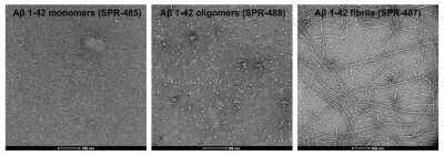
Electron Microscopy: beta Amyloid 42 Peptide [NBP3-18318] - TEM of amyloid beta 1-42 monomers (NBP3-18318, left), oligomers (middle) and fibrils (right). Negative stain transmission electron microscopy images acquired at 80 Kv on carbon coated 400 mesh copper grids using phosphotungstic acid and uranyl acetate stain. Scale bar = 100 nm.
Formulation, Preparation, and Storage
NBP3-18318
| Formulation | Dry powder. |
| Preservative | No Preservative |
| Concentration | Please see the vial label for concentration. If unlisted please contact technical services. |
| Reconstitution | See Reconstitution Instructions PDF for detailed protocol |
| Shipping | The product is shipped with polar packs. Upon receipt, store it immediately at the temperature recommended below. |
| Stability & Storage | Store at -70C. Avoid freeze-thaw cycles. |
Calculators
Background: beta Amyloid 42
Alternate Names
AAA, Abeta, ABPP, AD1, alpha-sAPP, Amyloid-beta precursor protein, APPI, Beta-Amyloid Peptide(1-42), CTFgamma, CVAP, PN2, PreA4
Gene Symbol
APP
Additional beta Amyloid 42 Products
Product Documents for beta Amyloid 42 Peptide
Certificate of Analysis
To download a Certificate of Analysis, please enter a lot or batch number in the search box below.
Product Specific Notices for beta Amyloid 42 Peptide
This product is for research use only and is not approved for use in humans or in clinical diagnosis. This product is guaranteed for 1 year from date of receipt.
Customer Reviews for beta Amyloid 42 Peptide
There are currently no reviews for this product. Be the first to review beta Amyloid 42 Peptide and earn rewards!
Have you used beta Amyloid 42 Peptide?
Submit a review and receive an Amazon gift card!
$25/€18/£15/$25CAN/¥2500 Yen for a review with an image
$10/€7/£6/$10CAN/¥1110 Yen for a review without an image
Submit a review
Protocols
Find general support by application which include: protocols, troubleshooting, illustrated assays, videos and webinars.
- Antigen Retrieval Protocol (PIER)
- Antigen Retrieval for Frozen Sections Protocol
- Appropriate Fixation of IHC/ICC Samples
- Cellular Response to Hypoxia Protocols
- Chromogenic IHC Staining of Formalin-Fixed Paraffin-Embedded (FFPE) Tissue Protocol
- Chromogenic Immunohistochemistry Staining of Frozen Tissue
- ClariTSA™ Fluorophore Kits
- Detection & Visualization of Antibody Binding
- Fluorescent IHC Staining of Frozen Tissue Protocol
- Graphic Protocol for Heat-induced Epitope Retrieval
- Graphic Protocol for the Preparation and Fluorescent IHC Staining of Frozen Tissue Sections
- Graphic Protocol for the Preparation and Fluorescent IHC Staining of Paraffin-embedded Tissue Sections
- Graphic Protocol for the Preparation of Gelatin-coated Slides for Histological Tissue Sections
- IHC Sample Preparation (Frozen sections vs Paraffin)
- Immunofluorescent IHC Staining of Formalin-Fixed Paraffin-Embedded (FFPE) Tissue Protocol
- Immunohistochemistry (IHC) and Immunocytochemistry (ICC) Protocols
- Immunohistochemistry Frozen Troubleshooting
- Immunohistochemistry Paraffin Troubleshooting
- Preparing Samples for IHC/ICC Experiments
- Preventing Non-Specific Staining (Non-Specific Binding)
- Primary Antibody Selection & Optimization
- Protocol for Heat-Induced Epitope Retrieval (HIER)
- Protocol for Making a 4% Formaldehyde Solution in PBS
- Protocol for VisUCyte™ HRP Polymer Detection Reagent
- Protocol for the Preparation & Fixation of Cells on Coverslips
- Protocol for the Preparation and Chromogenic IHC Staining of Frozen Tissue Sections
- Protocol for the Preparation and Chromogenic IHC Staining of Frozen Tissue Sections - Graphic
- Protocol for the Preparation and Chromogenic IHC Staining of Paraffin-embedded Tissue Sections
- Protocol for the Preparation and Chromogenic IHC Staining of Paraffin-embedded Tissue Sections - Graphic
- Protocol for the Preparation and Fluorescent IHC Staining of Frozen Tissue Sections
- Protocol for the Preparation and Fluorescent IHC Staining of Paraffin-embedded Tissue Sections
- Protocol for the Preparation of Gelatin-coated Slides for Histological Tissue Sections
- R&D Systems Quality Control Western Blot Protocol
- TUNEL and Active Caspase-3 Detection by IHC/ICC Protocol
- The Importance of IHC/ICC Controls
- Troubleshooting Guide: Immunohistochemistry
- Troubleshooting Guide: Western Blot Figures
- Western Blot Conditions
- Western Blot Protocol
- Western Blot Protocol for Cell Lysates
- Western Blot Troubleshooting
- Western Blot Troubleshooting Guide
- View all Protocols, Troubleshooting, Illustrated assays and Webinars
Loading...