beta-III Tubulin Antibody - BSA Free
Novus Biologicals | Catalog # NB100-1612
![Western Blot: beta-III Tubulin Antibody [NB100-1612]](https://resources.rndsystems.com/images/products/beta-III-Tubulin-Antibody-Western-Blot-NB100-1612-img0006.jpg "Western Blot: beta-III Tubulin Antibody [NB100-1612]")
Key Product Details
Species Reactivity
Validated:
Cited:
Applications
Validated:
Cited:
Label
Antibody Source
Format
Product Specifications
Immunogen
Marker
Clonality
Host
Isotype
Theoretical MW
Disclaimer note: The observed molecular weight of the protein may vary from the listed predicted molecular weight due to post translational modifications, post translation cleavages, relative charges, and other experimental factors.
Scientific Data Images for beta-III Tubulin Antibody - BSA Free
Western Blot: beta-III Tubulin Antibody [NB100-1612]
beta-III-Tubulin-Antibody-Western-Blot-NB100-1612-img0006.jpg![Immunocytochemistry/ Immunofluorescence: beta-III Tubulin Antibody [NB100-1612]](https://resources.rndsystems.com/images/products/beta-III-Tubulin-Antibody-Immunocytochemistry-Immunofluorescence-NB100-1612-img0011.jpg "Immunocytochemistry/ Immunofluorescence: beta-III Tubulin Antibody [NB100-1612]")
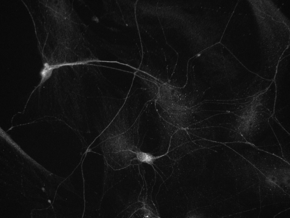
Immunocytochemistry/ Immunofluorescence: beta-III Tubulin Antibody [NB100-1612]
Immunocytochemistry/Immunofluorescence: beta-III Tubulin Antibody [NB100-1612] - Beta-III Tubulin in rat primary motor neurons. Ab dilution 1:1000 in PBST (0.1% Triton X-100) + 10% GS O/N at 4C. ICC/IF image submitted by a verified customer review.![Western Blot: beta-III Tubulin Antibody [NB100-1612]](https://resources.rndsystems.com/images/products/beta-III-Tubulin-Antibody-Western-Blot-NB100-1612-img0007.jpg "Western Blot: beta-III Tubulin Antibody [NB100-1612]")
Western Blot: beta-III Tubulin Antibody [NB100-1612]
beta-III-Tubulin-Antibody-Western-Blot-NB100-1612-img0007.jpg![beta-III Tubulin Antibody Western Blot: beta-III Tubulin Antibody [NB100-1612]](https://resources.rndsystems.com/images/products/antibody/nb100-1612_chicken-polyclonal-beta-iii-tubulin-antibody-245202393341..jpg "Western Blot: beta-III Tubulin Antibody [NB100-1612]")
Western Blot: beta-III Tubulin Antibody [NB100-1612]
beta-III Tubulin Western Blot using homogenates of adult mouse brain (1:3000).
Western Blot: beta-III Tubulin Antibody [NB100-1612] -
Western Blot: beta-III Tubulin Antibody [NB100-1612] - The levels of glial fibrillary acidic protein (GFAP) & S100 beta as astrocytic protein marker proteins in the hippocampal area. (A) Western blot images using GFAP, tubulin beta‐3, & beta ‐actin antibodies to total protein lysate in hippocampal area. The statistical results of internal standardized GFAP/ beta ‐tubulin ratio by beta ‐actin levels relative to young adult wild‐type C57BL/6J. White & black bars indicate the wild‐type C57BL/6J & Tet‐mev‐1 mice, respectively. Data are expressed as mean ± SD; *P < 0.05; **P < 0.01; n = >12 in each group. (B) Western blot images using S100 beta, tubulin beta‐3, & beta ‐actin antibodies to total protein lysate in hippocampal area. The statistical results of internal standardized S100 beta / beta ‐tubulin ratio by beta ‐actin levels relative to young adult wild‐type C57BL/6J. White & black bars indicate the wild‐type C57BL/6J & Tet‐mev‐1 mice, respectively. Data are expressed as mean ± SD; *P < 0.05; **P < 0.01; n = >12 in each group. (C) Micrographs of immunohistochemical analysis on paraffined hippocampal tissue sections using GFAP & S100 beta antibody. Brown cells indicate GFAP‐stained astrocytes. Scale bar = 100 μm. Image collected & cropped by CiteAb from the following publication (https://pubmed.ncbi.nlm.nih.gov/27623715), licensed under a CC-BY license. Not internally tested by Novus Biologicals.Applications for beta-III Tubulin Antibody - BSA Free
Immunocytochemistry/ Immunofluorescence
Immunohistochemistry
Western Blot
Reviewed Applications
Read 2 reviews rated 4.5 using NB100-1612 in the following applications:
Flow Cytometry Panel Builder
Bio-Techne Knows Flow Cytometry
Save time and reduce costly mistakes by quickly finding compatible reagents using the Panel Builder Tool.
Advanced Features
- Spectra Viewer - Custom analysis of spectra from multiple fluorochromes
- Spillover Popups - Visualize the spectra of individual fluorochromes
- Antigen Density Selector - Match fluorochrome brightness with antigen density

Formulation, Preparation, and Storage
Purification
Formulation
Format
Preservative
Concentration
Shipping
Stability & Storage
Background: beta-III Tubulin
Alternate Names
Gene Symbol
UniProt
Additional beta-III Tubulin Products
Product Documents for beta-III Tubulin Antibody - BSA Free
Certificate of Analysis
To download a Certificate of Analysis, please enter a lot or batch number in the search box below.
Product Specific Notices for beta-III Tubulin Antibody - BSA Free
Chicken products cannot be exported to Canada.
Purification Notes
After repeated injections, immune eggs were collected, and the IgY fractions were purified from the yolks. These IgY fractions were then affinity-purified using a peptide column, and the concentrations of the eluates adjusted to 300 ug/ml. Finally, equal volumes of each of the three affinity-purified anti-peptide antibodies were mixed, and the preparation was filter-sterilized.
Storage Notes
Store at 4C in the dark. Under these conditions, the antibodies should have a shelf life of at least 12 months (provided they remain sterile). Do not freeze these antibodies unless you want to store them for longer periods of time. Note, however, that each time an antibody preparation is frozen, about half of its binding activity is lost.
This product is for research use only and is not approved for use in humans or in clinical diagnosis. Primary Antibodies are guaranteed for 1 year from date of receipt.
Related Research Areas
Citations for beta-III Tubulin Antibody - BSA Free
 Powered by Bioz
Customer Reviews for beta-III Tubulin Antibody - BSA Free (2)
Have you used beta-III Tubulin Antibody - BSA Free?
Submit a review and receive an Amazon gift card!
$25/€18/£15/$25CAN/¥2500 Yen for a review with an image
$10/€7/£6/$10CAN/¥1110 Yen for a review without an image
Submit a review
Customer Images
-(025-ml)_NB100-1612_7716.jpg)
-
Application: ImmunocytochemistrySample Tested: Rat Primary Motor NeuronsSpecies: RatVerified Customer | Posted 02/19/2020beta-III Tubulin in rat primary motor neurons. Ab dilution 1:1000 in PBST (0.1% triton) + 10% GS O/N 4ºC.Fixed in PFA 4% + Sacharose 4% in PBS 20 min RT. Ab dilution 1:1000 in PBST (0.1% triton) + 10% GS O/N 4ºC.

-
Application: ImmunocytochemistrySample Tested:Species: RatVerified Customer | Posted 05/19/2014E18 rat primary hippocampal neurons 8 DIV. beta tubulin III
There are no reviews that match your criteria.
Protocols
View specific protocols for beta-III Tubulin Antibody - BSA Free (NB100-1612):
Citrate Buffer Antigen Retrieval Protocol
Background: Formaldehyde fixation (2% or 4%, or as a component of 10% formalin) produces protein cross-links in tissues that tends to interfere with antibody penetration. This seems to be particularly true of paraffin- embedded formaldehyde-fixed tissue. Since chicken IgY antibodies are larger than rabbit or mouse IgG's, "extra steps" may be necessary to compensate for their larger size.
The citrate-based "antigen retrieval" protocol outlined below has been shown to improve chicken IgY antibody penetration into 4% formalde- hyde-fixed paraffin-embedded sections, and can increase the degree and intensity of immunoreactivity and immunostaining.
Reagents (NOTE: You can use either the Sodium Citrate or Citric Acid Buffers in step #3, below)
"Sodium Citrate Buffer" (10mM Sodium Citrate, 0.05% Tween 20, pH 6.0)
Weigh out 2.94 grams of trisodium citrate (dihydrate). Dissolve in approximately 900 mls of deionized, distilled water. Adjust the pH to 6.00 with 1.0 N HCl. Add
0.5 ml of Tween-20. Mix. Bring up the volume to 1.0 litres with water. Store this solution at room temperature for 3 months or at 4C for longer periods.
"Citric Acid Buffer" (10mM Citric Acid, 0.05% Tween 20, pH 6.0)
Weigh out 1.92 grams of citric acid (anhydrous). Dissolve in approximately 900 mls of deionized, distilled water. Adjust the pH to 6.0 with 1.0 N NaOH. Add
0.5 ml of Tween-20. Mix. Bring up the volume to 1.0 litres with water. Store this solution at room temperature for 3 months or at 4C for longer periods.
"Phosphate-Buffered Saline" [PBS, 10 mM Sodium phosphate-buffered (pH 7.2) isotonic (0.9%, w/v) saline solution] PBS Tween (0.05% Tween 20 in PBS)
Ethanol (80%, 90%, 95%, 100%) diluted with water.
Xylene
Procedure (for use with paraffin-embedded sections):
1 Deparaffinize tissue sections in 2 changes of xylene (5 minutes each).
2. Hydrate in 2 changes of 100% ethanol (3 minutes each), 95% ethanol (1 minute), 90% ethanol (1 minute), 80% ethanol (1 minute). Rinse in distilled water.
3. Pre-heat steamer or water bath with staining dish containing either Sodium Citrate Buffer or Citrate Buffer. Wait until temperature reaches 95-100 degrees C.
NOTE: Microwave or pressure cooker can be used as an alternative as a heating source.
4. Immerse slides in the staining dish. Place the lid loosely on the staining dish and incubate for 20-40 minutes (optimal incubation times will vary).
5. Remove the staining dish, and allow it to cool to room temperature (for 20 minutes or so).
6. Rinse sections in PBS Tween twice for 2 minutes each time.
NOTE: The remainder of this protocol is meant to be a suggestion, and can be substituted with your regular immunostaining protocol.
7. Block sections for 30 minutes with Blocking buffer diluted 1:10 with water.
8. Incubate sections with primary antibody at appropriate dilution in antibody dilution buffer overnight at 4 degrees C. Since chicken IgY antibodies are larger than mammalian IgG's, this overnight incubation allows more time for antibody penetration into tissue sections.
9. Rinse sections with PBS Tween 20 twice for 5 minutes each time.
10. Incubate sections with labeled secondary antibody (see NOTE, below) at appropriate dilution (for one hour at room temperature) in a 1:100 dilution of blocking buffer (diluted in PBS).
11. Rinse with PBS Tween 20 for three times for 5 minutes each time.
NOTE: This protocol may use HRP- or fluorescently-labeled secondary antibodies produced in goats or rabbits.
Find general support by application which include: protocols, troubleshooting, illustrated assays, videos and webinars.
- 7-Amino Actinomycin D (7-AAD) Cell Viability Flow Cytometry Protocol
- Antigen Retrieval Protocol (PIER)
- Antigen Retrieval for Frozen Sections Protocol
- Appropriate Fixation of IHC/ICC Samples
- Cellular Response to Hypoxia Protocols
- Chromogenic IHC Staining of Formalin-Fixed Paraffin-Embedded (FFPE) Tissue Protocol
- Chromogenic Immunohistochemistry Staining of Frozen Tissue
- ClariTSA™ Fluorophore Kits
- Detection & Visualization of Antibody Binding
- Extracellular Membrane Flow Cytometry Protocol
- Flow Cytometry Protocol for Cell Surface Markers
- Flow Cytometry Protocol for Staining Membrane Associated Proteins
- Flow Cytometry Staining Protocols
- Flow Cytometry Troubleshooting Guide
- Fluorescent IHC Staining of Frozen Tissue Protocol
- Graphic Protocol for Heat-induced Epitope Retrieval
- Graphic Protocol for the Preparation and Fluorescent IHC Staining of Frozen Tissue Sections
- Graphic Protocol for the Preparation and Fluorescent IHC Staining of Paraffin-embedded Tissue Sections
- Graphic Protocol for the Preparation of Gelatin-coated Slides for Histological Tissue Sections
- ICC Cell Smear Protocol for Suspension Cells
- ICC Immunocytochemistry Protocol Videos
- ICC for Adherent Cells
- IHC Sample Preparation (Frozen sections vs Paraffin)
- Immunocytochemistry (ICC) Protocol
- Immunocytochemistry Troubleshooting
- Immunofluorescence of Organoids Embedded in Cultrex Basement Membrane Extract
- Immunofluorescent IHC Staining of Formalin-Fixed Paraffin-Embedded (FFPE) Tissue Protocol
- Immunohistochemistry (IHC) and Immunocytochemistry (ICC) Protocols
- Immunohistochemistry Frozen Troubleshooting
- Immunohistochemistry Paraffin Troubleshooting
- Intracellular Flow Cytometry Protocol Using Alcohol (Methanol)
- Intracellular Flow Cytometry Protocol Using Detergents
- Intracellular Nuclear Staining Flow Cytometry Protocol Using Detergents
- Intracellular Staining Flow Cytometry Protocol Using Alcohol Permeabilization
- Intracellular Staining Flow Cytometry Protocol Using Detergents to Permeabilize Cells
- Preparing Samples for IHC/ICC Experiments
- Preventing Non-Specific Staining (Non-Specific Binding)
- Primary Antibody Selection & Optimization
- Propidium Iodide Cell Viability Flow Cytometry Protocol
- Protocol for Heat-Induced Epitope Retrieval (HIER)
- Protocol for Liperfluo
- Protocol for Making a 4% Formaldehyde Solution in PBS
- Protocol for VisUCyte™ HRP Polymer Detection Reagent
- Protocol for the Characterization of Human Th22 Cells
- Protocol for the Characterization of Human Th9 Cells
- Protocol for the Fluorescent ICC Staining of Cell Smears - Graphic
- Protocol for the Fluorescent ICC Staining of Cultured Cells on Coverslips - Graphic
- Protocol for the Preparation & Fixation of Cells on Coverslips
- Protocol for the Preparation and Chromogenic IHC Staining of Frozen Tissue Sections
- Protocol for the Preparation and Chromogenic IHC Staining of Frozen Tissue Sections - Graphic
- Protocol for the Preparation and Chromogenic IHC Staining of Paraffin-embedded Tissue Sections
- Protocol for the Preparation and Chromogenic IHC Staining of Paraffin-embedded Tissue Sections - Graphic
- Protocol for the Preparation and Fluorescent ICC Staining of Cells on Coverslips
- Protocol for the Preparation and Fluorescent ICC Staining of Non-adherent Cells
- Protocol for the Preparation and Fluorescent ICC Staining of Stem Cells on Coverslips
- Protocol for the Preparation and Fluorescent IHC Staining of Frozen Tissue Sections
- Protocol for the Preparation and Fluorescent IHC Staining of Paraffin-embedded Tissue Sections
- Protocol for the Preparation of Gelatin-coated Slides for Histological Tissue Sections
- Protocol for the Preparation of a Cell Smear for Non-adherent Cell ICC - Graphic
- Protocol: Annexin V and PI Staining by Flow Cytometry
- Protocol: Annexin V and PI Staining for Apoptosis by Flow Cytometry
- R&D Systems Quality Control Western Blot Protocol
- TUNEL and Active Caspase-3 Detection by IHC/ICC Protocol
- The Importance of IHC/ICC Controls
- Troubleshooting Guide: Fluorokine Flow Cytometry Kits
- Troubleshooting Guide: Immunohistochemistry
- Troubleshooting Guide: Western Blot Figures
- Western Blot Conditions
- Western Blot Protocol
- Western Blot Protocol for Cell Lysates
- Western Blot Troubleshooting
- Western Blot Troubleshooting Guide
- View all Protocols, Troubleshooting, Illustrated assays and Webinars
FAQs for beta-III Tubulin Antibody - BSA Free
-
Q: Do you know if the beta-III tubulin antibody (Cat NB100-1612) has been successfully used for ELISA?
A: Our tubulin beta 3 antibody with catalogue number NB100-1612 has not yet been validated for ELISA.
-
Q: I would like to find out the isotype information for Catalog Number NB100-1612. Is it IgG or IgY? What kind of secondary antibody should I use?
A: Our tubulin beta 3 antibody with catalogue number NB100-1612 is a chicken polyclonal, of the isotype IgY. You can see our relevant secondary antibodies for the detection of chicken IgY at the following link: View Secondary Antibodies
-
Q: Do you know if the beta-III tubulin antibody (Cat NB100-1612) has been successfully used for ELISA?
A: Our tubulin beta 3 antibody with catalogue number NB100-1612 has not yet been validated for ELISA.
-
Q: I would like to find out the isotype information for Catalog Number NB100-1612. Is it IgG or IgY? What kind of secondary antibody should I use?
A: Our tubulin beta 3 antibody with catalogue number NB100-1612 is a chicken polyclonal, of the isotype IgY. You can see our relevant secondary antibodies for the detection of chicken IgY at the following link: View Secondary Antibodies