![Western Blot: BRD2 Antibody [NBP1-30475]](https://resources.rndsystems.com/images/products/BRD2-Antibody-Western-Blot-NBP1-30475-img0024.jpg "Western Blot: BRD2 Antibody [NBP1-30475]")
Loading...
Key Product Details
Validated by
Independent Antibodies
Species Reactivity
Validated:
Human, Mouse
Cited:
Human
Predicted:
Bovine (100%), Canine (100%). Backed by our 100% Guarantee.
Applications
Validated:
Immunohistochemistry, Immunohistochemistry-Paraffin, Western Blot, Immunoprecipitation
Cited:
Western Blot, Proximity Ligation Assay
Label
Unconjugated
Antibody Source
Polyclonal Rabbit IgG
Loading...
Product Specifications
Immunogen
The immunogen for this product maps to a region between residue 675 and 725 of human bromodomain containing 2 using the numbering given in entry NP_005095.1 (GeneID 6046).
Clonality
Polyclonal
Host
Rabbit
Isotype
IgG
Scientific Data Images for BRD2 Antibody
Western Blot: BRD2 Antibody [NBP1-30475]
Western Blot: BRD2 Antibody [NBP1-30475] - Whole cell lysate (50 ug) from NIH 3T3 and TCMK-1 cells prepared using NETN lysis buffer. Antibody: Affinity purified rabbit anti-BRD2 antibody used for WB at 0.1 ug/ml. Detection:Chemiluminescence with an exposure time of 30 seconds.![Western Blot: BRD2 Antibody [NBP1-30475]](https://resources.rndsystems.com/images/products/BRD2-Antibody-Western-Blot-NBP1-30475-img0023.jpg "Western Blot: BRD2 Antibody [NBP1-30475]")
Western Blot: BRD2 Antibody [NBP1-30475]
Western Blot: BRD2 Antibody [NBP1-30475] - Whole cell lysate (50 ug) from HEK293T, HeLa, and Jurkat cells prepared using NETN lysis buffer. Antibody: Affinity purified rabbit anti-BRD2 antibodyused for WB at 0.04 ug/ml. Detection: Chemiluminescence with an exposure time of 75 seconds.
NEIL2 Antibody [NBP3-13112] -
U87-MG whole cell and nuclear extracts (30 μg) were separated by 10% SDS-PAGE, and the membrane was blotted with NEIL2 antibody diluted at 1:1000. The HRP-conjugated anti-rabbit IgG antibody (NBP2-19301) was used to detect the primary antibody.![Immunoprecipitation: BRD2 Antibody [NBP1-30475]](https://resources.rndsystems.com/images/products/BRD2-Antibody-Immunoprecipitation-NBP1-30475-img0022.jpg "Immunoprecipitation: BRD2 Antibody [NBP1-30475]")
Immunoprecipitation: BRD2 Antibody [NBP1-30475]
Immunoprecipitation: BRD2 Antibody [NBP1-30475] - Detection of human BRD2 by western blot of immunoprecipitates. Samples: Whole cell lysate (1.0 mg per IP reaction; 20% of IP loaded) from HEK293T cells prepared using NETN lysis buffer. Antibodies: Affinity purified rabbit anti-BRD2 antibody NBP1-30475 used for IP at 6 ug per reaction. BRD2 was also immunoprecipitated by rabbit anti-BRD2 antibody NBP1-30474 and rabbit anti-BRD2 recombinant monoclonal antibody [BL-167-2A2] (NBP2-76410). For blotting immunoprecipitated BRD2, NBP2-76410 was used at 1:1000. Detection: Chemiluminescence with an exposure time time of 3 minutes.Applications for BRD2 Antibody
Application
Recommended Usage
Immunohistochemistry
1:100 - 1:500
Immunohistochemistry-Paraffin
1:100 - 1:500
Immunoprecipitation
2-10 ug/mg lysate
Western Blot
1:2000 - 1:10000
Application Notes
Epitope retrieval with citrate buffer pH 6.0 is recommended for FFPE tissue sections.
Reviewed Applications
Read 1 review rated 5 using NBP1-30475 in the following applications:
Formulation, Preparation, and Storage
Purification
Immunogen affinity purified
Formulation
TBS, 0.1% BSA
Preservative
0.09% Sodium Azide
Concentration
0.2 mg/ml
Shipping
The product is shipped with polar packs. Upon receipt, store it immediately at the temperature recommended below.
Stability & Storage
Store at 4C. Do not freeze.
Background: BRD2
BRD2 and the other BET proteins have been implicated in a variety of diseases and pathologies. The BET proteins are known drivers of cancer through mutation and over-expression (1). Recently, in studies examining the role of Type 2 diabetes and obesity in breast cancer progression, the BET proteins have been shown to be critical regulators of metabolism and metastasis and are co-activators for the transcription of genes that encode pro-inflammatory cytokines in immune cells infiltrating the breast cancer microenvironment (1). Accordingly, knockdown of Brd2 in mice protected the animals from developing Type 2 diabetes and stopped the inflammatory response typically elicited by obesity (4). BRD2 is also highly expressed in the brain and the gene has been shown to play a role in juvenile myoclonic epilepsy, a common form of epilepsy that typically reveals itself during adolescence (5). In addition to the brain, BRD2 is highly expressed in the bone marrow and consequently its kinase activity has been shown to increase upon cellular proliferation and is significantly elevated in the peripheral blood lymphocytes of lymphoma patients (2, 3).
Research has been done to better understand protein interactions with severe acute respiratory syndrome coronavirus-2 (SARS-CoV-2), the causative agent of the novel coronavirus disease 2019 (COVID-19), as possible targets for drug therapies. It was recently described that that the transmembrane envelope protein (E) of SARS-CoV-2 binds to both BRD2 and BRD4, suggesting that bromodomain inhibitors could be a potential drug target (6). More specifically, the bromodomain inhibitors could be relevant regarding the secondary immune-related consequences that arise from SARS-CoV-2 infection (6). Bromodomain inhibitors are currently the focus of multiple clinical trials as a potential therapeutic in cancer and pulmonary arterial hypertension (6).
References
1. Andrieu, G.P., Shafran, J.S., Deeney, J.T., Bharadwaj, K.R., Rangarajan, A., & Denis, G.V. (2018). BET proteins in abnormal metabolism, inflammation, and the breast cancer microenvironment. J Leukoc Biol. https://doi:10.1002/JLB.5RI0917-380RR
2. BRD2 bromodomain 2 (human), NCBI
3. Taniguchi, Y. (2016). The Bromodomain and Extra-Terminal Domain (BET) Family: Functional Anatomy of BET Paralogous Proteins. Int J Mol Sci. https://doi:10.3390/ijms17111849
4. Wang, F., Deeney, J.T., & Denis, G.V. (2013). Brd2 gene disruption causes "metabolically healthy" obesity: epigenetic and chromatin-based mechanisms that uncouple obesity from type 2 diabetes. Vitam Horm. https://doi:10.1016/B978-0-12-407766-9.00003-1
5. Gilsoul, M., Grisar, T., Delgado-Escueta, A.V., de Nijs, L., & Lakaye, B. (2019). Subtle Brain Developmental Abnormalities in the Pathogenesis of Juvenile Myoclonic Epilepsy. Front Cell Neurosci. https://doi:10.3389/fncel.2019.00433
6. Harrison, C. (2020). Drug researchers pursue new lines of attack against COVID-19. Nat Biotechnol. https://doi.org/10.1038/d41587-020-00013-z
Long Name
Bromodomain Containing 2
Alternate Names
FSRG1, RING3, RNF3
Entrez Gene IDs
6046 (Human)
Gene Symbol
BRD2
UniProt
Additional BRD2 Products
Product Documents for BRD2 Antibody
Certificate of Analysis
To download a Certificate of Analysis, please enter a lot or batch number in the search box below.
Product Specific Notices for BRD2 Antibody
This product is for research use only and is not approved for use in humans or in clinical diagnosis. Primary Antibodies are guaranteed for 1 year from date of receipt.
Citations for BRD2 Antibody
 Powered by Bioz
Customer Reviews for BRD2 Antibody (1)
5 out of 5
1 Customer Rating
Have you used BRD2 Antibody?
Submit a review and receive an Amazon gift card!
$25/€18/£15/$25CAN/¥2500 Yen for a review with an image
$10/€7/£6/$10CAN/¥1110 Yen for a review without an image
Submit a review
Customer Images
Showing
1
-
1 of
1 review
Showing All
Filter By:
-
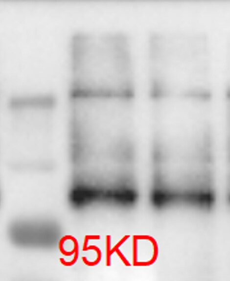
Application: Western BlotSample Tested: human glioma cellsSpecies: HumanVerified Customer | Posted 08/29/2020western blot analysis of glioma whole cell lysates with BRD2 antibody

There are no reviews that match your criteria.
Protocols
Find general support by application which include: protocols, troubleshooting, illustrated assays, videos and webinars.
- Antigen Retrieval Protocol (PIER)
- Antigen Retrieval for Frozen Sections Protocol
- Appropriate Fixation of IHC/ICC Samples
- Cellular Response to Hypoxia Protocols
- Chromogenic IHC Staining of Formalin-Fixed Paraffin-Embedded (FFPE) Tissue Protocol
- Chromogenic Immunohistochemistry Staining of Frozen Tissue
- ClariTSA™ Fluorophore Kits
- Detection & Visualization of Antibody Binding
- Fluorescent IHC Staining of Frozen Tissue Protocol
- Graphic Protocol for Heat-induced Epitope Retrieval
- Graphic Protocol for the Preparation and Fluorescent IHC Staining of Frozen Tissue Sections
- Graphic Protocol for the Preparation and Fluorescent IHC Staining of Paraffin-embedded Tissue Sections
- Graphic Protocol for the Preparation of Gelatin-coated Slides for Histological Tissue Sections
- IHC Sample Preparation (Frozen sections vs Paraffin)
- Immunofluorescent IHC Staining of Formalin-Fixed Paraffin-Embedded (FFPE) Tissue Protocol
- Immunohistochemistry (IHC) and Immunocytochemistry (ICC) Protocols
- Immunohistochemistry Frozen Troubleshooting
- Immunohistochemistry Paraffin Troubleshooting
- Immunoprecipitation Protocol
- Preparing Samples for IHC/ICC Experiments
- Preventing Non-Specific Staining (Non-Specific Binding)
- Primary Antibody Selection & Optimization
- Protocol for Heat-Induced Epitope Retrieval (HIER)
- Protocol for Making a 4% Formaldehyde Solution in PBS
- Protocol for VisUCyte™ HRP Polymer Detection Reagent
- Protocol for the Preparation & Fixation of Cells on Coverslips
- Protocol for the Preparation and Chromogenic IHC Staining of Frozen Tissue Sections
- Protocol for the Preparation and Chromogenic IHC Staining of Frozen Tissue Sections - Graphic
- Protocol for the Preparation and Chromogenic IHC Staining of Paraffin-embedded Tissue Sections
- Protocol for the Preparation and Chromogenic IHC Staining of Paraffin-embedded Tissue Sections - Graphic
- Protocol for the Preparation and Fluorescent IHC Staining of Frozen Tissue Sections
- Protocol for the Preparation and Fluorescent IHC Staining of Paraffin-embedded Tissue Sections
- Protocol for the Preparation of Gelatin-coated Slides for Histological Tissue Sections
- R&D Systems Quality Control Western Blot Protocol
- TUNEL and Active Caspase-3 Detection by IHC/ICC Protocol
- The Importance of IHC/ICC Controls
- Troubleshooting Guide: Immunohistochemistry
- Troubleshooting Guide: Western Blot Figures
- Western Blot Conditions
- Western Blot Protocol
- Western Blot Protocol for Cell Lysates
- Western Blot Troubleshooting
- Western Blot Troubleshooting Guide
- View all Protocols, Troubleshooting, Illustrated assays and Webinars
Loading...