Calnexin Antibody - BSA Free
Novus Biologicals | Catalog # NB100-1965
![Western Blot: Calnexin AntibodyBSA Free [NB100-1965]](https://resources.rndsystems.com/images/products/Calnexin-Antibody-Western-Blot-NB100-1965-img0011.jpg "Western Blot: Calnexin AntibodyBSA Free [NB100-1965]")
Key Product Details
Validated by
Biological Validation
Species Reactivity
Validated:
Human, Mouse, Rat, Porcine, Avian, Bovine, Chicken, Drosophila, Guinea Pig, Rabbit, Sheep, Xenopus, Zebrafish
Cited:
Human, Mouse, Porcine, Bovine, Fish - Danio rerio (Zebrafish), Hamster - Cricetulus (Chinese Hamster)
Predicted:
Canine (100%). Backed by our 100% Guarantee.
Applications
Validated:
Immunohistochemistry, Immunohistochemistry-Paraffin, Western Blot, Flow Cytometry, Immunocytochemistry/ Immunofluorescence, Simple Western, Immunoprecipitation
Cited:
Western Blot, Immunoblotting, Immunocytochemistry/ Immunofluorescence, Simple Western, IF/IHC
Label
Unconjugated
Antibody Source
Polyclonal Rabbit IgG
Format
BSA Free
Loading...
Product Specifications
Immunogen
A synthetic peptide made to an internal region of the canine Calnexin protein (within residues 25-100). [Swiss-Prot P24643]
Reactivity Notes
Quail. Predicted to react with dog based on 100% sequence homology.
Localization
Endoplasmic reticulum
Marker
Endoplasmic Reticulum Membrane Marker
Clonality
Polyclonal
Host
Rabbit
Isotype
IgG
Theoretical MW
97 kDa.
Disclaimer note: The observed molecular weight of the protein may vary from the listed predicted molecular weight due to post translational modifications, post translation cleavages, relative charges, and other experimental factors.
Disclaimer note: The observed molecular weight of the protein may vary from the listed predicted molecular weight due to post translational modifications, post translation cleavages, relative charges, and other experimental factors.
Scientific Data Images for Calnexin Antibody - BSA Free
Western Blot: Calnexin AntibodyBSA Free [NB100-1965]
Western Blot: Calnexin Antibody [NB100-1965] - WB analysis of CANX in cell lysates as noted.![Western Blot: Calnexin AntibodyBSA Free [NB100-1965]](https://resources.rndsystems.com/images/products/Calnexin-Antibody-Western-Blot-NB100-1965-img0015.jpg "Western Blot: Calnexin AntibodyBSA Free [NB100-1965]")
Western Blot: Calnexin AntibodyBSA Free [NB100-1965]
Calnexin-Antibody-Western-Blot-NB100-1965-img0015.jpg![Immunohistochemistry: Calnexin Antibody - BSA Free [NB100-1965]](https://resources.rndsystems.com/images/products/Calnexin-Antibody-Immunohistochemistry-NB100-1965-img0016.jpg "Immunohistochemistry: Calnexin Antibody - BSA Free [NB100-1965]")
![Immunocytochemistry/ Immunofluorescence: Calnexin Antibody - BSA Free [NB100-1965]](https://resources.rndsystems.com/images/products/Calnexin-Antibody-Immunocytochemistry-Immunofluorescence-NB100-1965-img0019.jpg "Immunocytochemistry/ Immunofluorescence: Calnexin Antibody - BSA Free [NB100-1965]")
Immunocytochemistry/ Immunofluorescence: Calnexin Antibody - BSA Free [NB100-1965]
Immunocytochemistry/Immunofluorescence: Calnexin Antibody [NB100-1965] - HeLa cells were fixed in 4% paraformaldehyde for 10 minutes and permeabilized in 0.05% Triton X-100 in PBS for 5 minutes. The cells were incubated with (NB100-1965) at 1 ug/ml overnight at 4C and detected with an anti-rabbit DyLight 488 (Green) at a 1:1000 dilution for 60 minutes. Nuclei were counterstained with DAPI (Blue). Cells were imaged using a 100X objective and digitally deconvolved.![Immunocytochemistry/ Immunofluorescence: Calnexin Antibody - BSA Free [NB100-1965]](https://resources.rndsystems.com/images/products/Calnexin-Antibody-Immunocytochemistry-Immunofluorescence-NB100-1965-img0018.jpg "Immunocytochemistry/ Immunofluorescence: Calnexin Antibody - BSA Free [NB100-1965]")
Immunocytochemistry/ Immunofluorescence: Calnexin Antibody - BSA Free [NB100-1965]
Immunocytochemistry/Immunofluorescence: Calnexin Antibody [NB100-1965] - NIH3T3 cells were fixed in 4% paraformaldehyde for 10 minutes and permeabilized in 0.05% Triton X-100 in PBS for 5 minutes. The cells were incubated with (NB100-1965) at 1 ug/ml overnight at 4C and detected with an anti-rabbit DyLight 488 (Green) at a 1:1000 dilution for 60 minutes. Nuclei were counterstained with DAPI (Blue). Cells were imaged using a 100X objective and digitally deconvolved.![Immunocytochemistry/ Immunofluorescence: Calnexin Antibody - BSA Free [NB100-1965]](https://resources.rndsystems.com/images/products/Calnexin-Antibody-Immunocytochemistry-Immunofluorescence-NB100-1965-img0020.jpg "Immunocytochemistry/ Immunofluorescence: Calnexin Antibody - BSA Free [NB100-1965]")
Immunocytochemistry/ Immunofluorescence: Calnexin Antibody - BSA Free [NB100-1965]
Immunocytochemistry/Immunofluorescence: Calnexin Antibody [NB100-1965] - Rat FR cells were fixed in 4% paraformaldehyde for 10 minutes and permeabilized in 0.05% Triton X-100 in PBS for 5 minutes. The cells were incubated with Calnexin Antibody (NB100-1965) at 1 ug/ml overnight at 4C and detected with an anti-rabbit DyLight 488 (Green) at a 1:1000 dilution for 60 minutes. Nuclei were counterstained with DAPI (Blue). Cells were imaged using a 100X objective and digitally deconvolved.![Western Blot: Calnexin AntibodyBSA Free [NB100-1965]](https://resources.rndsystems.com/images/products/Calnexin-Antibody---BSA-Free-Western-Blot-NB100-1965-img0024.jpg "Western Blot: Calnexin AntibodyBSA Free [NB100-1965]")
Western Blot: Calnexin AntibodyBSA Free [NB100-1965]
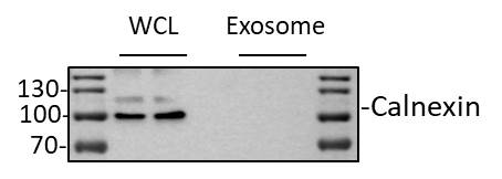
Western Blot: Calnexin Antibody - BSA Free [NB100-1965] - Whole cell lysate or exosome from MDA-MD-231 cells was loaded with 20 ug/lane. 10% SDS-PAGE. Calnexin Antibody (NB100-1965) was used for primary antibody: 1:3000, 4C, overnight. Image from verified customer review.![Flow Cytometry: Calnexin Antibody - BSA Free [NB100-1965]](https://resources.rndsystems.com/images/products/Calnexin-Antibody-Flow-Cytometry-NB100-1965-img0023.jpg "Flow Cytometry: Calnexin Antibody - BSA Free [NB100-1965]")
Flow Cytometry: Calnexin Antibody - BSA Free [NB100-1965]
Flow Cytometry: Calnexin Antibody [NB100-1965] - An intracellular stain was performed on rat FR cells with Calnexin Antibody NB100-1965 (blue) and a matched isotype control NBP2-24891 (orange). Cells were fixed with 4% PFA and then permeabilized with 0.1% saponin. Cells were incubated in an antibody dilution of 1.0 ug/mL for 30 minutes at room temperature, followed by Rabbit IgG (H+L) Cross-Adsorbed Secondary Antibody, Dylight 550 (SA5-10033, Thermo Fisher).![Western Blot: Calnexin AntibodyBSA Free [NB100-1965]](https://resources.rndsystems.com/images/products/Calnexin-Antibody-Western-Blot-NB100-1965-img0008.jpg "Western Blot: Calnexin AntibodyBSA Free [NB100-1965]")
Western Blot: Calnexin AntibodyBSA Free [NB100-1965]
Western Blot: Calnexin Antibody [NB100-1965] - A dilution of 1/2000 used in TBS with 0.05% Tween-20. Bands visualized by ECL using goat anti-rabbit HRP at 1:2000.![Immunohistochemistry: Calnexin Antibody - BSA Free [NB100-1965]](https://resources.rndsystems.com/images/products/Calnexin-Antibody-Immunohistochemistry-NB100-1965-img0010.jpg "Immunohistochemistry: Calnexin Antibody - BSA Free [NB100-1965]")
Immunohistochemistry: Calnexin Antibody - BSA Free [NB100-1965]
Immunohistochemistry: Calnexin Antibody [NB100-1965] - IHC analysis of CANX in mouse prostate using DAB with hematoxylin counterstain.![Flow Cytometry: Calnexin Antibody - BSA Free [NB100-1965]](https://resources.rndsystems.com/images/products/Calnexin-Antibody-Flow-Cytometry-NB100-1965-img0021.jpg "Flow Cytometry: Calnexin Antibody - BSA Free [NB100-1965]")
Flow Cytometry: Calnexin Antibody - BSA Free [NB100-1965]
Flow Cytometry: Calnexin Antibody [NB100-1965] - An intracellular stain was performed on Daudi cells with Calnexin Antibody NB100-1965 (blue) and a matched isotype control NBP2-24891 (orange). Cells were fixed with 4% PFA and then permeabilized with 0.1% saponin. Cells were incubated in an antibody dilution of 1.0 ug/mL for 30 minutes at room temperature, followed by Rabbit IgG (H+L) Cross-Adsorbed Secondary Antibody, Dylight 550 (SA5-10033, Thermo Fisher).![Flow Cytometry: Calnexin Antibody - BSA Free [NB100-1965]](https://resources.rndsystems.com/images/products/Calnexin-Antibody-Flow-Cytometry-NB100-1965-img0022.jpg "Flow Cytometry: Calnexin Antibody - BSA Free [NB100-1965]")
Flow Cytometry: Calnexin Antibody - BSA Free [NB100-1965]
Flow Cytometry: Calnexin Antibody [NB100-1965] - An intracellular stain was performed on NIH3T3 cells with Calnexin Antibody NB100-1965 (blue) and a matched isotype control NBP2-24891 (orange). Cells were fixed with 4% PFA and then permeabilized with 0.1% saponin. Cells were incubated in an antibody dilution of 1.0 ug/mL for 30 minutes at room temperature, followed by Rabbit IgG (H+L) Cross-Adsorbed Secondary Antibody, Dylight 550 (SA5-10033, Thermo Fisher).![Simple Western: Calnexin AntibodyBSA Free [NB100-1965]](https://resources.rndsystems.com/images/products/Calnexin-Antibody-Simple-Western-NB100-1965-img0014.jpg "Simple Western: Calnexin AntibodyBSA Free [NB100-1965]")
Simple Western: Calnexin AntibodyBSA Free [NB100-1965]
Simple Western: Calnexin Antibody [NB100-1965] - Simple Western lane view shows a specific band for Calnexin in 1.0 mg/ml of HeLa lysate. This experiment was performed under reducing conditions using the 12-230 kDa separation system.
Western Blot: Calnexin Antibody - BSA Free [NB100-1965] -
Western Blot: Calnexin Antibody - BSA Free [NB100-1965] - Hepatic apoB levels (A), liver function tests (B), & relative hepatic levels of selected transcripts encoding proteins involved in activation of fibrosis & ER stress (C) in male WT & Tm6sf2−/− mice described in Fig. 3A.A, immunoblotting analysis was performed on liver protein (50 μg) using a rabbit anti-mouse apoB polyclonal antibody (1:1,000; Abcam) & ECL (SuperSignal West Pico Kit, Thermo Scientific). The ECL signal was visualized using a LI-COR imager (Odyssey Fc imager) & analyzed using LI-COR Image Studio software. B, plasma levels of aspartate aminotransferase (AST) & alanine aminotransferase (ALT) in chow-fed male mice. C, RNA levels were detected using quantitative real-time PCR (qRT-PCR), normalized to levels of 36B4 & expressed relative to the levels in the WT animals (n = 5). COL1A1, collagen alpha-1(I) chain; ACTA2, actin, aortic smooth muscle; XBP1s/u, X-box-binding protein 1 spliced/unspliced; ATF4, activating transcription factor 4; EDEM, ER degradation enhancer, mannosidase alpha -like 1; CHOP(DDIT3), C/EBP-homologous protein. Values are means ± S.E. (error bars). *, p < 0.05. AU, arbitrary units. Image collected & cropped by CiteAb from the following publication (https://pubmed.ncbi.nlm.nih.gov/27013658), licensed under a CC-BY license. Not internally tested by Novus Biologicals.![Calnexin Antibody - BSA Free Simple Western: Calnexin Antibody - BSA Free [NB100-1965]](https://resources.rndsystems.com/images/products/antibody/nb100-1965_rabbit-polyclonal-calnexin-antibody-simple-western-218202522394.jpg "Simple Western: Calnexin Antibody - BSA Free [NB100-1965]")
Simple Western: Calnexin Antibody - BSA Free [NB100-1965]
Simple Western: Calnexin Antibody - BSA Free [NB100-1965] - (F) Detection of exosome marker proteins in cell supernatant and nanoparticle-enriched fluid after purification by Exodus using Wes Protein Simple."M" denotes Marker, "1" represents nanoparticle-enriched fluid after purification of MSC-XF supernatant by Exodus, "2" represents nanoparticle-enriched fluid after purification of MSC-EV supernatant by Exodus. Exosome-specific positively expressed proteins include CD9, CD63, CD81, TSG101, and Alix, while Calnexin is a negative exosomal protein marker. Image collected and cropped by CiteAb from the following publication (https://pubmed.ncbi.nlm.nih.gov/40496140) licensed under a CC-BY license.Applications for Calnexin Antibody - BSA Free
Application
Recommended Usage
Flow Cytometry
1-5 ug/ml
Immunocytochemistry/ Immunofluorescence
1-5 ug/ml
Immunohistochemistry
1:40
Immunohistochemistry-Paraffin
1:40
Immunoprecipitation
1:100
Simple Western
1:25
Western Blot
2 ug/ml
Application Notes
This Calnexin antibody is useful for Immunocytochemistry/Immunofluorescence, Immunohistochemistry-paraffin embedded sections, Immunoprecipitation and Western Blot. In Western blot a band is observed approx. 97 kDa. Prior to immunostaining paraffin tissues, antigen retrieval with sodium citrate buffer (pH 6.0) is recommended.
In Simple Western only 10 - 15 uL of the recommended dilution is used per data point.
See Simple Western Antibody Database for Simple Western validation: Tested in HOK-16B, NOK-SI, UM-SCC-104, UM-SCC-105, UM-SCC-118, UM-SCC-17A, UM-SCC-38, UM-SCC-47, UM-SCC-92, UPCI-SCC-152; separated by Size; apparent MW was 115 kDa. Separated by Size-Wes, Sally Sue/Peggy Sue.
Reviewed Applications
Read 4 reviews rated 4.3 using NB100-1965 in the following applications:
Flow Cytometry Panel Builder
Bio-Techne Knows Flow Cytometry
Save time and reduce costly mistakes by quickly finding compatible reagents using the Panel Builder Tool.
Advanced Features
- Spectra Viewer - Custom analysis of spectra from multiple fluorochromes
- Spillover Popups - Visualize the spectra of individual fluorochromes
- Antigen Density Selector - Match fluorochrome brightness with antigen density

Formulation, Preparation, and Storage
Purification
Immunogen affinity purified
Formulation
PBS
Format
BSA Free
Preservative
0.02% Sodium Azide
Concentration
1 mg/ml
Shipping
The product is shipped with polar packs. Upon receipt, store it immediately at the temperature recommended below.
Stability & Storage
Store at 4C short term. Aliquot and store at -20C long term. Avoid freeze-thaw cycles.
Background: Calnexin
Alternate Names
calnexin, CNX, IP90FLJ26570, Major histocompatibility complex class I antigen-binding protein p88, P90
Entrez Gene IDs
29144 (Rat)
Gene Symbol
CANX
UniProt
Additional Calnexin Products
Product Documents for Calnexin Antibody - BSA Free
Certificate of Analysis
To download a Certificate of Analysis, please enter a lot or batch number in the search box below.
Product Specific Notices for Calnexin Antibody - BSA Free
This product is for research use only and is not approved for use in humans or in clinical diagnosis. Primary Antibodies are guaranteed for 1 year from date of receipt.
Citations for Calnexin Antibody - BSA Free
 Powered by Bioz
Customer Reviews for Calnexin Antibody - BSA Free (4)
4.3 out of 5
4 Customer Ratings
Have you used Calnexin Antibody - BSA Free?
Submit a review and receive an Amazon gift card!
$25/€18/£15/$25CAN/¥2500 Yen for a review with an image
$10/€7/£6/$10CAN/¥1110 Yen for a review without an image
Submit a review
Customer Images

Showing
1
-
4 of
4 reviews
Showing All
Filter By:
-
Application: Western BlotSample Tested: MDA MB 231 whole cell lysates and conditioned mediumSpecies: HumanVerified Customer | Posted 10/21/2022Western Blot: whole cell lysate or exosome from MDA-MD-231 cells was loaded with 20 ug/lane. 10% SDS-PAGE. Calnexin Antibody (NB100-1965) was used for primary antibody: 1:3000, 4℃, overnight.

-
Application: ImmunoprecipitationSample Tested: B cell lymphomaSpecies: HumanVerified Customer | Posted 03/14/20225million cells/well, immunoprecipitation with anti-Calnexin (Novus) and blotted for a given protein. No protein detection, not recommended for IP.
Bio-Techne ResponseThank you for reviewing our product. We are sorry to hear that this product did not perform as expected. We have been in touch with the customer to resolve this issue according to our Product Guarantee and to the customer’s satisfaction.
-
Application: Western BlotSample Tested: 293t HEK and U2OS cellsSpecies: HumanVerified Customer | Posted 02/16/2022Lane 1: WCL of 5 x 10^6 HEK cells Lane 2: Membrane enrichment of 5 x 10^6 HEK cells Size is around 75 kDA. One clear and specific band.Lane 1: WCL of 5 x 10^6 HEK cells Lane 2: Membrane enrichment of 5 x 10^6 HEK cells Size is around 75 kDA. One clear and specific band.
-
Application: Western BlotSample Tested: RAW 264.7 mouse monocyte/macrophage cell lineSpecies: MouseVerified Customer | Posted 06/13/2018Lane 1 is the 100 kDa MWM overlay image. Lane 2 is 20 ug of RAW 264.7 whole cell lysate, positive for calnexin. Lane 3 is 20 ug of exosome lysate from RAW 264.7 cells, negative for calnexin. Imaged using iBright imager.20 ug of whole cell lysate and exosomal lysate were loaded. Primary was used @ 2 ug/mL. Donkey anti-rabbit-HRP secondary was used @ 1:5000. Entry level (ug sensitivity) ECL reagents were used.
There are no reviews that match your criteria.
Protocols
View specific protocols for Calnexin Antibody - BSA Free (NB100-1965):
Culture cells to appropriate density in 35 mm culture dishes or 6-well plates.
1. Remove culture medium and add 10% formalin to the dish. Fix at room temperature for 30 minutes.
2. Remove the formalin and add ice cold methanol. Incubate for 5-10 minutes.
3. Remove methanol and add washing solution (i.e. PBS). Be sure to not let the specimen dry out. Wash three times for 10 minutes.
4. To block nonspecific antibody binding incubate in 10% normal goat serum from 1 hour to overnight at room temperature.
5. Add primary antibody at appropriate dilution and incubate at room temperature from 2 hours to overnight at room temperature.
6. Remove primary antibody and replace with washing solution. Wash three times for 10 minutes.
7. Add secondary antibody at appropriate dilution. Incubate for 1 hour at room temperature.
8. Remove antibody and replace with wash solution, then wash for 10 minutes. Add Hoechst 33258 to wash solution at 1:25,0000 and incubate for 10 minutes. Wash a third time for 10 minutes.
9. Cells can be viewed directly after washing. The plates can also be stored in PBS containing Azide covered in Parafilm (TM). Cells can also be cover-slipped using Fluoromount, with appropriate sealing.
*The above information is only intended as a guide. The researcher should determine what protocol best meets their needs. Please follow safe laboratory procedures.
Antigen Unmasking:
Bring slides to a boil in 10 mM sodium citrate buffer (pH 6.0) then maintain at a sub-boiling temperature for 10 minutes. Cool slides on bench-top for 30 minutes.
Staining:
1. Wash sections in deionized water three times for 5 minutes each.
2. Wash sections in wash buffer for 5 minutes.
3. Block each section with 100-400 ul blocking solution for 1 hour at room temperature.
4. Remove blocking solution and add 100-400 ul diluted primary antibody. Incubate overnight at 4C.
5. Remove antibody solution and wash sections in wash buffer three times for 5 minutes each.
6. Add 100-400 ul biotinylated diluted secondary antibody. Incubate 30 minutes at room temperature.
7. Remove secondary antibody solution and wash sections three times with wash buffer for 5 minutes each.
8. Add 100-400 ul Streptavidin-HRP reagent to each section and incubate for 30 minutes at room temperature.
9. Wash sections three times in wash buffer for 5 minutes each.
10. Add 100-400 ul DAB substrate to each section and monitor staining closely.
11. As soon as the sections develop, immerse slides in deionized water.
12. Counterstain sections in hematoxylin.
13. Wash sections in deionized water two times for 5 minutes each.
14. Dehydrate sections.
15. Mount coverslips.
1. Perform SDS-PAGE on samples to be analyzed, loading 40 ug of total protein per lane.
2. Transfer proteins to membrane according to the instructions provided by the manufacturer of the membrane and transfer apparatus.
3. Stain according to standard Ponceau S procedure (or similar product) to assess transfer success, and mark molecular weight standards where appropriate.
4. Rinse the blot.
5. Block the membrane using standard blocking buffer for at least 1 hour.
6. Wash the membrane in wash buffer three times for 10 minutes each.
7. Dilute primary antibody in blocking buffer and incubate 1 hour at room temperature.
8. Wash the membrane in wash buffer three times for 10 minutes each.
9. Apply the diluted HRP conjugated secondary antibody in blocking buffer (as per manufacturers instructions) and incubate 1 hour at room temperature.
10. Wash the blot in wash buffer three times for 10 minutes each (this step can be repeated as required to reduce background).
11. Apply the detection reagent of choice in accordance with the manufacturers instructions.
Note: Tween-20 can be added to the blocking or antibody dilution buffer at a final concentration of 0.05-0.2%.
Find general support by application which include: protocols, troubleshooting, illustrated assays, videos and webinars.
- 7-Amino Actinomycin D (7-AAD) Cell Viability Flow Cytometry Protocol
- Antigen Retrieval Protocol (PIER)
- Antigen Retrieval for Frozen Sections Protocol
- Appropriate Fixation of IHC/ICC Samples
- Cellular Response to Hypoxia Protocols
- Chromogenic IHC Staining of Formalin-Fixed Paraffin-Embedded (FFPE) Tissue Protocol
- Chromogenic Immunohistochemistry Staining of Frozen Tissue
- ClariTSA™ Fluorophore Kits
- Detection & Visualization of Antibody Binding
- Extracellular Membrane Flow Cytometry Protocol
- Flow Cytometry Protocol for Cell Surface Markers
- Flow Cytometry Protocol for Staining Membrane Associated Proteins
- Flow Cytometry Staining Protocols
- Flow Cytometry Troubleshooting Guide
- Fluorescent IHC Staining of Frozen Tissue Protocol
- Graphic Protocol for Heat-induced Epitope Retrieval
- Graphic Protocol for the Preparation and Fluorescent IHC Staining of Frozen Tissue Sections
- Graphic Protocol for the Preparation and Fluorescent IHC Staining of Paraffin-embedded Tissue Sections
- Graphic Protocol for the Preparation of Gelatin-coated Slides for Histological Tissue Sections
- ICC Cell Smear Protocol for Suspension Cells
- ICC Immunocytochemistry Protocol Videos
- ICC for Adherent Cells
- IHC Sample Preparation (Frozen sections vs Paraffin)
- Immunocytochemistry (ICC) Protocol
- Immunocytochemistry Troubleshooting
- Immunofluorescence of Organoids Embedded in Cultrex Basement Membrane Extract
- Immunofluorescent IHC Staining of Formalin-Fixed Paraffin-Embedded (FFPE) Tissue Protocol
- Immunohistochemistry (IHC) and Immunocytochemistry (ICC) Protocols
- Immunohistochemistry Frozen Troubleshooting
- Immunohistochemistry Paraffin Troubleshooting
- Immunoprecipitation Protocol
- Intracellular Flow Cytometry Protocol Using Alcohol (Methanol)
- Intracellular Flow Cytometry Protocol Using Detergents
- Intracellular Nuclear Staining Flow Cytometry Protocol Using Detergents
- Intracellular Staining Flow Cytometry Protocol Using Alcohol Permeabilization
- Intracellular Staining Flow Cytometry Protocol Using Detergents to Permeabilize Cells
- Preparing Samples for IHC/ICC Experiments
- Preventing Non-Specific Staining (Non-Specific Binding)
- Primary Antibody Selection & Optimization
- Propidium Iodide Cell Viability Flow Cytometry Protocol
- Protocol for Heat-Induced Epitope Retrieval (HIER)
- Protocol for Liperfluo
- Protocol for Making a 4% Formaldehyde Solution in PBS
- Protocol for VisUCyte™ HRP Polymer Detection Reagent
- Protocol for the Characterization of Human Th22 Cells
- Protocol for the Characterization of Human Th9 Cells
- Protocol for the Fluorescent ICC Staining of Cell Smears - Graphic
- Protocol for the Fluorescent ICC Staining of Cultured Cells on Coverslips - Graphic
- Protocol for the Preparation & Fixation of Cells on Coverslips
- Protocol for the Preparation and Chromogenic IHC Staining of Frozen Tissue Sections
- Protocol for the Preparation and Chromogenic IHC Staining of Frozen Tissue Sections - Graphic
- Protocol for the Preparation and Chromogenic IHC Staining of Paraffin-embedded Tissue Sections
- Protocol for the Preparation and Chromogenic IHC Staining of Paraffin-embedded Tissue Sections - Graphic
- Protocol for the Preparation and Fluorescent ICC Staining of Cells on Coverslips
- Protocol for the Preparation and Fluorescent ICC Staining of Non-adherent Cells
- Protocol for the Preparation and Fluorescent ICC Staining of Stem Cells on Coverslips
- Protocol for the Preparation and Fluorescent IHC Staining of Frozen Tissue Sections
- Protocol for the Preparation and Fluorescent IHC Staining of Paraffin-embedded Tissue Sections
- Protocol for the Preparation of Gelatin-coated Slides for Histological Tissue Sections
- Protocol for the Preparation of a Cell Smear for Non-adherent Cell ICC - Graphic
- Protocol: Annexin V and PI Staining by Flow Cytometry
- Protocol: Annexin V and PI Staining for Apoptosis by Flow Cytometry
- R&D Systems Quality Control Western Blot Protocol
- TUNEL and Active Caspase-3 Detection by IHC/ICC Protocol
- The Importance of IHC/ICC Controls
- Troubleshooting Guide: Fluorokine Flow Cytometry Kits
- Troubleshooting Guide: Immunohistochemistry
- Troubleshooting Guide: Western Blot Figures
- Western Blot Conditions
- Western Blot Protocol
- Western Blot Protocol for Cell Lysates
- Western Blot Troubleshooting
- Western Blot Troubleshooting Guide
- View all Protocols, Troubleshooting, Illustrated assays and Webinars
Loading...