CD36 Antibody - BSA Free
Novus Biologicals | Catalog # NB400-144
![Immunohistochemistry: CD36 Antibody - BSA Free [NB400-144]](https://resources.rndsystems.com/images/products/CD36-SR-B3-Antibody-Immunohistochemistry-NB400-144-img0016.jpg "Immunohistochemistry: CD36 Antibody - BSA Free [NB400-144]")
Key Product Details
Validated by
Orthogonal Validation, Biological Validation
Species Reactivity
Validated:
Human, Mouse, Rat, Porcine, Avian, Bovine, Primate, Rabbit
Cited:
Human, Mouse, Rat, Porcine, Bovine, Rabbit
Applications
Validated:
Immunohistochemistry, Immunohistochemistry-Paraffin, Immunohistochemistry-Frozen, Western Blot, Immunocytochemistry/ Immunofluorescence
Cited:
Immunohistochemistry-Paraffin, Immunohistochemistry-Frozen, Western Blot, ELISA, Immunofluorescence, Immunocytochemistry/ Immunofluorescence, Proximity Ligation Assay, IF/IHC
Label
Unconjugated
Antibody Source
Polyclonal Rabbit IgG
Format
BSA Free
Loading...
Product Specifications
Immunogen
This CD36 Antibody was developed against a synthetic peptide mapping to a region of human CD36 between residues 100-200 [Uniprot# P16671]
Reactivity Notes
Use in Mouse reported in scientific literature (PMID:34974159) Porcine reactivity reported in scientific literature (PMID: 23727393). Rat reactivity reported in scientific literature (PMID: 25635851). Rabbit reactivity reported in scientific literature (PMID: 30105261). Avian reactivity reported from a verified customer review.
Localization
Integral membrane
Marker
Endothelial Cell Marker
Clonality
Polyclonal
Host
Rabbit
Isotype
IgG
Theoretical MW
110 kDa.
Disclaimer note: The observed molecular weight of the protein may vary from the listed predicted molecular weight due to post translational modifications, post translation cleavages, relative charges, and other experimental factors.
Disclaimer note: The observed molecular weight of the protein may vary from the listed predicted molecular weight due to post translational modifications, post translation cleavages, relative charges, and other experimental factors.
Scientific Data Images for CD36 Antibody - BSA Free
![Western Blot: CD36 AntibodyBSA Free [NB400-144]](https://resources.rndsystems.com/images/products/CD36-SR-B3-Antibody-Western-Blot-NB400-144-img0009.jpg "Western Blot: CD36 AntibodyBSA Free [NB400-144]")
Western Blot: CD36 AntibodyBSA Free [NB400-144]
Western Blot: CD36 Antibody [NB400-144] - Total protein from Human Skin and Adipose tissue, Mouse Adipose and Rat Adipose tissue was separated on a 12% gel by SDS-PAGE, transferred to PVDF membrane and blocked in 5% non-fat milk in TBST. The membrane was probed with 2.0 ug/ml anti-CD36 in 5% non-fat milk in TBST and detected with an anti-rabbit HRP secondary antibody using chemiluminescence.![Immunohistochemistry-Paraffin: CD36 Antibody - BSA Free [NB400-144]](https://resources.rndsystems.com/images/products/CD36-SR-B3-Antibody-Immunohistochemistry-Paraffin-NB400-144-img0005.jpg "Immunohistochemistry-Paraffin: CD36 Antibody - BSA Free [NB400-144]")
Immunohistochemistry-Paraffin: CD36 Antibody - BSA Free [NB400-144]
Immunohistochemistry-Paraffin: CD36 Antibody [NB400-144] - Staining of CD36 in human adipocytes.![Western Blot: CD36 AntibodyBSA Free [NB400-144]](https://resources.rndsystems.com/images/products/CD36-SR-B3-Antibody-Western-Blot-NB400-144-img0011.jpg "Western Blot: CD36 AntibodyBSA Free [NB400-144]")
Western Blot: CD36 AntibodyBSA Free [NB400-144]
Western Blot: CD36 Antibody [NB400-144] - Mouse primary muscle cell lysate. Western blot image submitted by a verified customer review.![Immunocytochemistry/ Immunofluorescence: CD36 Antibody - BSA Free [NB400-144]](https://resources.rndsystems.com/images/products/CD36-SR-B3-Antibody-Immunocytochemistry-Immunofluorescence-NB400-144-img0017.jpg "Immunocytochemistry/ Immunofluorescence: CD36 Antibody - BSA Free [NB400-144]")
Immunocytochemistry/ Immunofluorescence: CD36 Antibody - BSA Free [NB400-144]
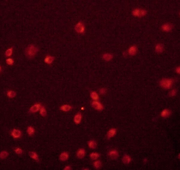
Immunocytochemistry/Immunofluorescence: CD36 Antibody [NB400-144] - Bone marrrow derived mouse macrophages stained with CD36 antibody. ICC/IF image submitted by a verified customer review.![Western Blot: CD36 AntibodyBSA Free [NB400-144]](https://resources.rndsystems.com/images/products/CD36-SR-B3-Antibody-Western-Blot-NB400-144-img0004.jpg "Western Blot: CD36 AntibodyBSA Free [NB400-144]")
Western Blot: CD36 AntibodyBSA Free [NB400-144]
Western Blot: CD36 Antibody [NB400-144] - Detection of CD36 in human adipocyte extract (30 ug). Lane 1: 0.5 ug/ml NB 400-144; lane 2: 2 ug/ml NB 400-144. ECL: 3 second exposure.
Western Blot: CD36 Antibody - BSA Free [NB400-144] -
ATM activation by chloroquine alleviates senescence.(a) Immunoblots showing protein levels of ATM, NBS1, and RAP80 in human skin fibroblasts (HSFs). A gradually increased level of p16 indicates cellular senescence, while elevated gamma H2AX level indicates accumulated DNA damage. (b) Immunoblots showing protein levels of ATM, NBS1, and RAP80 in mouse embryonic fibroblasts (MEFs). (c) Immunoblots showing protein levels of ATM, NBS1, and RAP80 in brain tissues isolated from 3-, 10-, and 18-month-old male mice. (d) SA-beta -Gal staining in HSFs treated with sh-ATM or scramble shRNA. Scale bar, 100 µm. (e) Quantification of SA-beta -Gal-positive staining of (d) from five views randomly captured for each group. Data represent means ± SEM. ***p<0.001. (f) Immunoblots showing increased gamma H2AX and unaffected LC3I/II in HSFs treated with sh-ATM or scramble shRNA. (g) Immunoblots showing protein levels of pS1981 ATM, gamma H2AX, and cleaved caspase-3 in HSFs treated with 10 μM of CQ for indicated time. (h) SA-beta -Gal staining in HSFs expressing either scramble or ATM shRNA treated with 1 μM CQ or DMSO (12 hr). Scale bar, 100 µm. (i) Quantification of SA-beta -Gal-positive staining of (h) from five views randomly captured for each group. Data represent means ± SEM. ***p<0.001; ‘N.S.’ indicates no significant difference. (j) HSFs at passage 20 were continuously cultured with 1 μM CQ or DMSO, and cell number was calculated at each passage. Data represent means ± SEM. ***p<0.01. (k) Immunoblots showing protein levels of gamma H2AX, p62, and LC3 in MEFs treated with 1 μM CQ or DMSO. Note that CQ had little effect on the expression levels of p62 and LC3. (l) MEFs at passage one were continuously cultured in 20% O2 with 1 μM CQ or DMSO, and cell number was determined at each passage. Data represent means ± SEM. ***p<0.01.10.7554/eLife.34836.006Figure 1—source data 1.Statistical analysis for SA-beta -Gal positive staining.10.7554/eLife.34836.007Figure 1—source data 2.Statistical analysis for EdU positive staining.Sta
Western Blot: Rabbit Polyclonal CD36 Antibody [NB400-144] -
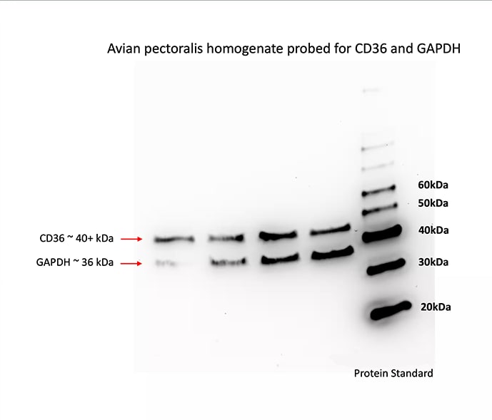
Western Blot: Rabbit Polyclonal CD36 Antibody [NB400-144] - The western blot was conducted on avian (passerine) skeletal muscle tissue homogenate. 6ug of total protein was loaded and GAPDH was probed to ensure total protein loads were similar across samples. Rabbit polyclonal CD36 antibody (NB400-144) was used at 1:5000 dilution (diluted in 5% milk). CD36 and GAPDH were probed sequentially - not multiplexed. CD36 produced a band above 40kDa and GAPDH at 36kDa. Image from a verified customer review.
Immunocytochemistry/ Immunofluorescence: CD36 Antibody - BSA Free [NB400-144] -
Immunocytochemistry/ Immunofluorescence: CD36 Antibody - BSA Free [NB400-144] - Timp4-deficiency results in defective lipid digestion & absorption. Fecal triglyceride (TG) content (Ai) in chow- or HFD-fed WT & Timp4−/− mice. Fecal lipid analysis by thin layer chromatography in chow-fed (Aii) & HFD-fed (iii) WT & Timp4−/− mice. (B) Immunostaining for CD36 in small intestine (proximal region) of chow-fed & HFD-fed WT & Timp4−/− mice. Western blot (C) & mRNA (D) levels for CD36 in enterocyte fraction of chow-fed & HFD-fed WT & Timp4−/− mice (collected from the proximal small intestine). (E) Triglyceride levels in enterocytes collected from the proximal small intestine. Averaged data are presented as mean ± S.E.M & analyzed by ANOVA. * indicates significance (p < 0.05). Image collected & cropped by CiteAb from the following publication (https://www.nature.com/articles/s41598-017-05951-4), licensed under a CC-BY license. Not internally tested by Novus Biologicals.
Western Blot: CD36 Antibody - BSA Free [NB400-144] -
Western Blot: CD36 Antibody - BSA Free [NB400-144] - Timp4-deficiency results in defective lipid digestion & absorption. Fecal triglyceride (TG) content (Ai) in chow- or HFD-fed WT & Timp4−/− mice. Fecal lipid analysis by thin layer chromatography in chow-fed (Aii) & HFD-fed (iii) WT & Timp4−/− mice. (B) Immunostaining for CD36 in small intestine (proximal region) of chow-fed & HFD-fed WT & Timp4−/− mice. Western blot (C) & mRNA (D) levels for CD36 in enterocyte fraction of chow-fed & HFD-fed WT & Timp4−/− mice (collected from the proximal small intestine). (E) Triglyceride levels in enterocytes collected from the proximal small intestine. Averaged data are presented as mean ± S.E.M & analyzed by ANOVA. * indicates significance (p < 0.05). Image collected & cropped by CiteAb from the following publication (https://www.nature.com/articles/s41598-017-05951-4), licensed under a CC-BY license. Not internally tested by Novus Biologicals.
Western Blot: CD36 Antibody - BSA Free [NB400-144] -
Western Blot: CD36 Antibody - BSA Free [NB400-144] - MiR-29a inhibits the expression of fatty acid translocase CD36 by targeting 3’ untranslated region (UTR). (A) qPCR analysis of Cd36 in live tissue. (B) Representative immunoblotting bands & densitometric results of CD36 in liver tissue. (C) qPCR analysis of cd36 expression of HepG2 cells in vitro after 48h transfection of scramble sequence or miR-29a-mimic. Data obtained from six independent experiments. (D) Upper panel, sequence information, & mutual pairing status of CD36-3’UTR, mmu-miR29a, & CD36-3’UTR Mut. Note that red characters represent mismatching sites. HepG2 was first transfected with CD36-3’UTR or CD36-3’UTR mutant luciferase reporter construct then treated with control medium (ctrl), miRNA-scramble, or miR-29a mimic, & finally lysed to detect the luciferase signal. Data are expressed as mean ± SE. * p < 0.05, ** p < 0.01, & *** p < 0.001 between the indicated groups. WT, wild type mice. HFD, high-fat diet. miR-29a, mice harboring overexpression of miR-29a. mmu-miR29a, mouse-origin miR-29a. ctrl, control. Mut, mutant. UTR, untranslated region. Image collected & cropped by CiteAb from the following publication (https://pubmed.ncbi.nlm.nih.gov/31652636), licensed under a CC-BY license. Not internally tested by Novus Biologicals.
Immunohistochemistry: CD36 Antibody - BSA Free [NB400-144] -
Immunohistochemistry: CD36 Antibody - BSA Free [NB400-144] - Integrin beta 3 modulates SMC transdifferentiation. a–c Mice were fed a HFD for 6 or 16 weeks as indicated, & then transverse aortic root sections were stained. In a, b sections from ApoE(−/−), SMMHC-CreERT2, ROSA26R(mTmG/+) mice were stained for SMA, GFP (fate marker), nuclei (DAPI), & either integrin beta 3 (a) or CD36 (b). Dashed yellow lines separate cap from core (a, b) & core from media (b). n = 5. In c sections from ApoE(−/−) mice that were also wild type or null for Itgb3 were stained for SMMHC, CD68, & nuclei (DAPI). n = 3. Boxed regions (a, c) are shown as close-ups on right; in c CD68+SMMHC+ cells in the media (arrowheads) & plaque (arrows) of the Itgb3 null atherosclerotic aorta are indicated. Med, tunica media; Lu, lumen; Pl, plaque. Scale bars, 25 μm. d–h Aortic SMCs were isolated from ApoE(−/−) mice & then subjected to siRNA-mediated knockdown with si-Itgb3 vs. scrambled (Scr; d–f) or with si-Itgb3, si-Tlr4 vs. si-Itgb3 (g, h). Levels of indicated transcripts from qRT-PCR are relative to Gapdh & normalized to either Scr in d, f or to si-Itgb3 in g, h. For d, g, n = 4–5 in duplicate. In e silenced SMCs were cultured with DiI-conjugated ox-LDL for 10 h & stained with DAPI; n = 5. In f, h silenced SMCs were exposed to soluble cholesterol:methyl-beta -cyclodextrin complexes for 3 days, & then mRNA levels were assessed; n = 4–7 in duplicate. *, **, ***, ^, **** vs. control (Scr in d, f & si-Itgb3 in g, h), p < 0.05, p < 0.01, p < 0.005, p < 0.001, & p < 0.0005, respectively. NS, not significant. Student’s t-test was used, & error bars represent standard deviations. i Schematic of the effect of beta 3 reduction via TLR4 & CD36 in SMCs on transdifferentiation Image collected & cropped by CiteAb from the following publication (https://www.nature.com/articles/s41467-018-04447-7), licensed under a CC-BY license. Not internally tested by Novus Biologicals.
Western Blot: CD36 Antibody - BSA Free [NB400-144] -
Western Blot: CD36 Antibody - BSA Free [NB400-144] - The increase in CD36 levels caused by lipids in Sirt3-deficient hepatocytes is mediated by Nrf2. VLDLR mRNA abundance (a) & protein levels of VLDLR & NQO1, an Nrf-2-target gene, (b) were assessed in Huh-7 cells incubated with fatty acid free-BSA or BSA-palmitate (0.3 mM) & exposed to either vehicle or the Sirt3 inhibitor AAPBO (100 μM) for 16 h. a, p < 0.05 vs. CT. b, p < 0.05 vs. CT cells incubated with palmitate. c, p < 0.05 vs. CT cells treated with AAPBO. c, fatty acid uptake in Huh-7 cells incubated with fatty acid free-BSA or BSA-palmitate (0.3 mM) & exposed to either vehicle or the Sirt3 inhibitor AAPBO (100 μM) for 16 h was measured by the uptake of BODIPY-C16. a, p < 0.05 vs. CT. b, p < 0.05 vs. CT cells incubated with palmitate. c, p < 0.05 vs. CT cells treated with AAPBO. mRNA abundance (d) & protein levels of VLDLR (e) in Huh-7 cells transfected with control (CT) or SIRT3 siRNA & incubated in the presence or absence 0.3 mM palmitate (Pal) for 24 h. Protein levels of CD36 (f), NQO1 (g) & PPAR gamma (h) in Huh-7 cells transfected with control (CT) or SIRT3 siRNA & incubated in the presence or absence 0.3 mM palmitate (Pal) or the Nrf2 inhibitor ML385 (10 μM) for 24 h. a, p < 0.05 vs. CT siRNA cells. b, p < 0.05 vs. CT siRNA cells incubated with palmitate. c, p < 0.05 vs. SIRT3 siRNA cells. d, p < 0.05 vs. CT siRNA cells incubated with palmitate & ML385 Image collected & cropped by CiteAb from the following publication (https://pubmed.ncbi.nlm.nih.gov/32912335), licensed under a CC-BY license. Not internally tested by Novus Biologicals.
Western Blot: CD36 Antibody - BSA Free [NB400-144] -
Western Blot: CD36 Antibody - BSA Free [NB400-144] - The increase in CD36 levels caused by lipids in Sirt3-deficient hepatocytes is mediated by Nrf2. VLDLR mRNA abundance (a) & protein levels of VLDLR & NQO1, an Nrf-2-target gene, (b) were assessed in Huh-7 cells incubated with fatty acid free-BSA or BSA-palmitate (0.3 mM) & exposed to either vehicle or the Sirt3 inhibitor AAPBO (100 μM) for 16 h. a, p < 0.05 vs. CT. b, p < 0.05 vs. CT cells incubated with palmitate. c, p < 0.05 vs. CT cells treated with AAPBO. c, fatty acid uptake in Huh-7 cells incubated with fatty acid free-BSA or BSA-palmitate (0.3 mM) & exposed to either vehicle or the Sirt3 inhibitor AAPBO (100 μM) for 16 h was measured by the uptake of BODIPY-C16. a, p < 0.05 vs. CT. b, p < 0.05 vs. CT cells incubated with palmitate. c, p < 0.05 vs. CT cells treated with AAPBO. mRNA abundance (d) & protein levels of VLDLR (e) in Huh-7 cells transfected with control (CT) or SIRT3 siRNA & incubated in the presence or absence 0.3 mM palmitate (Pal) for 24 h. Protein levels of CD36 (f), NQO1 (g) & PPAR gamma (h) in Huh-7 cells transfected with control (CT) or SIRT3 siRNA & incubated in the presence or absence 0.3 mM palmitate (Pal) or the Nrf2 inhibitor ML385 (10 μM) for 24 h. a, p < 0.05 vs. CT siRNA cells. b, p < 0.05 vs. CT siRNA cells incubated with palmitate. c, p < 0.05 vs. SIRT3 siRNA cells. d, p < 0.05 vs. CT siRNA cells incubated with palmitate & ML385 Image collected & cropped by CiteAb from the following publication (https://pubmed.ncbi.nlm.nih.gov/32912335), licensed under a CC-BY license. Not internally tested by Novus Biologicals.Applications for CD36 Antibody - BSA Free
Application
Recommended Usage
Immunocytochemistry/ Immunofluorescence
1:50-1:200
Immunohistochemistry
1:200-1:400
Immunohistochemistry-Frozen
reported in scientific literature (PMID 24531551)
Immunohistochemistry-Paraffin
1:200-1:400
Western Blot
1:500-1:2000
Application Notes
In Western Blot, a band is seen ~75-80 kDa. The theoretical molecular weight of CD36 is ~53 kDa. The difference in theoretical MW and actual MW as seen in Western blot is most likely due to the heavy glycosylation and palmitoylation of this protein. The observed molecular weight of the protein may vary from the listed predicted molecular weight due to post translational modifications, post translation cleavages, relative charges, and other experimental factors.
Reviewed Applications
Read 10 reviews rated 3.8 using NB400-144 in the following applications:
Formulation, Preparation, and Storage
Purification
Immunogen affinity purified
Formulation
PBS
Format
BSA Free
Preservative
0.02% Sodium Azide
Concentration
1 mg/ml
Shipping
The product is shipped with polar packs. Upon receipt, store it immediately at the temperature recommended below.
Stability & Storage
Store at 4C short term. Aliquot and store at -20C long term. Avoid freeze-thaw cycles.
Background: CD36
The expression of CD36 has been reported in platelets, erythrocytes, monocytes, differentiated adipocytes, skeletal muscle, mammary epithelial cells, spleen cells, some skin microdermal endothelial cells, and in cancer. Circulating levels of soluble CD36 (sCD36) has also been reported in chronic inflammatory disease such as type 2 diabetes and chronic kidney disease. CD36 participates in angiogenesis, innate immunity, and the clearance of apoptotic phagocytes. In lipid metabolism, CD36 functions as a macrophage receptor for oxidized LDL and as an adipocyte receptor/transporter for long-chain FFAs. Plasmodium falciparum, the parasite that causes malaria, binds CD36 via PfEMP1 proteins, tethering parasite-infected erythrocytes to endothelial receptors (5). Anti-CD36 isoantibodies have been detected in Type 1 CD36-deficient mothers and is implicated as the cause of fetal/neonatal alloimmune thrombocytopenia (6).
References
1) Febbraio, M., Hajjar, D. P., & Silverstein, R. L. (2001). CD36: a class B scavenger receptor involved in angiogenesis, atherosclerosis, inflammation, and lipid metabolism. The Journal of clinical investigation, 108(6), 785-791. PMID: 11560944
2) Silverstein RL, Febbraio M. (2000) CD36 and atherosclerosis. Curr Opin Lipidol. 2000 11(5):483-91. PMID: 11048891.
3) Endemann G, Stanton LW, Madden KS, Bryant CM, White RT, Protter AA. (1993) CD36 is a receptor for oxidized low density lipoprotein. J Biol Chem. 268(16):11811-6. PMID: 7685021.
4) Wang, J., & Li, Y. (2019). CD36 tango in cancer: signaling pathways and functions. Theranostics, 9(17), 4893-4908. PMID: 31410189
5) Hsieh FL, Turner L, Bolla JR, Robinson CV, Lavstsen T, Higgins MK. (2016) The structural basis for CD36 binding by the malaria parasite. Nat Commun. 7:12837. PMID: 27667267
6) Gruarin P, Ulliers D, Thorne RF, Alessio M. (2000) Methionine 156 in the immunodominant domain of CD36 contributes to define the epitope recognized by the NL07 MoAb. Mol Cell Biochem 214, 115-121. PMID: 11195795.
Alternate Names
BDPLT10, CD36 antigen (collagen type I receptor, thrombospondin receptor), CD36 molecule (thrombospondin receptor), CHDS7, cluster determinant 36, FAT, Fatty acid translocase, Glycoprotein IIIb, GP3B, GP4, GPIIIB, GPIV, leukocyte differentiation antigen CD36, PAS IV, PAS-4 protein, PASIV, platelet glycoprotein 4, platelet glycoprotein IV, SCARB3, scavenger receptor class B, member 3
Gene Symbol
CD36
Additional CD36 Products
Product Documents for CD36 Antibody - BSA Free
Certificate of Analysis
To download a Certificate of Analysis, please enter a lot or batch number in the search box below.
Product Specific Notices for CD36 Antibody - BSA Free
This product is for research use only and is not approved for use in humans or in clinical diagnosis. Primary Antibodies are guaranteed for 1 year from date of receipt.
Citations for CD36 Antibody - BSA Free
 Powered by Bioz
Customer Reviews for CD36 Antibody - BSA Free (10)
3.8 out of 5
10 Customer Ratings
Have you used CD36 Antibody - BSA Free?
Submit a review and receive an Amazon gift card!
$25/€18/£15/$25CAN/¥2500 Yen for a review with an image
$10/€7/£6/$10CAN/¥1110 Yen for a review without an image
Submit a review
Customer Images


Showing
1
-
5 of
10 reviews
Showing All
Filter By:
-
Application: Western BlotSample Tested: Whole Skeletal Muscle LysateSpecies: Avian and PasserineVerified Customer | Posted 02/23/2024Western blot to probe CD36 levels in Passerine (avian) skeletal muscle tissueThe western blot was conducted on avian (passerine) skeletal muscle tissue homogenate. 6ug of total protein was loaded and GAPDH was probed to ensure total protein loads were similar across samples. Novus biologicals Rabbit polyclonal CD36 antibody (NB400-144) was used at 1:5000 dilution (diluted in 5% milk). CD36 and GAPDH were probed sequentially - not multiplexed. CD36 produced a band above 40kDa and GAPDH at 36kDa.
 Bio-Techne ResponseThis review was submitted through the legacy Novus Innovators Program, reflecting a new species or application tested on a primary antibody.
 -
Application: Simple WesternSample Tested: Mouse adipose tissue, rat adipose tissue and mouse liverSpecies: Rat and MouseVerified Customer | Posted 02/16/2022Simple Western lane shows lysates from mouse and rat adipose and liver, loaded at 0.4 mg/ml. CD36 88-kDa; bands did not appear using 100 ug/ml of CD36 antibody NB400-144; followed by an anti-rabbit secondary HRP antibody Bio-techne, 042-206.Western blot was performed using the protein simple WES system A 12-230 kDa plate kit was used and the protocol was performed as stated in the protein simple WES user guide. mouse and rat tissues were homogenized with T-PER lysis buffer all samples were at a protein concentration of 0.4 mg/ml before loading them onto the plate. CD36 antibody concentrate was diluted 1:10 in order to use 100 ug/ml on all samples
Bio-Techne ResponseThis review was submitted through the legacy Novus Innovators Program, reflecting a new species or application tested on a primary antibody.
-
Application: ImmunocytochemistrySample Tested: Bone Marrrow Derived MacrophagesSpecies: MouseVerified Customer | Posted 10/02/2020Works fine for IF staining in cells
-
Application: Western BlotSample Tested: Primary muscle cell lysateSpecies: MouseVerified Customer | Posted 03/13/2018
-
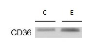
Application: Western BlotSample Tested: white adipose tissueSpecies: MouseVerified Customer | Posted 03/01/2018Alcohol feeding significantly up-regulated CD36 expression.
-
Application: ImmunohistochemistrySample Tested: mouse liverSpecies: MouseVerified Customer | Posted 05/11/2017
-
Application: Western BlotSample Tested: cardiac tissueSpecies: MouseVerified Customer | Posted 11/30/2016Used antibody at a 1:1000 dilution in insoluble fraction of infarct tissue. Antibody shows many bands especially in post-infarct tissue.
-
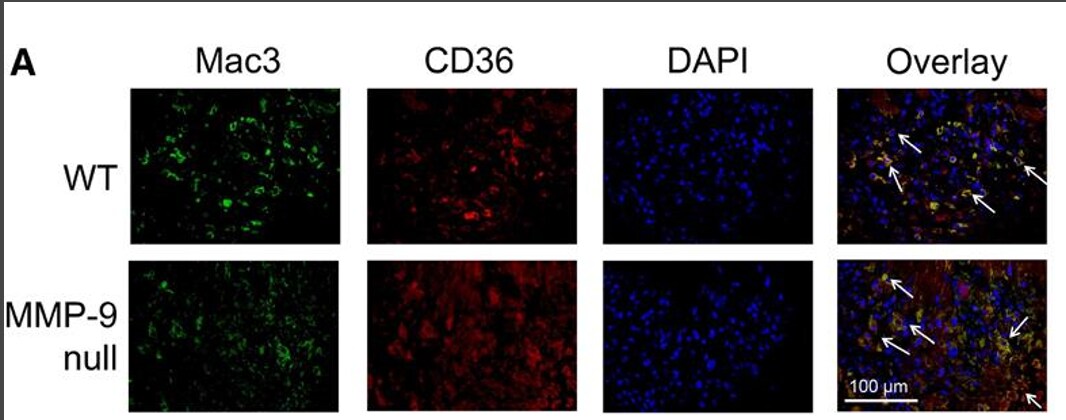
Application: ImmunofluorescenceSample Tested: cardiac tissueSpecies: MouseVerified Customer | Posted 11/30/2016Overlay analysis indicated CD36 was largely expressed by macrophages. Staining showed higher levels of CD36+ macrophages in the MMP-9 null mice at day 7 post-MI.Co-localization was performed using either an antibody for Mac-3 and CD36 to determine if macrophages were a major source of CD36. We used paraffin embedded infarct tissue.
-
Application: Western BlotSample Tested: See PMID 23727393Species: OtherVerified Customer | Posted 12/23/2014
-
Application: Western BlotSample Tested: Whole tissue lysate ApoE murine aorta, Sample Amount: 20ugSpecies: MouseVerified Customer | Posted 02/09/2011
There are no reviews that match your criteria.
Protocols
View specific protocols for CD36 Antibody - BSA Free (NB400-144):
Immunocytochemistry Protocol
Culture cells to appropriate density in 35 mm culture dishes or 6-well plates.
1. Remove culture medium and wash the cells briefly in PBS. Add 10% formalin to the dish and fix at room temperature for 10 minutes.
2. Remove the formalin and wash the cells in PBS.
3. Permeablize the cells with 0.1% Triton X100 or other suitable detergent for 10 min.
4. Remove the permeablization buffer and wash three times for 10 minutes each in PBS. Be sure to not let the specimen dry out.
5. To block nonspecific antibody binding, incubate in 10% normal goat serum from 1 hour to overnight at room temperature.
6. Add primary antibody at appropriate dilution and incubate overnight at 4C.
7. Remove primary antibody and replace with PBS. Wash three times for 10 minutes each.
8. Add secondary antibody at appropriate dilution. Incubate for 1 hour at room temperature.
9. Remove secondary antibody and replace with PBS. Wash three times for 10 minutes each.
10. Counter stain DNA with DAPi if required.
Immunohistochemistry-Paraffin Embedded Sections
Antigen Unmasking:
Bring slides to a boil in 10 mM sodium citrate buffer (pH 6.0) then maintain at a sub-boiling temperature for 10 minutes. Cool slides on bench-top for 30 minutes (keep slides in the sodium citrate buffer at all times).
Staining:
1. Wash sections in deionized water three times for 5 minutes each.
2. Wash sections in PBS for 5 minutes.
3. Block each section with 100-400 ul blocking solution (1% BSA in PBS) for 1 hour at room temperature.
4. Remove blocking solution and add 100-400 ul diluted primary antibody. Incubate overnight at 4 C.
5. Remove antibody solution and wash sections in wash buffer three times for 5 minutes each.
6. Add 100-400 ul HRP polymer conjugated secondary antibody. Incubate 30 minutes at room temperature.
7. Wash sections three times in wash buffer for 5 minutes each.
8. Add 100-400 ul DAB substrate to each section and monitor staining closely.
9. As soon as the sections develop, immerse slides in deionized water.
10. Counterstain sections in hematoxylin.
11. Wash sections in deionized water two times for 5 minutes each.
12. Dehydrate sections.
13. Mount coverslips.
Western Blot Protocol
1. Perform SDS-PAGE on samples to be analyzed, loading 10-25 ug of total protein per lane.
2. Transfer proteins to PVDF membrane according to the instructions provided by the manufacturer of the membrane and transfer apparatus.
3. Stain the membrane with Ponceau S (or similar product) to assess transfer success, and mark molecular weight standards where appropriate.
4. Rinse the blot TBS -0.05% Tween 20 (TBST).
5. Block the membrane in 5% Non-fat milk in TBST (blocking buffer) for at least 1 hour.
6. Wash the membrane in TBST three times for 10 minutes each.
7. Dilute primary antibody in blocking buffer and incubate overnight at 4C with gentle rocking.
8. Wash the membrane in TBST three times for 10 minutes each.
9. Incubate the membrane in diluted HRP conjugated secondary antibody in blocking buffer (as per manufacturer's instructions) for 1 hour at room temperature.
10. Wash the blot in TBST three times for 10 minutes each (this step can be repeated as required to reduce background).
11. Apply the detection reagent of choice in accordance with the manufacturer's instructions.
Find general support by application which include: protocols, troubleshooting, illustrated assays, videos and webinars.
- Antigen Retrieval Protocol (PIER)
- Antigen Retrieval for Frozen Sections Protocol
- Appropriate Fixation of IHC/ICC Samples
- Cellular Response to Hypoxia Protocols
- Chromogenic IHC Staining of Formalin-Fixed Paraffin-Embedded (FFPE) Tissue Protocol
- Chromogenic Immunohistochemistry Staining of Frozen Tissue
- ClariTSA™ Fluorophore Kits
- Detection & Visualization of Antibody Binding
- Fluorescent IHC Staining of Frozen Tissue Protocol
- Graphic Protocol for Heat-induced Epitope Retrieval
- Graphic Protocol for the Preparation and Fluorescent IHC Staining of Frozen Tissue Sections
- Graphic Protocol for the Preparation and Fluorescent IHC Staining of Paraffin-embedded Tissue Sections
- Graphic Protocol for the Preparation of Gelatin-coated Slides for Histological Tissue Sections
- ICC Cell Smear Protocol for Suspension Cells
- ICC Immunocytochemistry Protocol Videos
- ICC for Adherent Cells
- IHC Sample Preparation (Frozen sections vs Paraffin)
- Immunocytochemistry (ICC) Protocol
- Immunocytochemistry Troubleshooting
- Immunofluorescence of Organoids Embedded in Cultrex Basement Membrane Extract
- Immunofluorescent IHC Staining of Formalin-Fixed Paraffin-Embedded (FFPE) Tissue Protocol
- Immunohistochemistry (IHC) and Immunocytochemistry (ICC) Protocols
- Immunohistochemistry Frozen Troubleshooting
- Immunohistochemistry Paraffin Troubleshooting
- Preparing Samples for IHC/ICC Experiments
- Preventing Non-Specific Staining (Non-Specific Binding)
- Primary Antibody Selection & Optimization
- Protocol for Heat-Induced Epitope Retrieval (HIER)
- Protocol for Making a 4% Formaldehyde Solution in PBS
- Protocol for VisUCyte™ HRP Polymer Detection Reagent
- Protocol for the Fluorescent ICC Staining of Cell Smears - Graphic
- Protocol for the Fluorescent ICC Staining of Cultured Cells on Coverslips - Graphic
- Protocol for the Preparation & Fixation of Cells on Coverslips
- Protocol for the Preparation and Chromogenic IHC Staining of Frozen Tissue Sections
- Protocol for the Preparation and Chromogenic IHC Staining of Frozen Tissue Sections - Graphic
- Protocol for the Preparation and Chromogenic IHC Staining of Paraffin-embedded Tissue Sections
- Protocol for the Preparation and Chromogenic IHC Staining of Paraffin-embedded Tissue Sections - Graphic
- Protocol for the Preparation and Fluorescent ICC Staining of Cells on Coverslips
- Protocol for the Preparation and Fluorescent ICC Staining of Non-adherent Cells
- Protocol for the Preparation and Fluorescent ICC Staining of Stem Cells on Coverslips
- Protocol for the Preparation and Fluorescent IHC Staining of Frozen Tissue Sections
- Protocol for the Preparation and Fluorescent IHC Staining of Paraffin-embedded Tissue Sections
- Protocol for the Preparation of Gelatin-coated Slides for Histological Tissue Sections
- Protocol for the Preparation of a Cell Smear for Non-adherent Cell ICC - Graphic
- R&D Systems Quality Control Western Blot Protocol
- TUNEL and Active Caspase-3 Detection by IHC/ICC Protocol
- The Importance of IHC/ICC Controls
- Troubleshooting Guide: Immunohistochemistry
- Troubleshooting Guide: Western Blot Figures
- Western Blot Conditions
- Western Blot Protocol
- Western Blot Protocol for Cell Lysates
- Western Blot Troubleshooting
- Western Blot Troubleshooting Guide
- View all Protocols, Troubleshooting, Illustrated assays and Webinars
FAQs for CD36 Antibody - BSA Free
Showing
1
-
2 of
2 FAQs
Showing All
-
Q: Can you tell me if this product, NB400-144, requires antigen retrieval for hick on paraffin embedded tissues? If so, what type of antigen retrieval is recommended?
A: Yes, we recommend antigen retrieval for IHC-P with NB400-144 and in our testing we performed heat-mediated antigen retrieval with sodium citrate buffer (pH 6).
-
Q: We used the NB400-144 to detect CD36 in guinea pig by Western blot analysis. Is there a similar antibody recommended for IP?
A:
Here is a link to other CD36 antibodies which are validated for IP. The CD36 antibody (NB400-144) that you already have is one of our best selling products with over 15 PubMed citations. We have not tested this antibody in IP yet and I do not see any reason why it would not work in IP. If you would be interested in testing this novel application, please take a look at our Innovator's Reward program.
-
Q: Can you tell me if this product, NB400-144, requires antigen retrieval for hick on paraffin embedded tissues? If so, what type of antigen retrieval is recommended?
A: Yes, we recommend antigen retrieval for IHC-P with NB400-144 and in our testing we performed heat-mediated antigen retrieval with sodium citrate buffer (pH 6).
-
Q: We used the NB400-144 to detect CD36 in guinea pig by Western blot analysis. Is there a similar antibody recommended for IP?
A:
Here is a link to other CD36 antibodies which are validated for IP. The CD36 antibody (NB400-144) that you already have is one of our best selling products with over 15 PubMed citations. We have not tested this antibody in IP yet and I do not see any reason why it would not work in IP. If you would be interested in testing this novel application, please take a look at our Innovator's Reward program.
Loading...