CD47 Antibody (B6H12.2) - Azide and BSA Free
Novus Biologicals | Catalog # NBP2-31106
![Immunocytochemistry/ Immunofluorescence: CD47 Antibody (B6H12.2) - Azide and BSA Free [NBP2-31106]](https://resources.rndsystems.com/images/products/CD47-Antibody-B6H12-2-Azide-Free-Immunocytochemistry-Immunofluorescence-NBP2-31106-img0012.jpg "Immunocytochemistry/ Immunofluorescence: CD47 Antibody (B6H12.2) - Azide and BSA Free [NBP2-31106]")
Key Product Details
Species Reactivity
Validated:
Human, Mouse
Cited:
Human, Mouse
Applications
Validated:
Immunohistochemistry, Immunohistochemistry-Paraffin, Western Blot, Flow Cytometry, Flow (Cell Surface), Immunocytochemistry/ Immunofluorescence, Immunoprecipitation, CyTOF-ready
Cited:
Immunohistochemistry-Paraffin, Western Blot, Immunocytochemistry/ Immunofluorescence, Immunoprecipitation
Label
Unconjugated
Antibody Source
Monoclonal Mouse IgG1 Clone # B6H12.2
Format
Azide and BSA Free
Loading...
Product Specifications
Immunogen
This antibody was raised against CD47.
Reactivity Notes
Mouse reactivity reported in scientific literature (PMID: 30833751).
Clonality
Monoclonal
Host
Mouse
Isotype
IgG1
Scientific Data Images for CD47 Antibody (B6H12.2) - Azide and BSA Free
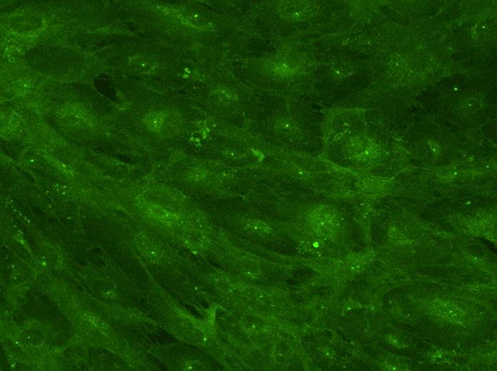
Immunocytochemistry/ Immunofluorescence: CD47 Antibody (B6H12.2) - Azide and BSA Free [NBP2-31106]
Immunocytochemistry/Immunofluorescence: CD47 Antibody (B6H12.2) - Azide Free [NBP2-31106] - A431 cells were fixed in 4% paraformaldehyde for 10 minutes and permeabilized in 0.05% Triton X-100 in PBS for 5 minutes. The cells were incubated with CD47 Antibody [B6H12.2] (NBP2-31106) at 1 ug/ml overnight at 4C and detected with an anti-mouse Dylight 488 (Green) at a 1:1000 dilution for 60 minutes. Nuclei were counterstained with DAPI (Blue). Cells were imaged using a 40X objective.![Western Blot: CD47 Antibody (B6H12.2)Azide and BSA Free [NBP2-31106]](https://resources.rndsystems.com/images/products/CD47-Antibody-B6H12-2-Azide-Free-Western-Blot-NBP2-31106-img0008.jpg "Western Blot: CD47 Antibody (B6H12.2)Azide and BSA Free [NBP2-31106]")
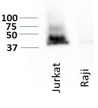
Western Blot: CD47 Antibody (B6H12.2)Azide and BSA Free [NBP2-31106]
Western Blot: CD47 Antibody (B6H12.2) - Azide Free [NBP2-31106] - Detection of CD47 expression on two hematological cancer cell lines, Jurkat and Raji. WB image submitted by a verified customer review.![Immunohistochemistry-Paraffin: CD47 Antibody (B6H12.2) - Azide and BSA Free [NBP2-31106]](https://resources.rndsystems.com/images/products/CD47-Antibody-B6H12-2-Azide-Free-Immunohistochemistry-Paraffin-NBP2-31106-img0010.jpg "Immunohistochemistry-Paraffin: CD47 Antibody (B6H12.2) - Azide and BSA Free [NBP2-31106]")
Immunohistochemistry-Paraffin: CD47 Antibody (B6H12.2) - Azide and BSA Free [NBP2-31106]
Immunohistochemistry-Paraffin: CD47 Antibody (B6H12.2) - Azide Free [NBP2-31106] - Human breast cancer tissue stained with CD47 Antibody (B6H12.2). IHC-P image submitted by a verified customer review.![Flow Cytometry: CD47 Antibody (B6H12.2) - Azide and BSA Free [NBP2-31106]](https://resources.rndsystems.com/images/products/CD47-Antibody-B6H12-2-Azide-Free-Flow-Cytometry-NBP2-31106-img0009.jpg "Flow Cytometry: CD47 Antibody (B6H12.2) - Azide and BSA Free [NBP2-31106]")
Flow Cytometry: CD47 Antibody (B6H12.2) - Azide and BSA Free [NBP2-31106]
Flow Cytometry: CD47 Antibody (B6H12.2) - Azide Free [NBP2-31106] - A surface stain was performed on A431 cells with CD47 Antibody [B6H12.2] NBP2-31106PCP (blue) and a matched isotype control (orange). Cells were incubated in an antibody dilution of 5 ug/mL for 20 minutes at room temperature. Both antibodies were conjugated to PerCP.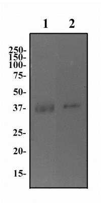
Western Blot: CD47 Antibody (B6H12.2) [Azide Free] [NBP2-31106] - Human brain (lane 1) and testis (lane 2) protein was separated on a 12% gel by SDS-PAGE. Protein was transferred to PVDF membrane, blocked and then probed with 2 ug/ml of anti-CD47. CD47 protein was detected using an anti-mouse HRP secondary antibody.
![Immunocytochemistry: CD47 Antibody (B6H12.2) - Azide and BSA Free [NBP2-31106]](https://resources.rndsystems.com/images/products/CD47-Antibody-B6H12-2-Azide-Free-Immunocytochemistry-NBP2-31106-img0007.jpg "Immunocytochemistry: CD47 Antibody (B6H12.2) - Azide and BSA Free [NBP2-31106]")
Immunocytochemistry: CD47 Antibody (B6H12.2) - Azide and BSA Free [NBP2-31106]
Immunocytochemistry: CD47 Antibody (B6H12.2) - Azide Free [NBP2-31106] - Human trabecular meshwork (primary ocular cells). Image from verified customer review.![Immunocytochemistry/ Immunofluorescence: CD47 Antibody (B6H12.2) - Azide and BSA Free [NBP2-31106]](https://resources.rndsystems.com/images/products/CD47-Antibody-B6H12-2-Azide-Free-Immunocytochemistry-Immunofluorescence-NBP2-31106-img0011.jpg "Immunocytochemistry/ Immunofluorescence: CD47 Antibody (B6H12.2) - Azide and BSA Free [NBP2-31106]")
Immunocytochemistry/ Immunofluorescence: CD47 Antibody (B6H12.2) - Azide and BSA Free [NBP2-31106]
Immunocytochemistry/Immunofluorescence: CD47 Antibody (B6H12.2) - Azide Free [NBP2-31106] - A431 cells were fixed in 4% paraformaldehyde for 10 minutes and permeabilized in 0.05% Triton X-100 in PBS for 5 minutes. The cells were incubated with CD47 Antibody [B6H12.2] conjugated to Biotin (NBP2-31106B) at 5 ug/ml for 1 hour at room temperature then detected with Streptavidin conjugated to DyLight 550. Nuclei were counterstained with DAPI (Blue). Cells were imaged using a 40X objective.![Immunohistochemistry-Paraffin: CD47 Antibody (B6H12.2) - Azide and BSA Free [NBP2-31106]](https://resources.rndsystems.com/images/products/CD47-Antibody-B6H12-2-Azide-Free-Immunohistochemistry-Paraffin-NBP2-31106-img0003.jpg "Immunohistochemistry-Paraffin: CD47 Antibody (B6H12.2) - Azide and BSA Free [NBP2-31106]")
Immunohistochemistry-Paraffin: CD47 Antibody (B6H12.2) - Azide and BSA Free [NBP2-31106]
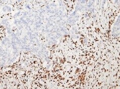
Immunohistochemistry-Paraffin: CD47 Antibody (B6H12.2) - Azide Free [NBP2-31106] - Tissue section of human normal breast using mouse monoclonal CD47 antibody (clone B6H12.2) at 0.5ug/ml concentration. The ductal/acinar epithelial cells in the breast section developed specific membrane-cytoplasmic staining.![Immunohistochemistry-Paraffin: CD47 Antibody (B6H12.2) - Azide and BSA Free [NBP2-31106]](https://resources.rndsystems.com/images/products/CD47-Antibody-B6H12-2-Azide-Free-Immunohistochemistry-Paraffin-NBP2-31106-img0002.jpg "Immunohistochemistry-Paraffin: CD47 Antibody (B6H12.2) - Azide and BSA Free [NBP2-31106]")
Immunohistochemistry-Paraffin: CD47 Antibody (B6H12.2) - Azide and BSA Free [NBP2-31106]
Immunohistochemistry-Paraffin: CD47 Antibody (B6H12.2) - Azide Free [NBP2-31106] - Tissue section of human normal breast using mouse monoclonal CD47 antibody (clone B6H12.2) at 0.5ug/ml concentration. The ductal/acinar epithelial cells in the breast section developed specific membrane-cytoplasmic staining.![Flow (Cell Surface): CD47 Antibody (B6H12.2) - Azide and BSA Free [NBP2-31106]](https://resources.rndsystems.com/images/products/CD47-Antibody-B6H12-2-Azide-Free-Flow-Cell-Surface-NBP2-31106-img0006.jpg "Flow (Cell Surface): CD47 Antibody (B6H12.2) - Azide and BSA Free [NBP2-31106]")
Flow (Cell Surface): CD47 Antibody (B6H12.2) - Azide and BSA Free [NBP2-31106]
Flow (Cell Surface): CD47 Antibody (B6H12.2) - Azide Free [NBP2-31106] - A surface stain was performed on human peripheral blood lymphocytes with CD47 (B6H12.2) antibody NBP2-31106 (blue) and a matched isotype control NBP2-27287 (orange). Cells were incubated in an antibody dilution of 1 ug/mL for 20 minutes at room temperature, followed by mouse F(ab)2 IgG (H+L) APC-conjugated secondary antibody [F0101B, R&D Systems].Applications for CD47 Antibody (B6H12.2) - Azide and BSA Free
Application
Recommended Usage
Flow Cytometry
1 ug/mL
Immunohistochemistry-Paraffin
0.5 ug/mL
Western Blot
1:1000
Application Notes
This antibody is CyTOF ready.
Reviewed Applications
Read 5 reviews rated 4.8 using NBP2-31106 in the following applications:
Flow Cytometry Panel Builder
Bio-Techne Knows Flow Cytometry
Save time and reduce costly mistakes by quickly finding compatible reagents using the Panel Builder Tool.
Advanced Features
- Spectra Viewer - Custom analysis of spectra from multiple fluorochromes
- Spillover Popups - Visualize the spectra of individual fluorochromes
- Antigen Density Selector - Match fluorochrome brightness with antigen density

Formulation, Preparation, and Storage
Purification
Protein G purified
Formulation
PBS
Format
Azide and BSA Free
Preservative
No Preservative
Concentration
1.0 mg/ml
Shipping
The product is shipped with polar packs. Upon receipt, store it immediately at the temperature recommended below.
Stability & Storage
Store at -20C. Avoid freeze-thaw cycles.
Background: CD47
Additional CD47 Products
Product Documents for CD47 Antibody (B6H12.2) - Azide and BSA Free
Certificate of Analysis
To download a Certificate of Analysis, please enter a lot or batch number in the search box below.
Product Specific Notices for CD47 Antibody (B6H12.2) - Azide and BSA Free
This product is for research use only and is not approved for use in humans or in clinical diagnosis. Primary Antibodies are guaranteed for 1 year from date of receipt.
Citations for CD47 Antibody (B6H12.2) - Azide and BSA Free
 Powered by Bioz
Customer Reviews for CD47 Antibody (B6H12.2) - Azide and BSA Free (5)
4.8 out of 5
5 Customer Ratings
Have you used CD47 Antibody (B6H12.2) - Azide and BSA Free?
Submit a review and receive an Amazon gift card!
$25/€18/£15/$25CAN/¥2500 Yen for a review with an image
$10/€7/£6/$10CAN/¥1110 Yen for a review without an image
Submit a review
Customer Images
Showing
1
-
5 of
5 reviews
Showing All
Filter By:
-
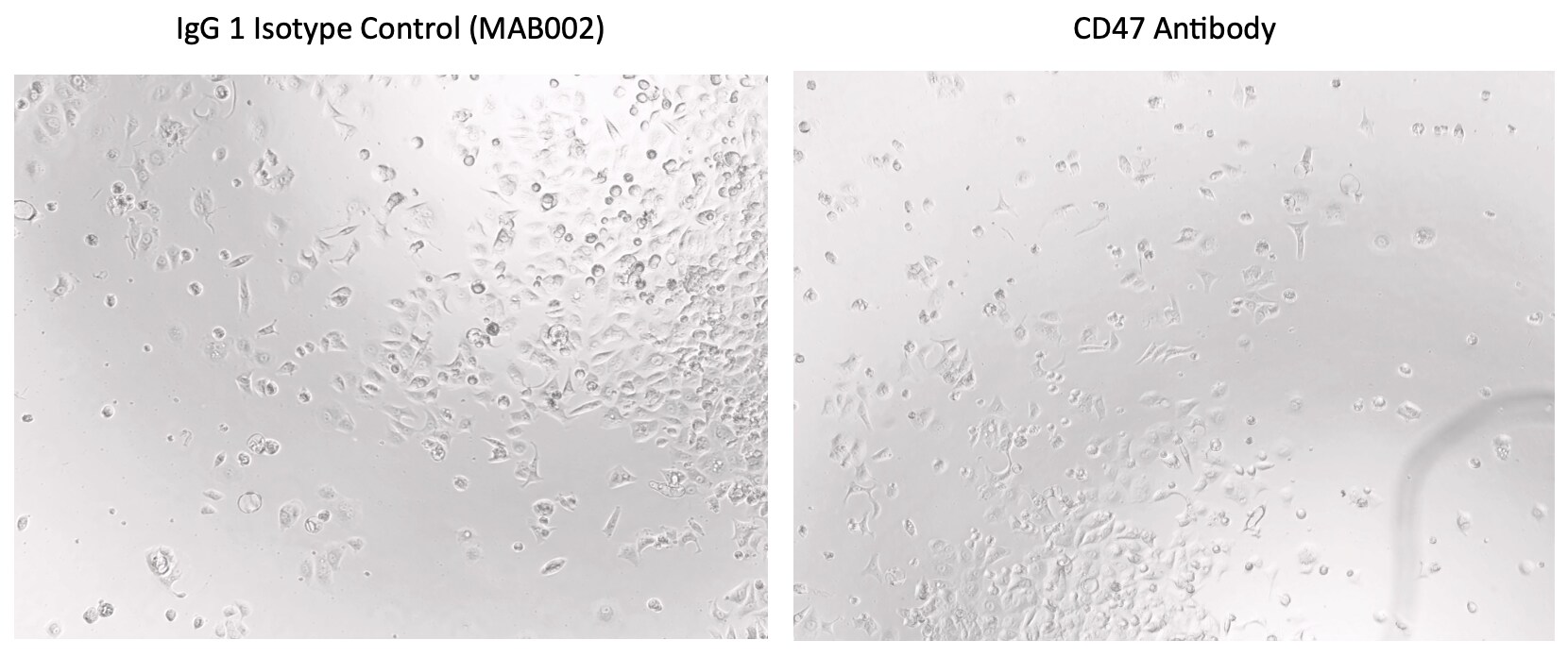
Application: Cell cultureSample Tested: MCF-7 human breast cancer cell lineSpecies: HumanVerified Customer | Posted 05/29/2025MCF7 treated with CD47 antibodyMCF7 cells were seeded on 96well plate with 10 ug/mL of CD47 and imaged after 24h
 Bio-Techne ResponseThis review reflects a new species or application tested on a primary antibody.
 -
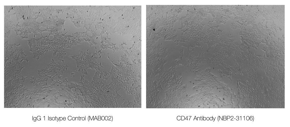
Application: Cell Culture treatmentSample Tested: HCT-116 human colorectal carcinoma cell lineSpecies: HumanVerified Customer | Posted 05/26/20255000 cells were treated with 10 ug/mL of CD47 or IgG1 isotope controlCell culture treatment - 5000 cells were treated with 10 ug/mL of antibody
Bio-Techne ResponseThis review reflects a new species or application tested on a primary antibody.
-
Application: Immunohistochemistry-ParaffinSample Tested: Breast cancer tissueSpecies: HumanVerified Customer | Posted 11/04/2021Breast cancer tissue4% formaldehyde fixed paraffin sections.
-
Application: Western BlotSample Tested: 20 ug whole cell lysateSpecies: HumanVerified Customer | Posted 05/19/2019Detection of CD47 expression on two hematological cancer cell lines, Jurkat and Raji.Samples were prepared in 4X lamelli buffer and were not boiled. Boiling causes loss of signal in WB.
-
Application: ImmunocytochemistrySample Tested: Human trabecular meshworkSpecies: HumanVerified Customer | Posted 04/25/2018
There are no reviews that match your criteria.
Protocols
View specific protocols for CD47 Antibody (B6H12.2) - Azide and BSA Free (NBP2-31106):
Protocol for Flow Cytometry Cell Surface Staining
Sample Preparation.
1. Grow cells to 60-85% confluency. Flow cytometry requires between 2 x 10⁵ and 1 x 10⁶ cells for optimal performance.
2. If cells are adherent, harvest gently by washing once with staining buffer and then scraping. Avoid using trypsin as this can disrupt certain epitopes of interest. If enzymatic harvest is required, use Accutase, Collagenase, or TrypLE Express for a less damaging option.
3. Reserve 100 uL for counting, then transfer cell volume into a 15 mL conical tube and centrifuge for 4 minutes at 400 RCF.
a. Count cells using a hemocytometer and a 1:1 trypan blue exclusion stain to determine cell viability before starting the flow protocol. If cells appear blue, do not proceed.
4. Re-suspend cells to a concentration of 1 x 10⁶ cells/mL in staining buffer (NBP2-26247).
5. Aliquot out 100 uL samples in accordance with your experimental samples.
Tip: When cell surface and intracellular staining are required in the same sample, it is advisable that the cell surface staining be performed first since the fixation and permeablization steps might reduce the availability of surface antigens.
Cell surface staining
1. Recommended: Block non-specific interactions using 0.5-1 ug of a species specific Fc-blocking reagent such as an anti-mouse CD16/CD32 antibody (NBP1-27946).
2. Add appropriate amount of each antibody (eg. 1 test or 1 ug per sample, as experimentally determined) to 100 uL of staining buffer (NBP2-26247) per sample (eg. use 1 mL of staining buffer for 10 samples).
3. Mix well and incubate at room temperature in dark for 20 minutes.
4. Add 1-2 mL of staining buffer and centrifuge at 400 RCF for 1 minute and discard supernatant.
5. Wash twice by re-suspending cells in staining buffer (2 mL for tubes or 200 uL for wells) and centrifuging at 400 RCF for 5 minutes. Discard supernatant.
6. Add appropriate amount of secondary antibody (as experimentally determined) to each sample.
7. Incubate at room temperature in dark for 20 minutes.
8. Add 1-2 mL of staining buffer and centrifuge at 400 RCF for 1 minute and discard supernatant.
9. Wash twice by re-suspending cells in staining buffer (2 mL for tubes or 200 uL for wells) and centrifuging at 400 RCF for 5 minutes. Discard supernatant.
10. Resuspend in an appropriate volume of staining buffer (usually 500 uL per sample) and proceed with analysis on your flow cytometer.
Immunocytochemistry Protocol
Culture cells to appropriate density in 35 mm culture dishes or 6-well plates.
1. Remove culture medium and wash the cells briefly in PBS. Add 10% formalin to the dish and fix at room temperature for 10 minutes.
2. Remove the formalin and wash the cells in PBS.
3. Permeablize the cells with 0.1% Triton X100 or other suitable detergent for 10 min.
4. Remove the permeablization buffer and wash three times for 10 minutes each in PBS. Be sure to not let the specimen dry out.
5. To block nonspecific antibody binding, incubate in 10% normal goat serum from 1 hour to overnight at room temperature.
6. Add primary antibody at appropriate dilution and incubate overnight at 4C.
7. Remove primary antibody and replace with PBS. Wash three times for 10 minutes each.
8. Add secondary antibody at appropriate dilution. Incubate for 1 hour at room temperature.
9. Remove secondary antibody and replace with PBS. Wash three times for 10 minutes each.
10. Counter stain DNA with DAPi if required.
Immunohistochemistry-Paraffin Embedded Sections
Antigen Unmasking:
Bring slides to a boil in 10 mM sodium citrate buffer (pH 6.0) then maintain at a sub-boiling temperature for 10 minutes. Cool slides on bench-top for 30 minutes (keep slides in the sodium citrate buffer all the time).
Staining:
1. Wash sections in deionized water three times for 5 minutes each.
2. Wash sections in PBS for 5 minutes.
3. Block each section with 100-400 ul blocking solution (1% BSA in PBS) for 1 hour at room temperature.
4. Remove blocking solution and add 100-400 ul diluted primary antibody. Incubate overnight at 4 C.
5. Remove antibody solution and wash sections in wash buffer three times for 5 minutes each.
6. Add 100-400 ul HRP polymer conjugated secondary antibody. Incubate 30 minutes at room temperature.
7. Wash sections three times in wash buffer for 5 minutes each.
8. Add 100-400 ul DAB substrate to each section and monitor staining closely.
9. As soon as the sections develop, immerse slides in deionized water.
10. Counterstain sections in hematoxylin.
11. Wash sections in deionized water two times for 5 minutes each.
12. Dehydrate sections.
13. Mount coverslips.
Western Blot Protocol
1. Perform SDS-PAGE on samples to be analyzed, loading 10-25 ug of total protein per lane.
2. Transfer proteins to PVDF membrane according to the instructions provided by the manufacturer of the membrane and transfer apparatus.
3. Stain the membrane with Ponceau S (or similar product) to assess transfer success, and mark molecular weight standards where appropriate.
4. Rinse the blot TBS -0.05% Tween 20 (TBST).
5. Block the membrane in 5% Non-fat milk in TBST (blocking buffer) for at least 1 hour.
6. Wash the membrane in TBST three times for 10 minutes each.
7. Dilute primary antibody in blocking buffer and incubate overnight at 4C with gentle rocking.
8. Wash the membrane in TBST three times for 10 minutes each.
9. Incubate the membrane in diluted HRP conjugated secondary antibody in blocking buffer (as per manufacturer's instructions) for 1 hour at room temperature.
10. Wash the blot in TBST three times for 10 minutes each (this step can be repeated as required to reduce background).
11. Apply the detection reagent of choice in accordance with the manufacturers instructions.
Find general support by application which include: protocols, troubleshooting, illustrated assays, videos and webinars.
- 7-Amino Actinomycin D (7-AAD) Cell Viability Flow Cytometry Protocol
- Antigen Retrieval Protocol (PIER)
- Antigen Retrieval for Frozen Sections Protocol
- Appropriate Fixation of IHC/ICC Samples
- Cellular Response to Hypoxia Protocols
- Chromogenic IHC Staining of Formalin-Fixed Paraffin-Embedded (FFPE) Tissue Protocol
- Chromogenic Immunohistochemistry Staining of Frozen Tissue
- ClariTSA™ Fluorophore Kits
- Detection & Visualization of Antibody Binding
- Extracellular Membrane Flow Cytometry Protocol
- Flow Cytometry Protocol for Cell Surface Markers
- Flow Cytometry Protocol for Staining Membrane Associated Proteins
- Flow Cytometry Staining Protocols
- Flow Cytometry Troubleshooting Guide
- Fluorescent IHC Staining of Frozen Tissue Protocol
- Graphic Protocol for Heat-induced Epitope Retrieval
- Graphic Protocol for the Preparation and Fluorescent IHC Staining of Frozen Tissue Sections
- Graphic Protocol for the Preparation and Fluorescent IHC Staining of Paraffin-embedded Tissue Sections
- Graphic Protocol for the Preparation of Gelatin-coated Slides for Histological Tissue Sections
- ICC Cell Smear Protocol for Suspension Cells
- ICC Immunocytochemistry Protocol Videos
- ICC for Adherent Cells
- IHC Sample Preparation (Frozen sections vs Paraffin)
- Immunocytochemistry (ICC) Protocol
- Immunocytochemistry Troubleshooting
- Immunofluorescence of Organoids Embedded in Cultrex Basement Membrane Extract
- Immunofluorescent IHC Staining of Formalin-Fixed Paraffin-Embedded (FFPE) Tissue Protocol
- Immunohistochemistry (IHC) and Immunocytochemistry (ICC) Protocols
- Immunohistochemistry Frozen Troubleshooting
- Immunohistochemistry Paraffin Troubleshooting
- Immunoprecipitation Protocol
- Intracellular Flow Cytometry Protocol Using Alcohol (Methanol)
- Intracellular Flow Cytometry Protocol Using Detergents
- Intracellular Nuclear Staining Flow Cytometry Protocol Using Detergents
- Intracellular Staining Flow Cytometry Protocol Using Alcohol Permeabilization
- Intracellular Staining Flow Cytometry Protocol Using Detergents to Permeabilize Cells
- Preparing Samples for IHC/ICC Experiments
- Preventing Non-Specific Staining (Non-Specific Binding)
- Primary Antibody Selection & Optimization
- Propidium Iodide Cell Viability Flow Cytometry Protocol
- Protocol for Heat-Induced Epitope Retrieval (HIER)
- Protocol for Liperfluo
- Protocol for Making a 4% Formaldehyde Solution in PBS
- Protocol for VisUCyte™ HRP Polymer Detection Reagent
- Protocol for the Characterization of Human Th22 Cells
- Protocol for the Characterization of Human Th9 Cells
- Protocol for the Fluorescent ICC Staining of Cell Smears - Graphic
- Protocol for the Fluorescent ICC Staining of Cultured Cells on Coverslips - Graphic
- Protocol for the Preparation & Fixation of Cells on Coverslips
- Protocol for the Preparation and Chromogenic IHC Staining of Frozen Tissue Sections
- Protocol for the Preparation and Chromogenic IHC Staining of Frozen Tissue Sections - Graphic
- Protocol for the Preparation and Chromogenic IHC Staining of Paraffin-embedded Tissue Sections
- Protocol for the Preparation and Chromogenic IHC Staining of Paraffin-embedded Tissue Sections - Graphic
- Protocol for the Preparation and Fluorescent ICC Staining of Cells on Coverslips
- Protocol for the Preparation and Fluorescent ICC Staining of Non-adherent Cells
- Protocol for the Preparation and Fluorescent ICC Staining of Stem Cells on Coverslips
- Protocol for the Preparation and Fluorescent IHC Staining of Frozen Tissue Sections
- Protocol for the Preparation and Fluorescent IHC Staining of Paraffin-embedded Tissue Sections
- Protocol for the Preparation of Gelatin-coated Slides for Histological Tissue Sections
- Protocol for the Preparation of a Cell Smear for Non-adherent Cell ICC - Graphic
- Protocol: Annexin V and PI Staining by Flow Cytometry
- Protocol: Annexin V and PI Staining for Apoptosis by Flow Cytometry
- R&D Systems Quality Control Western Blot Protocol
- TUNEL and Active Caspase-3 Detection by IHC/ICC Protocol
- The Importance of IHC/ICC Controls
- Troubleshooting Guide: Fluorokine Flow Cytometry Kits
- Troubleshooting Guide: Immunohistochemistry
- Troubleshooting Guide: Western Blot Figures
- Western Blot Conditions
- Western Blot Protocol
- Western Blot Protocol for Cell Lysates
- Western Blot Troubleshooting
- Western Blot Troubleshooting Guide
- View all Protocols, Troubleshooting, Illustrated assays and Webinars
Loading...
Associated Pathways
