CDK2 [p Tyr15] Antibody (SN72-04)
Novus Biologicals | Catalog # NBP2-67686
Recombinant Monoclonal Antibody
Loading...
Key Product Details
Species Reactivity
Human, Mouse, Rat
Applications
Immunohistochemistry, Immunohistochemistry-Paraffin, Western Blot, Immunoprecipitation
Label
Unconjugated
Antibody Source
Recombinant Monoclonal Rabbit IgG Clone # SN72-04 expressed in HEK293
Loading...
Product Specifications
Immunogen
Synthetic phospho-peptide corresponding to residues surrounding Tyr15 of human CDK2. (SwissProt: P24941 Human; SwissProt: P97377 Mouse; SwissProt: Q63699 Rat)
Modification
p Tyr15
Localization
Cytoplasm, Cytoskeleton, Endosome, Nucleus.
Clonality
Monoclonal
Host
Rabbit
Isotype
IgG
Scientific Data Images for CDK2 [p Tyr15] Antibody (SN72-04)
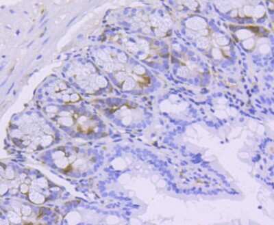
Immunohistochemistry-Paraffin: CDK2 [p Tyr15] Antibody (SN72-04) [NBP2-67686] - Analysis of paraffin-embedded mouse colon tissue using anti-Phospho-Cdk2(Y15) antibody. Counter stained with hematoxylin.
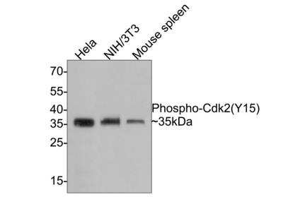
Western Blot: CDK2 [p Tyr15] Antibody (SN72-04) [NBP2-67686] - Lane 1: Hela cell lysate (10 ug/Lane)Lane 2: NIH/3T3 cell lysate (10 ug/Lane)Lane 3: Mouse spleen tissue lysate (20 ug/Lane)Predicted band size: 34 kDa Observed band size: 35 kDa Exposure time: 1 minute; 12% SDS-PAGE gel. Proteins were transferred to a PVDF membrane and blocked with 5% NFDM/TBST for 1 hour at room temperature. The primary antibody (ET1611-52) at 1/1,000 dilution was used in 5% NFDM/TBST at room temperature for 2 hours. Goat Anti-Rabbit IgG - HRP Secondary Antibody (HA1001) at 1:300,000 dilution was used for 1 hour at room temperature.
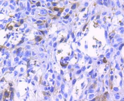
Immunohistochemistry-Paraffin: CDK2 [p Tyr15] Antibody (SN72-04) [NBP2-67686] - Analysis of paraffin-embedded human breast carcinoma tissue using anti-Phospho-Cdk2(Y15) antibody. Counter stained with hematoxylin.
Applications for CDK2 [p Tyr15] Antibody (SN72-04)
Application
Recommended Usage
Immunohistochemistry-Paraffin
1:200-1:1000
Western Blot
1:1000
Application Notes
IP: Use at an assay dependent concentration.
Formulation, Preparation, and Storage
Purification
Protein A purified
Formulation
TBS (pH7.4), 0.05% BSA, 40% Glycerol
Preservative
0.05% Sodium Azide
Concentration
1 mg/ml
Shipping
The product is shipped with polar packs. Upon receipt, store it immediately at the temperature recommended below.
Stability & Storage
Store at 4C short term. Aliquot and store at -20C long term. Avoid freeze-thaw cycles.
Background: CDK2
Long Name
Cyclin-dependent Kinase 2
Alternate Names
cdc2-related protein kinase, cell devision kinase 2, Cell division protein kinase 2, cyclin-dependent kinase 2, EC 2.7.11, EC 2.7.11.22, p33 protein kinase, p33(CDK2)
Gene Symbol
CDK2
Additional CDK2 Products
Product Documents for CDK2 [p Tyr15] Antibody (SN72-04)
Certificate of Analysis
To download a Certificate of Analysis, please enter a lot or batch number in the search box below.
Product Specific Notices for CDK2 [p Tyr15] Antibody (SN72-04)
This product is for research use only and is not approved for use in humans or in clinical diagnosis. Primary Antibodies are guaranteed for 1 year from date of receipt.
Related Research Areas
Customer Reviews for CDK2 [p Tyr15] Antibody (SN72-04)
There are currently no reviews for this product. Be the first to review CDK2 [p Tyr15] Antibody (SN72-04) and earn rewards!
Have you used CDK2 [p Tyr15] Antibody (SN72-04)?
Submit a review and receive an Amazon gift card!
$25/€18/£15/$25CAN/¥2500 Yen for a review with an image
$10/€7/£6/$10CAN/¥1110 Yen for a review without an image
Submit a review
Protocols
Find general support by application which include: protocols, troubleshooting, illustrated assays, videos and webinars.
- Antigen Retrieval Protocol (PIER)
- Antigen Retrieval for Frozen Sections Protocol
- Appropriate Fixation of IHC/ICC Samples
- Cellular Response to Hypoxia Protocols
- Chromogenic IHC Staining of Formalin-Fixed Paraffin-Embedded (FFPE) Tissue Protocol
- Chromogenic Immunohistochemistry Staining of Frozen Tissue
- ClariTSA™ Fluorophore Kits
- Detection & Visualization of Antibody Binding
- Fluorescent IHC Staining of Frozen Tissue Protocol
- Graphic Protocol for Heat-induced Epitope Retrieval
- Graphic Protocol for the Preparation and Fluorescent IHC Staining of Frozen Tissue Sections
- Graphic Protocol for the Preparation and Fluorescent IHC Staining of Paraffin-embedded Tissue Sections
- Graphic Protocol for the Preparation of Gelatin-coated Slides for Histological Tissue Sections
- IHC Sample Preparation (Frozen sections vs Paraffin)
- Immunofluorescent IHC Staining of Formalin-Fixed Paraffin-Embedded (FFPE) Tissue Protocol
- Immunohistochemistry (IHC) and Immunocytochemistry (ICC) Protocols
- Immunohistochemistry Frozen Troubleshooting
- Immunohistochemistry Paraffin Troubleshooting
- Immunoprecipitation Protocol
- Preparing Samples for IHC/ICC Experiments
- Preventing Non-Specific Staining (Non-Specific Binding)
- Primary Antibody Selection & Optimization
- Protocol for Heat-Induced Epitope Retrieval (HIER)
- Protocol for Making a 4% Formaldehyde Solution in PBS
- Protocol for VisUCyte™ HRP Polymer Detection Reagent
- Protocol for the Preparation & Fixation of Cells on Coverslips
- Protocol for the Preparation and Chromogenic IHC Staining of Frozen Tissue Sections
- Protocol for the Preparation and Chromogenic IHC Staining of Frozen Tissue Sections - Graphic
- Protocol for the Preparation and Chromogenic IHC Staining of Paraffin-embedded Tissue Sections
- Protocol for the Preparation and Chromogenic IHC Staining of Paraffin-embedded Tissue Sections - Graphic
- Protocol for the Preparation and Fluorescent IHC Staining of Frozen Tissue Sections
- Protocol for the Preparation and Fluorescent IHC Staining of Paraffin-embedded Tissue Sections
- Protocol for the Preparation of Gelatin-coated Slides for Histological Tissue Sections
- R&D Systems Quality Control Western Blot Protocol
- TUNEL and Active Caspase-3 Detection by IHC/ICC Protocol
- The Importance of IHC/ICC Controls
- Troubleshooting Guide: Immunohistochemistry
- Troubleshooting Guide: Western Blot Figures
- Western Blot Conditions
- Western Blot Protocol
- Western Blot Protocol for Cell Lysates
- Western Blot Troubleshooting
- Western Blot Troubleshooting Guide
- View all Protocols, Troubleshooting, Illustrated assays and Webinars
Loading...
Associated Pathways
