CDP/CUTL1 Antibody - BSA Free
Novus Biologicals | Catalog # NBP2-13883
![Western Blot: CDP/CUTL1 Antibody [NBP2-13883]](https://resources.rndsystems.com/images/products/CDP-CUTL1-Antibody-Western-Blot-NBP2-13883-img0007.jpg "Western Blot: CDP/CUTL1 Antibody [NBP2-13883]")
Loading...
Key Product Details
Species Reactivity
Validated:
Human
Cited:
Human, Mouse
Predicted:
Rat (96%). Backed by our 100% Guarantee.
Applications
Validated:
Immunohistochemistry-Frozen, Western Blot, Chromatin Immunoprecipitation-exo-Seq, Microarray
Cited:
Western Blot
Label
Unconjugated
Antibody Source
Polyclonal Rabbit IgG
Format
BSA Free
Loading...
Product Specifications
Immunogen
This antibody was developed against a recombinant protein corresponding to the amino acids: KSFQGEIDALSKRSKEAEAAFLNVYKRLIDVPDPVPALDLGQQLQLKVQRLHDIETENQKLRETLEEYNKEFAEVKNQEVTIKALKEKIREYEQTLKNQAETIALEKEQKLQNDFAEKERKLQETQMSTTSKLEEAEHKVQSLQTALE
Reactivity Notes
Mouse reactivity reported in scientific literature (PMID: 27471254) and verified customer review.
Clonality
Polyclonal
Host
Rabbit
Isotype
IgG
Scientific Data Images for CDP/CUTL1 Antibody - BSA Free
Western Blot: CDP/CUTL1 Antibody [NBP2-13883]
Western Blot: CDP/CUTL1 Antibody [NBP2-13883] - Analysis in human thyroid gland tissue.![Immunohistochemistry-Frozen: CDP/CUTL1 Antibody [NBP2-13883]](https://resources.rndsystems.com/images/products/CDP-CUTL1-Antibody-Immunohistochemistry-Frozen-NBP2-13883-img0004.jpg "Immunohistochemistry-Frozen: CDP/CUTL1 Antibody [NBP2-13883]")
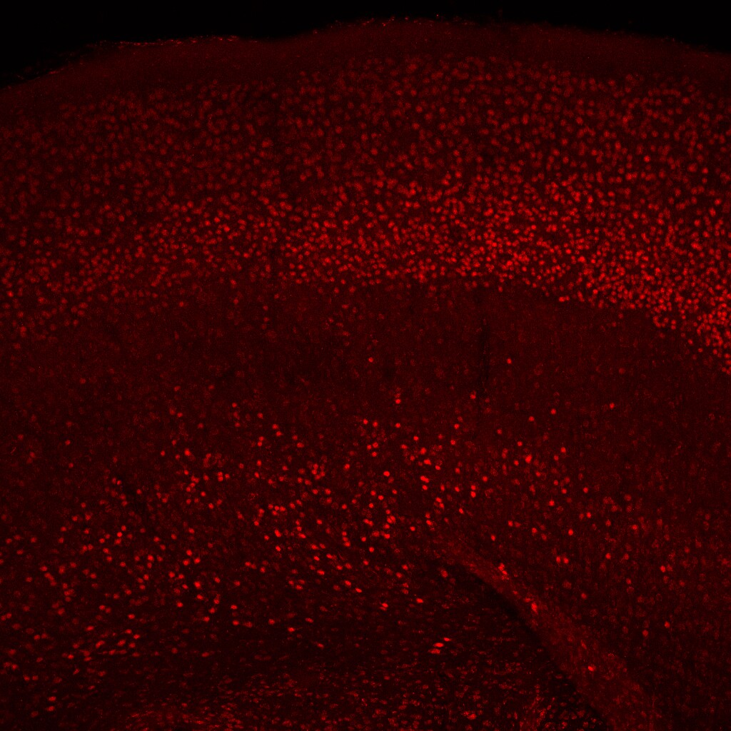
Immunohistochemistry-Frozen: CDP/CUTL1 Antibody [NBP2-13883]
Immunohistochemistry-Frozen: CDP/CUTL1 Antibody [NBP2-13883] - P15 mouse brain stained with CUTL1 antibody. IHC-Fr image submitted by a verified customer review.![CDP/CUTL1 Antibody - BSA Free Chromatin Immunoprecipitation-exo-Seq: CDP/CUTL1 Antibody - BSA Free [NBP2-13883]](https://resources.rndsystems.com/images/products/nbp2-13883_rabbit-polyclonal-cdp-cutl1-antibody-2562025913919.jpg "Chromatin Immunoprecipitation-exo-Seq: CDP/CUTL1 Antibody - BSA Free [NBP2-13883]")
Chromatin Immunoprecipitation-exo-Seq: CDP/CUTL1 Antibody - BSA Free [NBP2-13883]
ChIP-Exo-Seq composite graph for Anti-CUX1 (NBP2-13883) tested in K562 cells. Strand-specific reads (blue: forward, red: reverse) and IgG controls (black: forward, grey: reverse) are plotted against the distance from a composite set of reference binding sites. The antibody exhibits robust target enrichment compared to a non-specific IgG control and precisely reveals its structural organization around the binding site. Data generated by Prof. B. F. Pugh´s Lab at Cornell University.Applications for CDP/CUTL1 Antibody - BSA Free
Application
Recommended Usage
Chromatin Immunoprecipitation-exo-Seq
1-10ug per reaction
Immunohistochemistry-Frozen
Validated from a verified customer review.
Microarray
Validated from a verified customer review.
Western Blot
0.04 - 0.4 ug/ml
Reviewed Applications
Read 2 reviews rated 3.5 using NBP2-13883 in the following applications:
Formulation, Preparation, and Storage
Purification
Affinity purified
Formulation
PBS (pH 7.2) and 40% Glycerol
Format
BSA Free
Preservative
0.02% Sodium Azide
Concentration
Concentrations vary lot to lot. See vial label for concentration. If unlisted please contact technical services.
Shipping
The product is shipped with polar packs. Upon receipt, store it immediately at the temperature recommended below.
Stability & Storage
Store at 4C short term. Aliquot and store at -20C long term. Avoid freeze-thaw cycles.
Background: CDP/CUTL1
Long Name
CCAAT Displacement protein/CUT-like Homeobox Protein 1
Alternate Names
CASP, COY1, CUTL1, CUX, CUX1, GOLIM6
Gene Symbol
CUX1
Additional CDP/CUTL1 Products
Product Documents for CDP/CUTL1 Antibody - BSA Free
Certificate of Analysis
To download a Certificate of Analysis, please enter a lot or batch number in the search box below.
Product Specific Notices for CDP/CUTL1 Antibody - BSA Free
This product is for research use only and is not approved for use in humans or in clinical diagnosis. Primary Antibodies are guaranteed for 1 year from date of receipt.
Related Research Areas
Citations for CDP/CUTL1 Antibody - BSA Free
 Powered by Bioz
Customer Reviews for CDP/CUTL1 Antibody - BSA Free (2)
3.5 out of 5
2 Customer Ratings
Have you used CDP/CUTL1 Antibody - BSA Free?
Submit a review and receive an Amazon gift card!
$25/€18/£15/$25CAN/¥2500 Yen for a review with an image
$10/€7/£6/$10CAN/¥1110 Yen for a review without an image
Submit a review
Customer Images

Showing
1
-
2 of
2 reviews
Showing All
Filter By:
-
Application: MicroarraySample Tested: EDTA PlasmaSpecies: HumanVerified Customer | Posted 12/07/2020Antibody was printed on custom arrays and incubated with fluorescently labeled human EDTA plasma

-
Application: Immunohistochemistry-FrozenSample Tested: P15 Mouse BrainSpecies: MouseVerified Customer | Posted 03/13/2016Cux1 P15 mouse brain
There are no reviews that match your criteria.
Protocols
Find general support by application which include: protocols, troubleshooting, illustrated assays, videos and webinars.
- Antigen Retrieval Protocol (PIER)
- Antigen Retrieval for Frozen Sections Protocol
- Appropriate Fixation of IHC/ICC Samples
- Cellular Response to Hypoxia Protocols
- Chromogenic IHC Staining of Formalin-Fixed Paraffin-Embedded (FFPE) Tissue Protocol
- Chromogenic Immunohistochemistry Staining of Frozen Tissue
- ClariTSA™ Fluorophore Kits
- Detection & Visualization of Antibody Binding
- Fluorescent IHC Staining of Frozen Tissue Protocol
- Graphic Protocol for Heat-induced Epitope Retrieval
- Graphic Protocol for the Preparation and Fluorescent IHC Staining of Frozen Tissue Sections
- Graphic Protocol for the Preparation and Fluorescent IHC Staining of Paraffin-embedded Tissue Sections
- Graphic Protocol for the Preparation of Gelatin-coated Slides for Histological Tissue Sections
- IHC Sample Preparation (Frozen sections vs Paraffin)
- Immunofluorescent IHC Staining of Formalin-Fixed Paraffin-Embedded (FFPE) Tissue Protocol
- Immunohistochemistry (IHC) and Immunocytochemistry (ICC) Protocols
- Immunohistochemistry Frozen Troubleshooting
- Immunohistochemistry Paraffin Troubleshooting
- Preparing Samples for IHC/ICC Experiments
- Preventing Non-Specific Staining (Non-Specific Binding)
- Primary Antibody Selection & Optimization
- Protocol for Heat-Induced Epitope Retrieval (HIER)
- Protocol for Making a 4% Formaldehyde Solution in PBS
- Protocol for VisUCyte™ HRP Polymer Detection Reagent
- Protocol for the Preparation & Fixation of Cells on Coverslips
- Protocol for the Preparation and Chromogenic IHC Staining of Frozen Tissue Sections
- Protocol for the Preparation and Chromogenic IHC Staining of Frozen Tissue Sections - Graphic
- Protocol for the Preparation and Chromogenic IHC Staining of Paraffin-embedded Tissue Sections
- Protocol for the Preparation and Chromogenic IHC Staining of Paraffin-embedded Tissue Sections - Graphic
- Protocol for the Preparation and Fluorescent IHC Staining of Frozen Tissue Sections
- Protocol for the Preparation and Fluorescent IHC Staining of Paraffin-embedded Tissue Sections
- Protocol for the Preparation of Gelatin-coated Slides for Histological Tissue Sections
- R&D Systems Quality Control Western Blot Protocol
- TUNEL and Active Caspase-3 Detection by IHC/ICC Protocol
- The Importance of IHC/ICC Controls
- Troubleshooting Guide: Immunohistochemistry
- Troubleshooting Guide: Western Blot Figures
- Western Blot Conditions
- Western Blot Protocol
- Western Blot Protocol for Cell Lysates
- Western Blot Troubleshooting
- Western Blot Troubleshooting Guide
- View all Protocols, Troubleshooting, Illustrated assays and Webinars
Loading...