
CKIP-1 Antibody - BSA Free
Novus Biologicals | Catalog # NBP1-76238

![Western Blot: CKIP-1 AntibodyBSA Free [NBP1-76238]](https://resources.rndsystems.com/images/products/CKIP-1-Antibody-Western-Blot-NBP1-76238-img0001.jpg "Western Blot: CKIP-1 AntibodyBSA Free [NBP1-76238]")
Scientific images may be resized, cropped, or adjusted for brightness or contrast for presentation purposes. These updates are limited to presentation and do not affect the underlying scientific interpretation of the data. Where available, additional source data may be provided upon request.
Key Product Details
Species Reactivity
Validated:
Cited:
Applications
Validated:
Cited:
Label
Antibody Source
Format
Product Specifications
Immunogen
Reactivity Notes
Clonality
Host
Isotype
Scientific Data Images for CKIP-1 Antibody - BSA Free
Western Blot: CKIP-1 AntibodyBSA Free [NBP1-76238]
Western Blot: CKIP-1 Antibody [NBP1-76238] - Human lung tissue lysate with CKIP1 antibody at (A) 1 and (B) 2 ug/mL.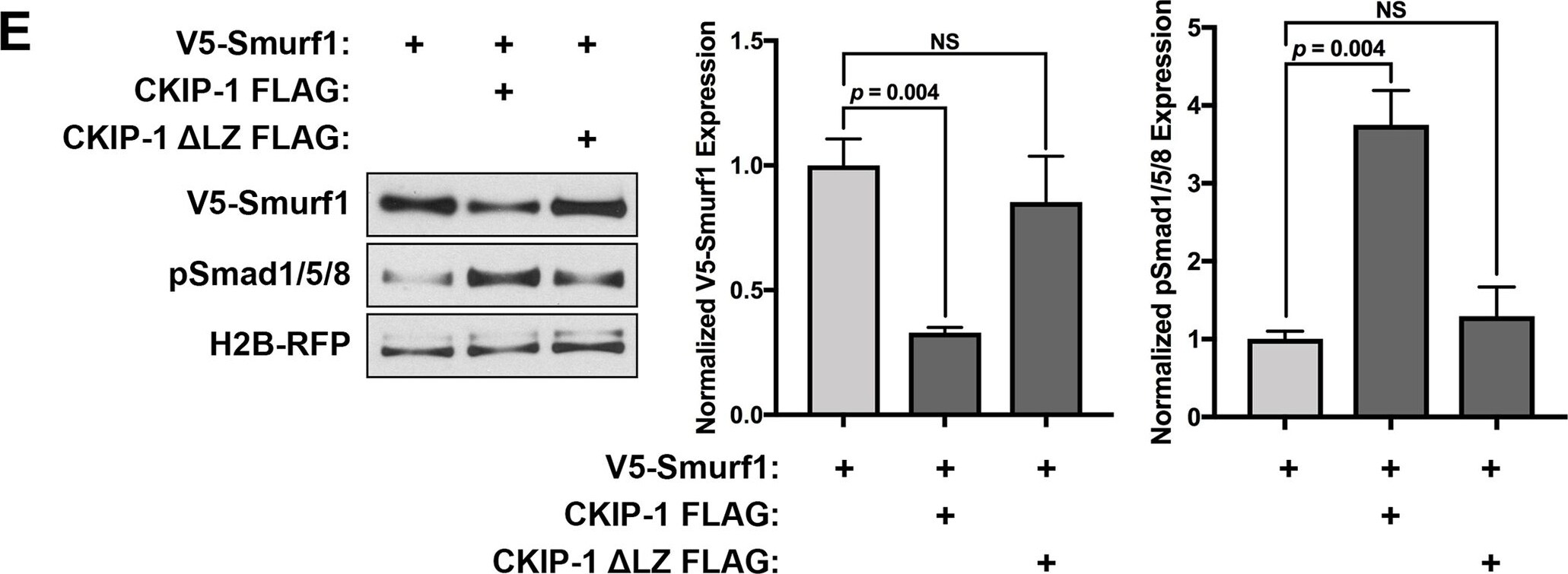
Western Blot: CKIP-1 Antibody - BSA Free [NBP1-76238] -
Western Blot: CKIP-1 Antibody - BSA Free [NBP1-76238] - CKIP-1 directly interacts with Smurf1 to promote Smurf1 degradation.DF-1 chicken fibroblasts were transfected with the indicated constructs & then immunostained using anti-FLAG & anti-V5 antibodies. (A) Localization of FLAG-tagged CKIP-1 & V5-tagged Smurf1 alone. (B) Schematics for CKIP-1 FLAG constructs highlighting the membrane-binding PH domain (red) & Smurf1-binding LZ (blue) followed by a C-terminal FLAG tag (yellow). (C) Full-length CKIP-1 FLAG was cotransfected with full-length V5-Smurf1 or V5-Smurf1 C700A dominant-negative mutant. In addition, CKIP-1 FLAG truncation mutants were cotransfected with full-length V5-Smurf1, showing that the PH domain is required for efficient membrane localization, & the LZ domain is required for Smurf1 colocalization. Arrows indicate membrane localization. Scale bars represent 10 μm. (D) Chick embryos were electroporated at HH4 with V5-Smurf1 & CKIP-1 FLAG & then stained for V5 or FLAG at HH8 & imaged in cryosection. Scale bars represent 25 μm. (E) Western blot analysis of HH7 whole-embryo lysates electroporated with constructs indicated & probed for V5, pSmad1/5/8, & RFP. Bar graphs represent normalized V5 & pSmad1/5/8 staining intensity means ± SEM from 3 replicates. Underlying data can be found in S1 Data. P values from two-tailed Student t test. CKIP-1, casein kinase interacting protein 1; HH, Hamburger-Hamilton stage; LZ, leucine zipper motif; NC; neural crest domain; NS, not significant; PH, pleckstrin homology; pSmad1/5/8, phospho-Smads 1/5/8; RFP, red fluorescent protein; Smurf1, Smad ubiquitin regulatory factor 1. Image collected & cropped by CiteAb from the following publication (https://pubmed.ncbi.nlm.nih.gov/29949573), licensed under a CC-BY license. Not internally tested by Novus Biologicals.Applications for CKIP-1 Antibody - BSA Free
ELISA
Western Blot
Formulation, Preparation, and Storage
Purification
Formulation
Format
Preservative
Concentration
Shipping
Stability & Storage
Background: CKIP-1
Alternate Names
Gene Symbol
UniProt
Additional CKIP-1 Products
Product Documents for CKIP-1 Antibody - BSA Free
Certificate of Analysis
To download a Certificate of Analysis, please enter a lot or batch number in the search box below.
Product Specific Notices for CKIP-1 Antibody - BSA Free
This product is for research use only and is not approved for use in humans or in clinical diagnosis. Primary Antibodies are guaranteed for 1 year from date of receipt.
Citations for CKIP-1 Antibody - BSA Free
 Powered by Bioz
Customer Reviews for CKIP-1 Antibody - BSA Free
There are currently no reviews for this product. Be the first to review CKIP-1 Antibody - BSA Free and earn rewards!
Have you used CKIP-1 Antibody - BSA Free?
Submit a review and receive an Amazon gift card!
$25/€18/£15/$25CAN/¥2500 for a review with an image
$10/€7/£6/$10CAN/¥1110 for a review without an image
Submit a review
Protocols
Find general support by application which include: protocols, troubleshooting, illustrated assays, videos and webinars.
- Cellular Response to Hypoxia Protocols
- ELISA Sample Preparation & Collection Guide
- ELISA Troubleshooting Guide
- How to Run an R&D Systems DuoSet ELISA
- How to Run an R&D Systems Quantikine ELISA
- How to Run an R&D Systems Quantikine™ QuicKit™ ELISA
- Quantikine HS ELISA Kit Assay Principle, Alkaline Phosphatase
- Quantikine HS ELISA Kit Principle, Streptavidin-HRP Polymer
- R&D Systems Quality Control Western Blot Protocol
- Sandwich ELISA (Colorimetric) – Biotin/Streptavidin Detection Protocol
- Sandwich ELISA (Colorimetric) – Direct Detection Protocol
- Troubleshooting Guide: ELISA
- Troubleshooting Guide: Western Blot Figures
- Western Blot Conditions
- Western Blot Protocol
- Western Blot Protocol for Cell Lysates
- Western Blot Troubleshooting
- Western Blot Troubleshooting Guide
- View all Protocols, Troubleshooting, Illustrated assays and Webinars