CREB Antibody - BSA Free
Novus Biologicals | Catalog # NBP1-90364
![Immunohistochemistry-Paraffin: CREB Antibody [NBP1-90364]](https://resources.rndsystems.com/images/products/CREB-Antibody-Immunohistochemistry-Paraffin-NBP1-90364-img0002.jpg "Immunohistochemistry-Paraffin: CREB Antibody [NBP1-90364]")
Key Product Details
Species Reactivity
Validated:
Cited:
Applications
Validated:
Cited:
Label
Antibody Source
Format
Product Specifications
Immunogen
Clonality
Host
Isotype
Scientific Data Images for CREB Antibody - BSA Free
Immunohistochemistry-Paraffin: CREB Antibody [NBP1-90364]
Immunohistochemistry-Paraffin: CREB Antibody [NBP1-90364] - Staining of human small intestine shows strong nuclear positivity in glandular cells.![CREB Antibody - BSA Free Chromatin Immunoprecipitation-exo-Seq: CREB Antibody - BSA Free [NBP1-90364]](https://resources.rndsystems.com/images/products/nbp1-90364_rabbit-polyclonal-creb-antibody-256202591432.jpg "Chromatin Immunoprecipitation-exo-Seq: CREB Antibody - BSA Free [NBP1-90364]")
Chromatin Immunoprecipitation-exo-Seq: CREB Antibody - BSA Free [NBP1-90364]
ChIP-Exo-Seq composite graph for Anti-CREB1 (NBP1-90364) tested in K562 cells. Strand-specific reads (blue: forward, red: reverse) and IgG controls (black: forward, grey: reverse) are plotted against the distance from a composite set of reference binding sites. The antibody exhibits robust target enrichment compared to a non-specific IgG control and precisely reveals its structural organization around the binding site. Data generated by Prof. B. F. Pugh´s Lab at Cornell University.![CREB Antibody - BSA Free Western Blot: CREB Antibody - BSA Free [NBP1-90364]](https://resources.rndsystems.com/images/products/nbp1-90364_rabbit-polyclonal-creb-antibody-3052025726383.jpg "Western Blot: CREB Antibody - BSA Free [NBP1-90364]")
Western Blot: CREB Antibody - BSA Free [NBP1-90364]
Lane 1: Marker [kDa] 230, 130, 95, 72, 56, 36, 28, 17, 11Lane 2: Human cell line RT-4
Lane 3: Human cell line U-251MG sp
![CREB Antibody - BSA Free Western Blot: CREB Antibody - BSA Free [NBP1-90364]](https://resources.rndsystems.com/images/products/nbp1-90364_rabbit-polyclonal-creb-antibody-3052025957016.jpg "Western Blot: CREB Antibody - BSA Free [NBP1-90364]")
Western Blot: CREB Antibody - BSA Free [NBP1-90364]
Lane 1: NIH-3T3 cell lysate (Mouse embryonic fibroblast cells)Lane 2: NBT-II cell lysate (Rat Wistar bladder tumour cells)
![Simple Western: CREB Antibody [NBP1-90364]](https://resources.rndsystems.com/images/products/CREB-Antibody-Simple-Western-NBP1-90364-img0008.jpg "Simple Western: CREB Antibody [NBP1-90364]")
Simple Western: CREB Antibody [NBP1-90364]
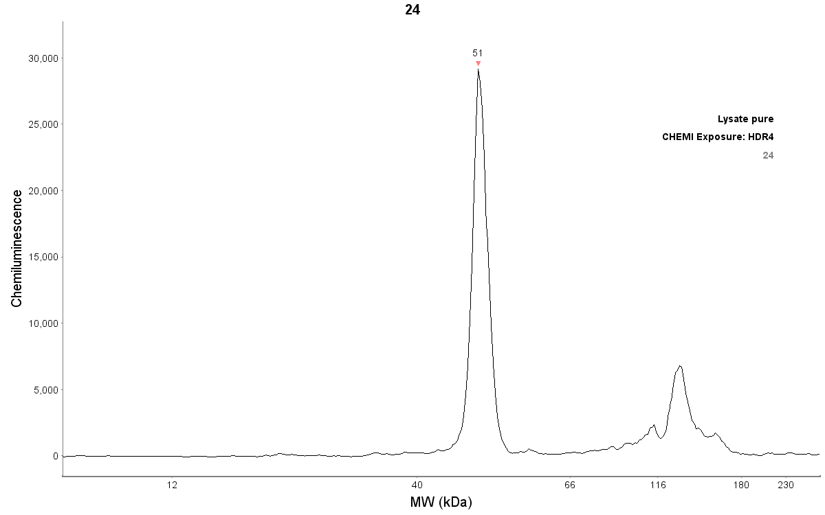
Simple Western: CREB Antibody [NBP1-90364] - Human foreskin cell lysate. CREB antibody dilution of 1:50. Protein concentration is 750 ug/mL. Detection is chemiluminescence. Simple Western image submitted by a verified customer review.![Simple Western: CREB Antibody [NBP1-90364]](https://resources.rndsystems.com/images/products/CREB-Antibody-Simple-Western-NBP1-90364-img0009.jpg "Simple Western: CREB Antibody [NBP1-90364]")
Simple Western: CREB Antibody [NBP1-90364]
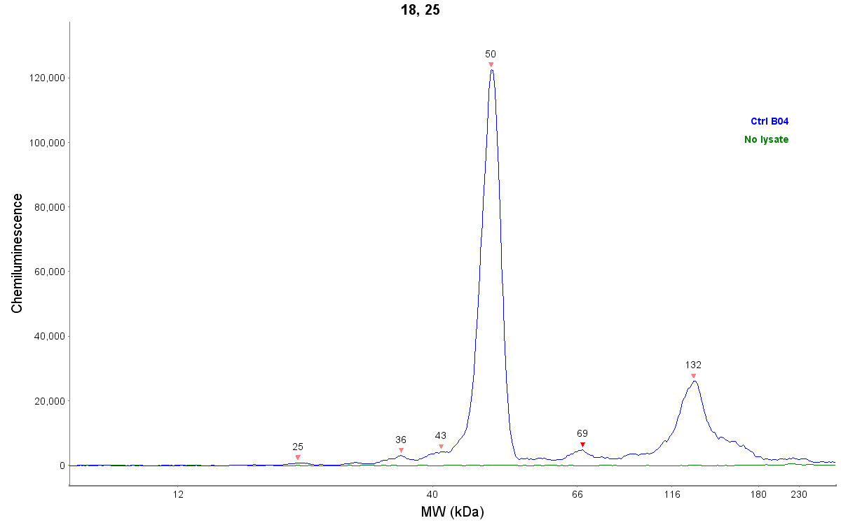
Simple Western: CREB Antibody [NBP1-90364] - Detection of CREB by Simple Western (JESS) in reconstructed human epidermis lysate (about 600 ug/mL protein concentration). Antibody at 1:50. Simple Western image submitted by a verified customer review.
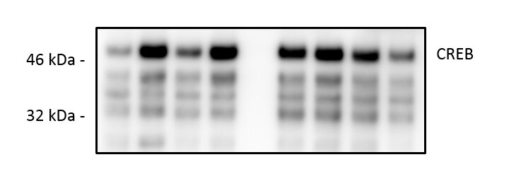
Western Blot: CREB Antibody - BSA Free [NBP1-90364] -
The effects of perampanel (PER) and GYKI 52,466 (GYKI) on total Ca2+/cAMP response element-binding protein (CREB) and its S133 phosphorylation in chronic epilepsy rats. Both AMPAR antagonists reduce CREB S133 phosphorylation in responders (Resp), but not non-responders (Non-Resp). (A) Representative images for Western blot of CREB and p-CREB levels in the hippocampal tissues. (B–F) Quantifications of CREB (B), p-CREB S133 (C), and p-CREB S133 ratio (D) levels in the hippocampal tissues. Open circles indicate each individual value. Horizontal bars indicate mean value. Error bars indicate SEM (*, # p < 0.05 vs. control and vehicle (Veh)-treated animals, respectively; one-way ANOVA with post hoc Bonferroni’s multiple comparison). Image collected and cropped by CiteAb from the following open publication (https://pubmed.ncbi.nlm.nih.gov/33348808), licensed under a CC-BY license. Not internally tested by Novus Biologicals.![CREB Antibody - BSA Free Immunocytochemistry/ Immunofluorescence: CREB Antibody - BSA Free [NBP1-90364]](https://resources.rndsystems.com/images/products/nbp1-90364_-immunocytochemistry-immunofluorescence-639174076824572858.jpg "Immunocytochemistry/ Immunofluorescence: CREB Antibody - BSA Free [NBP1-90364]")
Immunocytochemistry/ Immunofluorescence: CREB Antibody - BSA Free [NBP1-90364]
Staining of human cell line U-2 OS shows localization to nucleoplasm.Applications for CREB Antibody - BSA Free
Chromatin Immunoprecipitation-exo-Seq
Immunocytochemistry/ Immunofluorescence
Immunohistochemistry
Immunohistochemistry-Paraffin
Simple Western
Western Blot
Reviewed Applications
Read 3 reviews rated 4.7 using NBP1-90364 in the following applications:
Formulation, Preparation, and Storage
Purification
Formulation
Format
Preservative
Concentration
Shipping
Stability & Storage
Background: CREB
Long Name
Alternate Names
Gene Symbol
Additional CREB Products
Product Documents for CREB Antibody - BSA Free
Certificate of Analysis
To download a Certificate of Analysis, please enter a lot or batch number in the search box below.
Product Specific Notices for CREB Antibody - BSA Free
This product is for research use only and is not approved for use in humans or in clinical diagnosis. Primary Antibodies are guaranteed for 1 year from date of receipt.
Citations for CREB Antibody - BSA Free
 Powered by Bioz
Customer Reviews for CREB Antibody - BSA Free (3)
Have you used CREB Antibody - BSA Free?
Submit a review and receive an Amazon gift card!
$25/€18/£15/$25CAN/¥2500 Yen for a review with an image
$10/€7/£6/$10CAN/¥1110 Yen for a review without an image
Submit a review
Customer Images
-
Application: Western BlotSample Tested: EndothelialSpecies: MouseVerified Customer | Posted 10/11/2019

-
Application: Simple WesternSample Tested: Human foreskin, adult ski, engineered human skin, keratinocytes, HaCaT cellsSpecies: HumanVerified Customer | Posted 06/17/2019Detection of CREB by Simple Western (JESS) in reconstructed human epidermis lysate (about 600 ug/mL protein concentration). Antibody dilution: 1/50
-
Application: Simple WesternSample Tested: Human foreskin, adult ski, engineered human skin, keratinocytes, HaCaT cellsSpecies: HumanVerified Customer | Posted 05/24/2019CREB antibody dilution of 1:50. Protein concentration is 750 ug/mL. Detection is chemiluminescence.JESS default run settings.
There are no reviews that match your criteria.
Protocols
Find general support by application which include: protocols, troubleshooting, illustrated assays, videos and webinars.
- Antigen Retrieval Protocol (PIER)
- Antigen Retrieval for Frozen Sections Protocol
- Appropriate Fixation of IHC/ICC Samples
- Cellular Response to Hypoxia Protocols
- Chromogenic IHC Staining of Formalin-Fixed Paraffin-Embedded (FFPE) Tissue Protocol
- Chromogenic Immunohistochemistry Staining of Frozen Tissue
- ClariTSA™ Fluorophore Kits
- Detection & Visualization of Antibody Binding
- Fluorescent IHC Staining of Frozen Tissue Protocol
- Graphic Protocol for Heat-induced Epitope Retrieval
- Graphic Protocol for the Preparation and Fluorescent IHC Staining of Frozen Tissue Sections
- Graphic Protocol for the Preparation and Fluorescent IHC Staining of Paraffin-embedded Tissue Sections
- Graphic Protocol for the Preparation of Gelatin-coated Slides for Histological Tissue Sections
- ICC Cell Smear Protocol for Suspension Cells
- ICC Immunocytochemistry Protocol Videos
- ICC for Adherent Cells
- IHC Sample Preparation (Frozen sections vs Paraffin)
- Immunocytochemistry (ICC) Protocol
- Immunocytochemistry Troubleshooting
- Immunofluorescence of Organoids Embedded in Cultrex Basement Membrane Extract
- Immunofluorescent IHC Staining of Formalin-Fixed Paraffin-Embedded (FFPE) Tissue Protocol
- Immunohistochemistry (IHC) and Immunocytochemistry (ICC) Protocols
- Immunohistochemistry Frozen Troubleshooting
- Immunohistochemistry Paraffin Troubleshooting
- Preparing Samples for IHC/ICC Experiments
- Preventing Non-Specific Staining (Non-Specific Binding)
- Primary Antibody Selection & Optimization
- Protocol for Heat-Induced Epitope Retrieval (HIER)
- Protocol for Making a 4% Formaldehyde Solution in PBS
- Protocol for VisUCyte™ HRP Polymer Detection Reagent
- Protocol for the Fluorescent ICC Staining of Cell Smears - Graphic
- Protocol for the Fluorescent ICC Staining of Cultured Cells on Coverslips - Graphic
- Protocol for the Preparation & Fixation of Cells on Coverslips
- Protocol for the Preparation and Chromogenic IHC Staining of Frozen Tissue Sections
- Protocol for the Preparation and Chromogenic IHC Staining of Frozen Tissue Sections - Graphic
- Protocol for the Preparation and Chromogenic IHC Staining of Paraffin-embedded Tissue Sections
- Protocol for the Preparation and Chromogenic IHC Staining of Paraffin-embedded Tissue Sections - Graphic
- Protocol for the Preparation and Fluorescent ICC Staining of Cells on Coverslips
- Protocol for the Preparation and Fluorescent ICC Staining of Non-adherent Cells
- Protocol for the Preparation and Fluorescent ICC Staining of Stem Cells on Coverslips
- Protocol for the Preparation and Fluorescent IHC Staining of Frozen Tissue Sections
- Protocol for the Preparation and Fluorescent IHC Staining of Paraffin-embedded Tissue Sections
- Protocol for the Preparation of Gelatin-coated Slides for Histological Tissue Sections
- Protocol for the Preparation of a Cell Smear for Non-adherent Cell ICC - Graphic
- R&D Systems Quality Control Western Blot Protocol
- TUNEL and Active Caspase-3 Detection by IHC/ICC Protocol
- The Importance of IHC/ICC Controls
- Troubleshooting Guide: Immunohistochemistry
- Troubleshooting Guide: Western Blot Figures
- Western Blot Conditions
- Western Blot Protocol
- Western Blot Protocol for Cell Lysates
- Western Blot Troubleshooting
- Western Blot Troubleshooting Guide
- View all Protocols, Troubleshooting, Illustrated assays and Webinars
FAQs for CREB Antibody - BSA Free
-
Q: May I know which creb1 antibody is good for ChIP? I found a paper using your rabbit polyclonal creb1 antibody for ChIP but they didn't indicate the catalog number.
A:
We are not aware of any of our Creb1 antibodies being used in ChIP at this time. If you can provide us with the PMID of the paper you are referring to I can contact the author. Otherwise, you can use our Innovators Reward Program to try an antibody in a novel species and application.
Associated Pathways


