
Cytomegalovirus Antibody (DDG9 and CCH2)
Novus Biologicals | Catalog # NBP1-22532

![Immunohistochemistry-Paraffin: Cytomegalovirus Antibody (DDG9 and CCH2) [NBP1-22532]](https://resources.rndsystems.com/images/products/Cytomegalovirus-Antibody-DDG9-and-CCH2-Immunohistochemistry-Paraffin-NBP1-22532-img0004.jpg "Immunohistochemistry-Paraffin: Cytomegalovirus Antibody (DDG9 and CCH2) [NBP1-22532]")
Loading...
Key Product Details
Species Reactivity
Cytomegalovirus, Virus
Applications
Immunohistochemistry, Immunohistochemistry-Paraffin
Label
Unconjugated
Antibody Source
Monoclonal Mouse IgG1 Kappa/IgG2a Kappa Clone # DDG9 and CCH2
Loading...
Product Specifications
Immunogen
BALB/C mice were immunized with cytomegalovirus infected cell lysate. This antibody is a cocktail of DDG9 and CCH2.
Localization
Nuclear
Specificity
This cocktail antibody reacts with an immediate early and early CMV antigens. The antibody stains infected cells giving a nuclear staining pattern with an immediate early and an early antigen.
Clonality
Monoclonal
Host
Mouse
Isotype
IgG1 Kappa/IgG2a Kappa
Scientific Data Images for Cytomegalovirus Antibody (DDG9 and CCH2)
Immunohistochemistry-Paraffin: Cytomegalovirus Antibody (DDG9 and CCH2) [NBP1-22532]
Immunohistochemistry-Paraffin: Cytomegalovirus Antibody (DDG9 and CCH2) [NBP1-22532] - IHC analysis of a formalin fixed paraffin embedded tissue section of CMV infected lung using Cytomegalovirus antibody (clone DDG9 and CCH2).![Immunohistochemistry: Cytomegalovirus Antibody (DDG9 and CCH2) [NBP1-22532]](https://resources.rndsystems.com/images/products/Cytomegalovirus-Antibody-DDG9-and-CCH2-Immunohistochemistry-NBP1-22532-img0005.jpg "Immunohistochemistry: Cytomegalovirus Antibody (DDG9 and CCH2) [NBP1-22532]")
Immunohistochemistry: Cytomegalovirus Antibody (DDG9 and CCH2) [NBP1-22532]
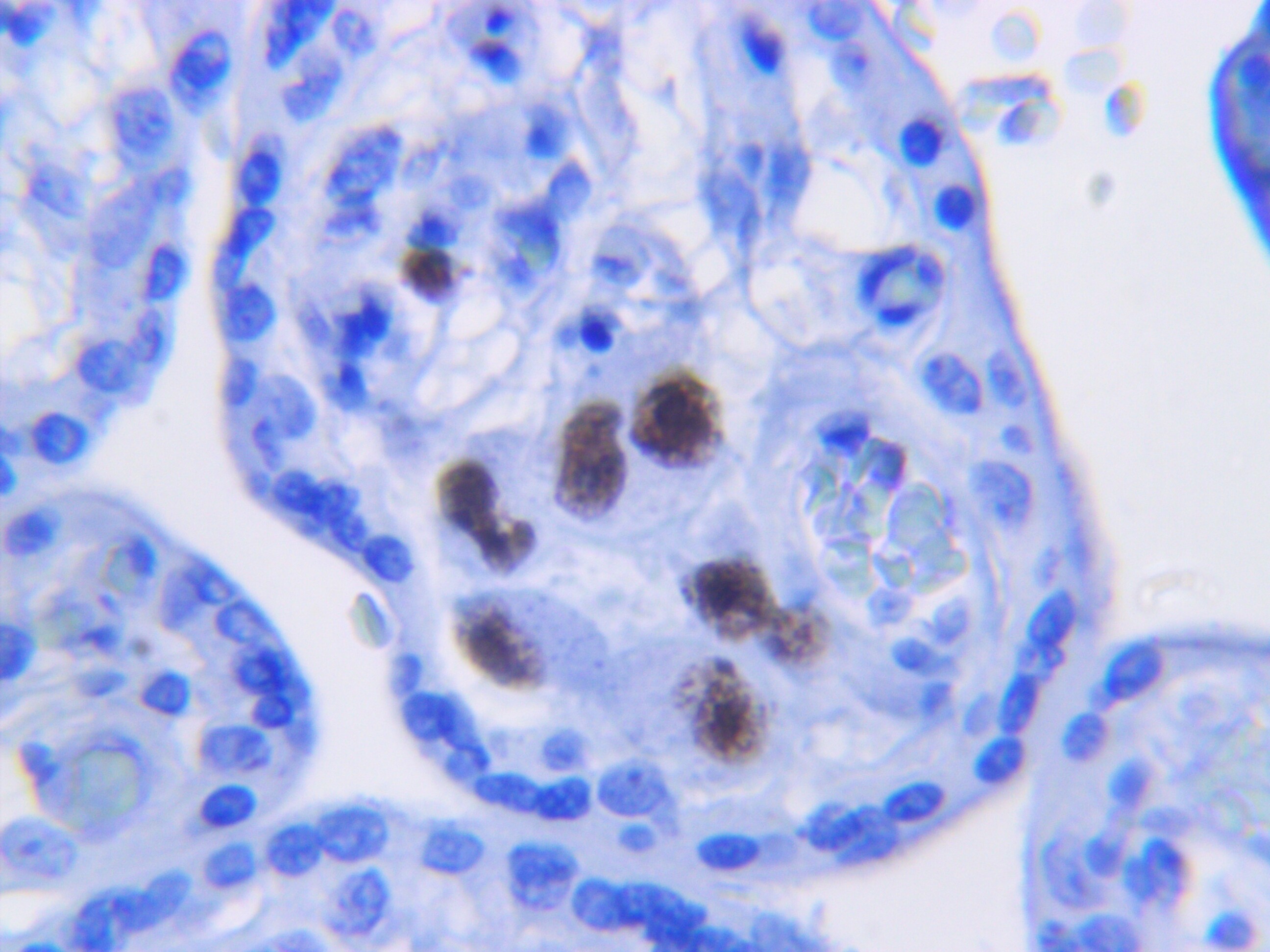
Immunohistochemistry: Cytomegalovirus Antibody (DDG9 and CCH2) [NBP1-22532] - Human Placenta CMV. HIER sodium citrate 6.0 - 30 minutes 1:10 dilution. This image was submitted via customer Review.![Immunohistochemistry-Paraffin: Cytomegalovirus Antibody (DDG9 and CCH2) [NBP1-22532]](https://resources.rndsystems.com/images/products/Cytomegalovirus-Antibody-DDG9-and-CCH2-Immunohistochemistry-Paraffin-NBP1-22532-img0002.jpg "Immunohistochemistry-Paraffin: Cytomegalovirus Antibody (DDG9 and CCH2) [NBP1-22532]")
Immunohistochemistry-Paraffin: Cytomegalovirus Antibody (DDG9 and CCH2) [NBP1-22532]
Immunohistochemistry-Paraffin: Cytomegalovirus Antibody (DDG9 and CCH2) [NBP1-22532] - IHC analysis of a formalin fixed paraffin embedded tissue section of CMV infected lung using Cytomegalovirus antibody (clone DDG9 and CCH2).![Immunohistochemistry-Paraffin: Cytomegalovirus Antibody (DDG9 and CCH2) [NBP1-22532]](https://resources.rndsystems.com/images/products/Cytomegalovirus-Antibody-DDG9-and-CCH2-Immunohistochemistry-Paraffin-NBP1-22532-img0003.jpg "Immunohistochemistry-Paraffin: Cytomegalovirus Antibody (DDG9 and CCH2) [NBP1-22532]")
Immunohistochemistry-Paraffin: Cytomegalovirus Antibody (DDG9 and CCH2) [NBP1-22532]
Immunohistochemistry-Paraffin: Cytomegalovirus Antibody (DDG9 and CCH2) [NBP1-22532] - IHC analysis of a formalin fixed paraffin embedded human salivary glands (CMV infected) using Cytomegalovirus antibody (clone DDG9 and CCH2).Applications for Cytomegalovirus Antibody (DDG9 and CCH2)
Application
Recommended Usage
Immunohistochemistry
1:10-1:25
Immunohistochemistry-Paraffin
1:10-1:25
Application Notes
IHC-P: recommended pretreatment of citrate buffer, pH 6.0. Recommended incubation time of 30 min at RT.
Reviewed Applications
Read 2 reviews rated 5 using NBP1-22532 in the following applications:
Formulation, Preparation, and Storage
Purification
Tissue culture supernatant
Formulation
Tissue culture supernatant
Preservative
0.05% Sodium Azide
Concentration
This product is unpurified. The exact concentration of antibody is not quantifiable.
Shipping
The product is shipped with polar packs. Upon receipt, store it immediately at the temperature recommended below.
Stability & Storage
Store at 4C. Do not freeze.
Background: Cytomegalovirus
Cytomegalovirus is a common virus, infecting ~60% of adults in developed countries and over 90% in developing countries, as indicated by presence of specific IgG antibodies (2-4). Although a majority of people infected remain asymptomatic, immunocompromised people, organ transplant recipients, and fetuses and newborns are more susceptible to HCMV-associated diseases (1-4). Annually, up to 2% of newborns are born with HCMV, making it the most common congenital infection (2-4). Cytomegalovirus is known to cause sensorineural hearing loss (SNHL), mental retardation, chorioretinitis, skin lesions, as well as end organ disease (3,5). There are a few CMV therapeutics approved by FDA including valganciclovir and letermovir; however, despite some benefits, there is the need for new antivirals and combination therapies (5). Potential new treatment options include monoclonal antibodies and sirtuins (5).
References
1. Connolly SA, Jardetzky TS, Longnecker R. The structural basis of herpesvirus entry. Nat Rev Microbiol. 2021;19(2):110-121. https://doi.org/10.1038/s41579-020-00448-w
2. Griffiths P, Reeves M. Pathogenesis of human cytomegalovirus in the immunocompromised host. Nat Rev Microbiol. 2021;19(12):759-773. https://doi.org/10.1038/s41579-021-00582-z
3. Krstanovic F, Britt WJ, Jonjic S, Brizic I. Cytomegalovirus Infection and Inflammation in Developing Brain. Viruses. 2021;13(6):1078. Published 2021 Jun 4. https://doi.org/10.3390/v13061078
4. Griffiths P, Baraniak I, Reeves M. The pathogenesis of human cytomegalovirus. J Pathol. 2015;235(2):288-297. https://doi.org/10.1002/path.4437
5. Acosta E, Bowlin T, Brooks J, et al. Advances in the Development of Therapeutics for Cytomegalovirus Infections. J Infect Dis. 2020;221(Suppl 1):S32-S44. https://doi.org/10.1093/infdis/jiz493
Alternate Names
CMV; HHV 5; HHV5; Human herpesvirus 5
Additional Cytomegalovirus Products
Product Documents for Cytomegalovirus Antibody (DDG9 and CCH2)
Certificate of Analysis
To download a Certificate of Analysis, please enter a lot or batch number in the search box below.
Product Specific Notices for Cytomegalovirus Antibody (DDG9 and CCH2)
This product is for research use only and is not approved for use in humans or in clinical diagnosis. Primary Antibodies are guaranteed for 1 year from date of receipt.
Customer Reviews for Cytomegalovirus Antibody (DDG9 and CCH2) (2)
5 out of 5
2 Customer Ratings
Have you used Cytomegalovirus Antibody (DDG9 and CCH2)?
Submit a review and receive an Amazon gift card!
$25/€18/£15/$25CAN/¥2500 for a review with an image
$10/€7/£6/$10CAN/¥1110 for a review without an image
Submit a review
Customer Images
Showing
1
-
2 of
2 reviews
Showing All
Filter By:
-
Application: ImmunohistochemistrySample Tested: 5um paraffin and Placental tissueSpecies: HumanVerified Customer | Posted 07/07/2017Human Placenta CMVHIER sodium citrate 6.0 - 30 minutes 1:10 dilution

-
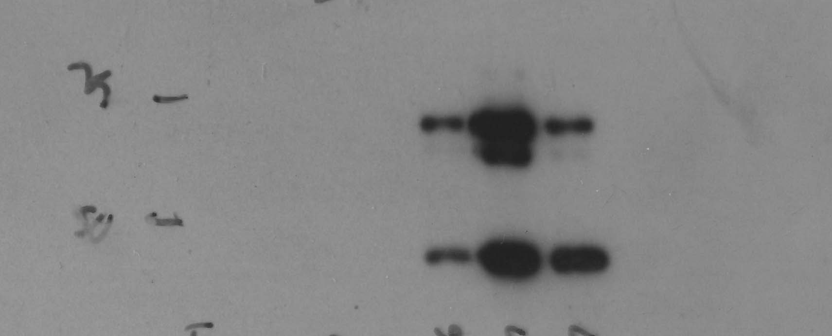
Application: Western BlotSample Tested: Gingival cells and Human Oral KeratinocyteSpecies: HumanVerified Customer | Posted 11/16/2016Keratinocytes Infected with CMV at Harvested at Day 6. 3 Samples were infected with CMV that express DDG9 and CCH2
There are no reviews that match your criteria.
Protocols
Find general support by application which include: protocols, troubleshooting, illustrated assays, videos and webinars.
- Antigen Retrieval Protocol (PIER)
- Antigen Retrieval for Frozen Sections Protocol
- Appropriate Fixation of IHC/ICC Samples
- Cellular Response to Hypoxia Protocols
- Chromogenic IHC Staining of Formalin-Fixed Paraffin-Embedded (FFPE) Tissue Protocol
- Chromogenic Immunohistochemistry Staining of Frozen Tissue
- ClariTSA™ Fluorophore Kits
- Detection & Visualization of Antibody Binding
- Fluorescent IHC Staining of Frozen Tissue Protocol
- Graphic Protocol for Heat-induced Epitope Retrieval
- Graphic Protocol for the Preparation and Fluorescent IHC Staining of Frozen Tissue Sections
- Graphic Protocol for the Preparation and Fluorescent IHC Staining of Paraffin-embedded Tissue Sections
- Graphic Protocol for the Preparation of Gelatin-coated Slides for Histological Tissue Sections
- IHC Sample Preparation (Frozen sections vs Paraffin)
- Immunofluorescent IHC Staining of Formalin-Fixed Paraffin-Embedded (FFPE) Tissue Protocol
- Immunohistochemistry (IHC) and Immunocytochemistry (ICC) Protocols
- Immunohistochemistry Frozen Troubleshooting
- Immunohistochemistry Paraffin Troubleshooting
- Preparing Samples for IHC/ICC Experiments
- Preventing Non-Specific Staining (Non-Specific Binding)
- Primary Antibody Selection & Optimization
- Protocol for Heat-Induced Epitope Retrieval (HIER)
- Protocol for Making a 4% Formaldehyde Solution in PBS
- Protocol for VisUCyte™ HRP Polymer Detection Reagent
- Protocol for the Preparation & Fixation of Cells on Coverslips
- Protocol for the Preparation and Chromogenic IHC Staining of Frozen Tissue Sections
- Protocol for the Preparation and Chromogenic IHC Staining of Frozen Tissue Sections - Graphic
- Protocol for the Preparation and Chromogenic IHC Staining of Paraffin-embedded Tissue Sections
- Protocol for the Preparation and Chromogenic IHC Staining of Paraffin-embedded Tissue Sections - Graphic
- Protocol for the Preparation and Fluorescent IHC Staining of Frozen Tissue Sections
- Protocol for the Preparation and Fluorescent IHC Staining of Paraffin-embedded Tissue Sections
- Protocol for the Preparation of Gelatin-coated Slides for Histological Tissue Sections
- TUNEL and Active Caspase-3 Detection by IHC/ICC Protocol
- The Importance of IHC/ICC Controls
- Troubleshooting Guide: Immunohistochemistry
- View all Protocols, Troubleshooting, Illustrated assays and Webinars
Loading...