![Live Imaging Microscopy: CyTRAK Orange (TM) [NBP2-81127]](https://resources.rndsystems.com/images/products/CyTRAK-Orange-TM-Live-Imaging-Microscopy-NBP2-81127-img0006.jpg "Live Imaging Microscopy: CyTRAK Orange (TM) [NBP2-81127]")
Loading...
Key Product Details
Applications
Flow Cytometry, Immunocytochemistry/ Immunofluorescence, Microscopy
Product Summary for CyTRAK Orange (TM)
Key Features of CyTRAK Orange (TM):
Orange fluorescent dye that preferentially stains the nucleus, but also defines the cytoplasmic area in both live and fixed preparations
Rapid staining and easy to use, without a wash or lyse step
This dual compartment label provides quick, single cell feature discrimination such as cell location, cell perimeter, cell shape and cell spread parameters
CyTRAK Orange (TM) co-excites at 488 nm with eGFP and FITC labels avoiding the need for laser alignment in confocal instruments when using this combination of probes. Since CyTRAK Orange (TM) is not excited at 633 or 647 nm, a separate series of parameters can be detected with red-exciting fluorophores such as APC, APC-Cy7.
Products are shipped at ambient temperature, but on receipt packs should be stored at 2-8C. DO NOT FREEZE. CyTRAK Orange (TM) can come out of solution when frozen and it is difficult (but not impossible) to get it back into solution
Loading...
Product Specifications
Applications
Flow Cytometry (1:500 - 1:1000)
Immunocytochemistry/ Immunofluorescence (1:1000)
Live Imaging Microscopy (1:1000)
Fluorescence Imaging (1:1000)
Immunocytochemistry/ Immunofluorescence (1:1000)
Live Imaging Microscopy (1:1000)
Fluorescence Imaging (1:1000)
Application Notes
CyTRAK Orange (TM) is supplied as a cranberry red aqueous solution at a concentration of 5mM: the 50 ul size covers 250 Imaging assays and 100 Flow Cytometry assays. The 200 ul size covers 1,000 Imaging assays, 6,600 HCS assays, and 400 Flow Cytometry assays.
Spectra Viewer
Plan Your Experiments
Use our spectra viewer to interactively plan your experiments, assessing multiplexing options. View the excitation and emission spectra for our fluorescent dye range and other commonly used dyes.
Spectra Viewer
Scientific Data Images for CyTRAK Orange (TM)
Live Imaging Microscopy: CyTRAK Orange (TM) [NBP2-81127]
Live Imaging Microscopy: CyTRAK Orange (TM) [NBP2-81127] - CyTRAK Orange was diluted in EMEM culture media and applied to live HeLa cells at 5 uM (1:1000) for 30 minutes at room temperature and protected from light. Imaging was done immediately after staining without washing the cells.![Flow Cytometry: CyTRAK Orange (TM) [NBP2-81127]](https://resources.rndsystems.com/images/products/CyTRAK-Orange-TM-Flow-Cytometry-NBP2-81127-img0005.jpg "Flow Cytometry: CyTRAK Orange (TM) [NBP2-81127]")
Flow Cytometry: CyTRAK Orange (TM) [NBP2-81127]
Flow Cytometry: CyTRAK Orange (TM) [NBP2-81127] - Nucleated cell gating by CyTRAK Orange (TM )intensity of intact, unlysed bone marrow with retained forward and side scatter characteristics.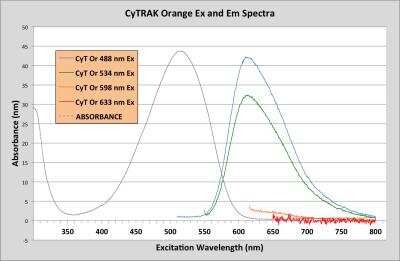
CyTRAK Orange (TM) [NBP2-81127] - Excitation and Emission Spectra for CyTRAK Orange (TM)
![Fluorescence Imaging: CyTRAK Orange (TM) [NBP2-81127]](https://resources.rndsystems.com/images/products/CyTRAK-Orange-TM-Immunocytochemistry-Immunofluorescence-NBP2-81127-img0002.jpg "Fluorescence Imaging: CyTRAK Orange (TM) [NBP2-81127]")
Fluorescence Imaging: CyTRAK Orange (TM) [NBP2-81127]
Fluorescence Imaging: CyTRAK Orange (TM) [NBP2-81127] - counterstaining of fixed U2OS cells, showing differential nuclear and cytoplasmic staining.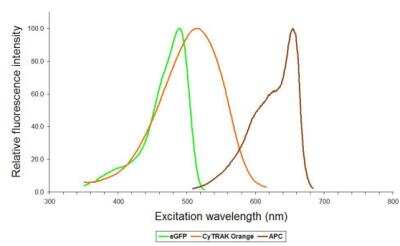
CyTRAK Orange (TM) [NBP2-81127] - Absorbance profile of CyTRAK Orange (TM)compared to eGFP and APC.
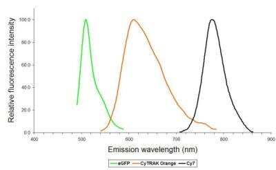
CyTRAK Orange (TM) [NBP2-81127] - Emission profile of CyTRAK Orange (TM)compared to eGFP and Cy7
Formulation, Preparation, and Storage
Purification
>97%
Concentration
Please see the protocols for proper use of this product. If no protocol is available, contact technical services for assistance.
Shipping
The product is shipped at ambient temperature. Upon receipt, store it immediately at the temperature recommended below.
Storage
Store at 4C in the dark. Do not freeze.
Background: CyTRAK Orange (TM)
The excitation wavelength maximum of CyTRAK Orange (TM) is 510 nm and its emission wavelength maximum is 610 nm. CyTRAK Orange (TM) is not excited by the UV or red laser.
Additional CyTRAK Orange (TM) Products
Product Documents for CyTRAK Orange (TM)
Certificate of Analysis
To download a Certificate of Analysis, please enter a lot or batch number in the search box below.
Product Specific Notices for CyTRAK Orange (TM)
CyTRAK Orange (TM) is a registered trademark of BioStatus Limited.
This product is for research use only and is not approved for use in humans or in clinical diagnosis. Support products are guaranteed for 6 months from date of receipt.
Customer Reviews for CyTRAK Orange (TM)
There are currently no reviews for this product. Be the first to review CyTRAK Orange (TM) and earn rewards!
Have you used CyTRAK Orange (TM)?
Submit a review and receive an Amazon gift card!
$25/€18/£15/$25CAN/¥2500 Yen for a review with an image
$10/€7/£6/$10CAN/¥1110 Yen for a review without an image
Submit a review
Protocols
Find general support by application which include: protocols, troubleshooting, illustrated assays, videos and webinars.
- 7-Amino Actinomycin D (7-AAD) Cell Viability Flow Cytometry Protocol
- Appropriate Fixation of IHC/ICC Samples
- Cellular Response to Hypoxia Protocols
- ClariTSA™ Fluorophore Kits
- Detection & Visualization of Antibody Binding
- Extracellular Membrane Flow Cytometry Protocol
- Flow Cytometry Protocol for Cell Surface Markers
- Flow Cytometry Protocol for Staining Membrane Associated Proteins
- Flow Cytometry Staining Protocols
- Flow Cytometry Troubleshooting Guide
- ICC Cell Smear Protocol for Suspension Cells
- ICC Immunocytochemistry Protocol Videos
- ICC for Adherent Cells
- Immunocytochemistry (ICC) Protocol
- Immunocytochemistry Troubleshooting
- Immunofluorescence of Organoids Embedded in Cultrex Basement Membrane Extract
- Immunohistochemistry (IHC) and Immunocytochemistry (ICC) Protocols
- Intracellular Flow Cytometry Protocol Using Alcohol (Methanol)
- Intracellular Flow Cytometry Protocol Using Detergents
- Intracellular Nuclear Staining Flow Cytometry Protocol Using Detergents
- Intracellular Staining Flow Cytometry Protocol Using Alcohol Permeabilization
- Intracellular Staining Flow Cytometry Protocol Using Detergents to Permeabilize Cells
- Preparing Samples for IHC/ICC Experiments
- Preventing Non-Specific Staining (Non-Specific Binding)
- Primary Antibody Selection & Optimization
- Propidium Iodide Cell Viability Flow Cytometry Protocol
- Protocol for Liperfluo
- Protocol for VisUCyte™ HRP Polymer Detection Reagent
- Protocol for the Characterization of Human Th22 Cells
- Protocol for the Characterization of Human Th9 Cells
- Protocol for the Fluorescent ICC Staining of Cell Smears - Graphic
- Protocol for the Fluorescent ICC Staining of Cultured Cells on Coverslips - Graphic
- Protocol for the Preparation and Fluorescent ICC Staining of Cells on Coverslips
- Protocol for the Preparation and Fluorescent ICC Staining of Non-adherent Cells
- Protocol for the Preparation and Fluorescent ICC Staining of Stem Cells on Coverslips
- Protocol for the Preparation of a Cell Smear for Non-adherent Cell ICC - Graphic
- Protocol: Annexin V and PI Staining by Flow Cytometry
- Protocol: Annexin V and PI Staining for Apoptosis by Flow Cytometry
- TUNEL and Active Caspase-3 Detection by IHC/ICC Protocol
- The Importance of IHC/ICC Controls
- Troubleshooting Guide: Fluorokine Flow Cytometry Kits
- View all Protocols, Troubleshooting, Illustrated assays and Webinars
Loading...