DENND5B Antibody - BSA Free
Novus Biologicals | Catalog # NBP2-31971
![Immunocytochemistry/ Immunofluorescence: DENND5B Antibody [NBP2-31971]](https://resources.rndsystems.com/images/products/DENND5B-Antibody-Immunocytochemistry-Immunofluorescence-NBP2-31971-img0007.jpg "Immunocytochemistry/ Immunofluorescence: DENND5B Antibody [NBP2-31971]")
Loading...
Key Product Details
Validated by
Knockout/Knockdown
Species Reactivity
Validated:
Human
Cited:
Mouse, Avian
Predicted:
Mouse (90%). Backed by our 100% Guarantee.
Applications
Validated:
Knockout Validated, Immunohistochemistry, Immunohistochemistry-Paraffin, Immunocytochemistry/ Immunofluorescence
Cited:
Immunohistochemistry-Frozen, Western Blot
Label
Unconjugated
Antibody Source
Polyclonal Rabbit IgG
Format
BSA Free
Loading...
Product Specifications
Immunogen
This antibody was developed against a recombinant protein corresponding to amino acids: FIWDFIEKVVAYFETTDQILDNEDDVLIQKSSCKTFCHYVNAINTAPRNIGK
Reactivity Notes
Rat (88%).
Clonality
Polyclonal
Host
Rabbit
Isotype
IgG
Scientific Data Images for DENND5B Antibody - BSA Free
Immunocytochemistry/ Immunofluorescence: DENND5B Antibody [NBP2-31971]
Immunocytochemistry/Immunofluorescence: DENND5B Antibody [NBP2-31971] - Staining of human cell line U-2 OS shows localization to nucleoli & microtubules. Antibody staining is shown in green.![Immunohistochemistry-Paraffin: DENND5B Antibody [NBP2-31971]](https://resources.rndsystems.com/images/products/DENND5B-Antibody-Immunohistochemistry-Paraffin-NBP2-31971-img0010.jpg "Immunohistochemistry-Paraffin: DENND5B Antibody [NBP2-31971]")
Immunohistochemistry-Paraffin: DENND5B Antibody [NBP2-31971]
Immunohistochemistry-Paraffin: DENND5B Antibody [NBP2-31971] - Staining of human fallopian tube shows moderate cytoplasmic positivity in glandular cells.![Immunohistochemistry-Paraffin: DENND5B Antibody [NBP2-31971]](https://resources.rndsystems.com/images/products/DENND5B-Antibody-Immunohistochemistry-Paraffin-NBP2-31971-img0004.jpg "Immunohistochemistry-Paraffin: DENND5B Antibody [NBP2-31971]")
Immunohistochemistry-Paraffin: DENND5B Antibody [NBP2-31971]
Immunohistochemistry-Paraffin: DENND5B Antibody [NBP2-31971] - Staining of human testis shows moderate cytoplasmic and membranous positivity in cells in seminiferus ducts.![Immunohistochemistry-Paraffin: DENND5B Antibody [NBP2-31971]](https://resources.rndsystems.com/images/products/DENND5B-Antibody-Immunohistochemistry-Paraffin-NBP2-31971-img0008.jpg "Immunohistochemistry-Paraffin: DENND5B Antibody [NBP2-31971]")
Immunohistochemistry-Paraffin: DENND5B Antibody [NBP2-31971]
Immunohistochemistry-Paraffin: DENND5B Antibody [NBP2-31971] - Staining of human testis shows moderate membranous and cytoplasmic positivity in cells in seminiferous ducts.![Immunohistochemistry-Paraffin: DENND5B Antibody [NBP2-31971]](https://resources.rndsystems.com/images/products/DENND5B-Antibody-Immunohistochemistry-Paraffin-NBP2-31971-img0009.jpg "Immunohistochemistry-Paraffin: DENND5B Antibody [NBP2-31971]")
Immunohistochemistry-Paraffin: DENND5B Antibody [NBP2-31971]
Immunohistochemistry-Paraffin: DENND5B Antibody [NBP2-31971] - Staining of human cerebral cortex shows strong positivity in neuropil.![Immunohistochemistry-Paraffin: DENND5B Antibody [NBP2-31971]](https://resources.rndsystems.com/images/products/DENND5B-Antibody-Immunohistochemistry-Paraffin-NBP2-31971-img0011.jpg "Immunohistochemistry-Paraffin: DENND5B Antibody [NBP2-31971]")
Immunohistochemistry-Paraffin: DENND5B Antibody [NBP2-31971]
Immunohistochemistry-Paraffin: DENND5B Antibody [NBP2-31971] - Staining of human rectum shows moderate cytoplasmic positivity in glandular cells.
Applications for DENND5B Antibody - BSA Free
Application
Recommended Usage
Immunocytochemistry/ Immunofluorescence
0.25-2 ug/ml
Immunohistochemistry
1:50 - 1:200
Immunohistochemistry-Paraffin
1:50 - 1:200
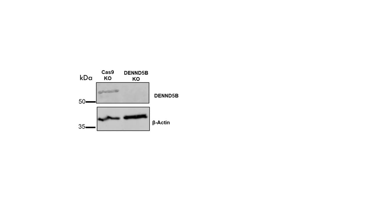
Knockout Validated
Knockout Validated from a verified customer review.
Application Notes
For IHC-Paraffin, HIER pH 6 retrieval is recommended. ICC/IF Fixation Permeabilization: Use PFA/Triton X-100.
Reviewed Applications
Read 2 reviews rated 4.5 using NBP2-31971 in the following applications:
Formulation, Preparation, and Storage
Purification
Affinity purified
Formulation
PBS (pH 7.2) and 40% Glycerol
Format
BSA Free
Preservative
0.02% Sodium Azide
Concentration
Concentrations vary lot to lot. See vial label for concentration. If unlisted please contact technical services.
Shipping
The product is shipped with polar packs. Upon receipt, store it immediately at the temperature recommended below.
Stability & Storage
Store at 4C short term. Aliquot and store at -20C long term. Avoid freeze-thaw cycles.
Background: DENND5B
Alternate Names
DENN domain-containing protein 5B, DENN/MADD domain containing 5B, DKFZp686P1174, FLJ41648, FLJ43333, MGC24039, Rab6IP1-like protein
Gene Symbol
DENND5B
UniProt
Additional DENND5B Products
Product Documents for DENND5B Antibody - BSA Free
Certificate of Analysis
To download a Certificate of Analysis, please enter a lot or batch number in the search box below.
Product Specific Notices for DENND5B Antibody - BSA Free
This product is for research use only and is not approved for use in humans or in clinical diagnosis. Primary Antibodies are guaranteed for 1 year from date of receipt.
Citations for DENND5B Antibody - BSA Free
 Powered by Bioz
Customer Reviews for DENND5B Antibody - BSA Free (2)
4.5 out of 5
2 Customer Ratings
Have you used DENND5B Antibody - BSA Free?
Submit a review and receive an Amazon gift card!
$25/€18/£15/$25CAN/¥2500 Yen for a review with an image
$10/€7/£6/$10CAN/¥1110 Yen for a review without an image
Submit a review
Customer Images
Showing
1
-
2 of
2 reviews
Showing All
Filter By:
-
Application: Western BlotSample Tested: Cell Culture SamplesSpecies: HumanVerified Customer | Posted 12/16/2025Control and knock out cells1:1000 dilution worked
 Bio-Techne ResponseThis review reflects a new species or application tested on a primary antibody.
 -
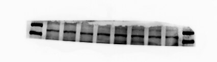
Application: Western BlotSample Tested: mouse primary cellSpecies: MouseVerified Customer | Posted 06/19/20206 percent of the gum, mouse cortex, in the region between 130 and 180, concentration about 1:200, consistent with the results in the primary cells.I will send you better pictures later.
There are no reviews that match your criteria.
Protocols
Find general support by application which include: protocols, troubleshooting, illustrated assays, videos and webinars.
- Antigen Retrieval Protocol (PIER)
- Antigen Retrieval for Frozen Sections Protocol
- Appropriate Fixation of IHC/ICC Samples
- Cellular Response to Hypoxia Protocols
- Chromogenic IHC Staining of Formalin-Fixed Paraffin-Embedded (FFPE) Tissue Protocol
- Chromogenic Immunohistochemistry Staining of Frozen Tissue
- ClariTSA™ Fluorophore Kits
- Detection & Visualization of Antibody Binding
- Fluorescent IHC Staining of Frozen Tissue Protocol
- Graphic Protocol for Heat-induced Epitope Retrieval
- Graphic Protocol for the Preparation and Fluorescent IHC Staining of Frozen Tissue Sections
- Graphic Protocol for the Preparation and Fluorescent IHC Staining of Paraffin-embedded Tissue Sections
- Graphic Protocol for the Preparation of Gelatin-coated Slides for Histological Tissue Sections
- ICC Cell Smear Protocol for Suspension Cells
- ICC Immunocytochemistry Protocol Videos
- ICC for Adherent Cells
- IHC Sample Preparation (Frozen sections vs Paraffin)
- Immunocytochemistry (ICC) Protocol
- Immunocytochemistry Troubleshooting
- Immunofluorescence of Organoids Embedded in Cultrex Basement Membrane Extract
- Immunofluorescent IHC Staining of Formalin-Fixed Paraffin-Embedded (FFPE) Tissue Protocol
- Immunohistochemistry (IHC) and Immunocytochemistry (ICC) Protocols
- Immunohistochemistry Frozen Troubleshooting
- Immunohistochemistry Paraffin Troubleshooting
- Preparing Samples for IHC/ICC Experiments
- Preventing Non-Specific Staining (Non-Specific Binding)
- Primary Antibody Selection & Optimization
- Protocol for Heat-Induced Epitope Retrieval (HIER)
- Protocol for Making a 4% Formaldehyde Solution in PBS
- Protocol for VisUCyte™ HRP Polymer Detection Reagent
- Protocol for the Fluorescent ICC Staining of Cell Smears - Graphic
- Protocol for the Fluorescent ICC Staining of Cultured Cells on Coverslips - Graphic
- Protocol for the Preparation & Fixation of Cells on Coverslips
- Protocol for the Preparation and Chromogenic IHC Staining of Frozen Tissue Sections
- Protocol for the Preparation and Chromogenic IHC Staining of Frozen Tissue Sections - Graphic
- Protocol for the Preparation and Chromogenic IHC Staining of Paraffin-embedded Tissue Sections
- Protocol for the Preparation and Chromogenic IHC Staining of Paraffin-embedded Tissue Sections - Graphic
- Protocol for the Preparation and Fluorescent ICC Staining of Cells on Coverslips
- Protocol for the Preparation and Fluorescent ICC Staining of Non-adherent Cells
- Protocol for the Preparation and Fluorescent ICC Staining of Stem Cells on Coverslips
- Protocol for the Preparation and Fluorescent IHC Staining of Frozen Tissue Sections
- Protocol for the Preparation and Fluorescent IHC Staining of Paraffin-embedded Tissue Sections
- Protocol for the Preparation of Gelatin-coated Slides for Histological Tissue Sections
- Protocol for the Preparation of a Cell Smear for Non-adherent Cell ICC - Graphic
- TUNEL and Active Caspase-3 Detection by IHC/ICC Protocol
- The Importance of IHC/ICC Controls
- Troubleshooting Guide: Immunohistochemistry
- View all Protocols, Troubleshooting, Illustrated assays and Webinars
Loading...