Desmocollin-1 Antibody - BSA Free
Novus Biologicals | Catalog # NBP1-88099
![Immunohistochemistry-Paraffin: Desmocollin-1 Antibody [NBP1-88099]](https://resources.rndsystems.com/images/products/Desmocollin-1-Antibody-Immunohistochemistry-Paraffin-NBP1-88099-img0012.jpg "Immunohistochemistry-Paraffin: Desmocollin-1 Antibody [NBP1-88099]")
Loading...
Key Product Details
Species Reactivity
Validated:
Human
Cited:
Human
Applications
Validated:
Immunohistochemistry, Immunohistochemistry-Paraffin, Simple Western
Cited:
Immunohistochemistry-Frozen, Western Blot, IF/IHC
Label
Unconjugated
Antibody Source
Polyclonal Rabbit IgG
Format
BSA Free
Loading...
Product Specifications
Immunogen
This antibody was developed against Recombinant Protein corresponding to amino acids: SCTGTLVVHLDDYNDHAPQIDKEVTICQNNEDFAVLKPVDPDGPENGPPFQFFLDNSASKNWNIEEKDGKTAILRQRQNLDYNYYSVPIQIKDRHGLVATHMLTVRVCDCSTPSECRMKDKSTR
Clonality
Polyclonal
Host
Rabbit
Isotype
IgG
Scientific Data Images for Desmocollin-1 Antibody - BSA Free
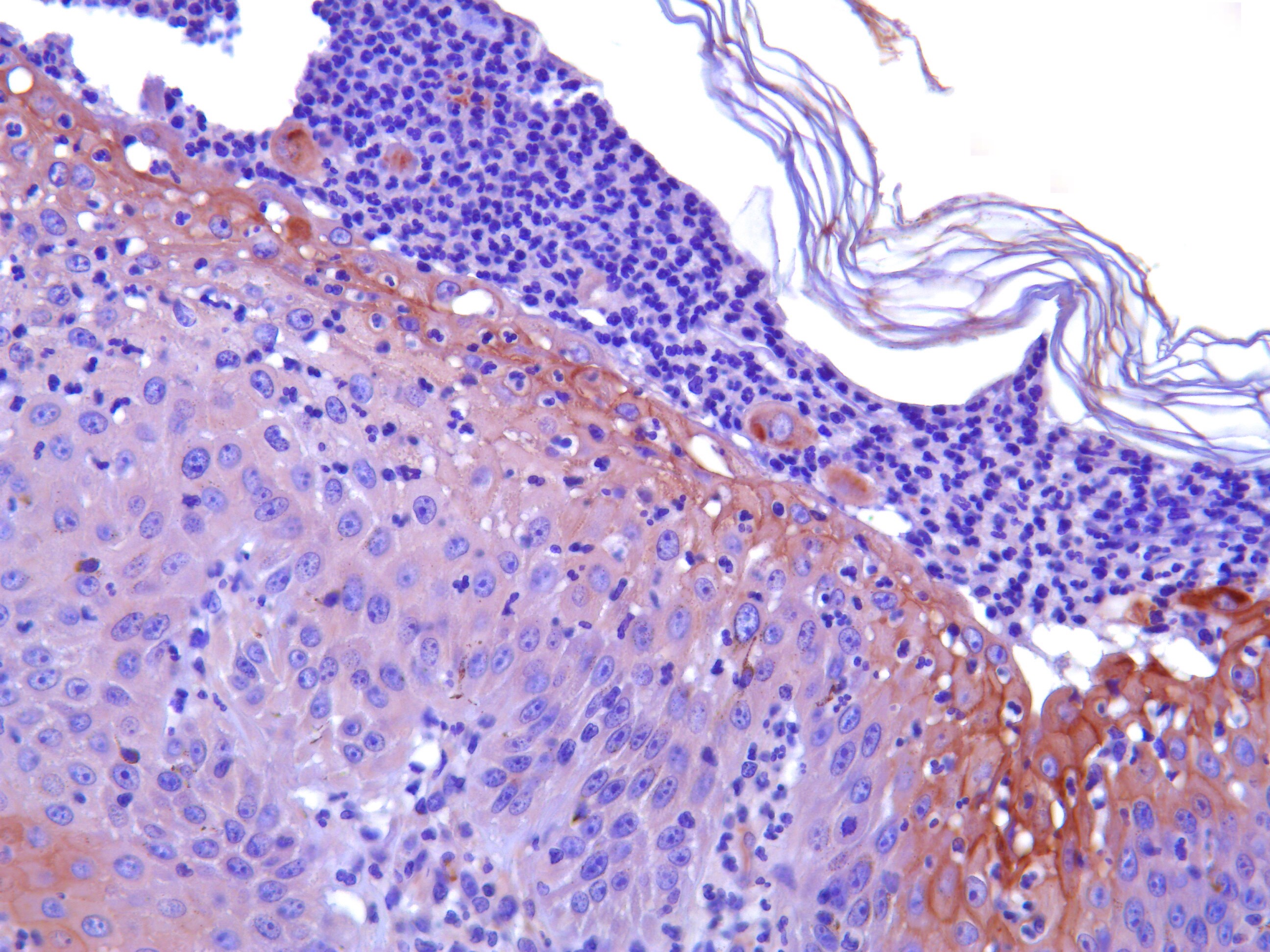
Immunohistochemistry-Paraffin: Desmocollin-1 Antibody [NBP1-88099]
Immunohistochemistry-Paraffin: Desmocollin-1 Antibody [NBP1-88099] - Staining of human skin shows moderate to strong membranous positivity in epidermal cells.![Immunohistochemistry-Paraffin: Desmocollin-1 Antibody [NBP1-88099]](https://resources.rndsystems.com/images/products/Desmocollin-1-Antibody-Immunohistochemistry-Paraffin-NBP1-88099-img0010.jpg "Immunohistochemistry-Paraffin: Desmocollin-1 Antibody [NBP1-88099]")
Immunohistochemistry-Paraffin: Desmocollin-1 Antibody [NBP1-88099]
Immunohistochemistry-Paraffin: Desmocollin-1 Antibody [NBP1-88099] - Staining of human fallopian tube shows very weak membranous positivity in glandular cells.![Immunohistochemistry-Paraffin: Desmocollin-1 Antibody [NBP1-88099]](https://resources.rndsystems.com/images/products/Desmocollin-1-Antibody-Immunohistochemistry-Paraffin-NBP1-88099-img0011.jpg "Immunohistochemistry-Paraffin: Desmocollin-1 Antibody [NBP1-88099]")
Immunohistochemistry-Paraffin: Desmocollin-1 Antibody [NBP1-88099]
Immunohistochemistry-Paraffin: Desmocollin-1 Antibody [NBP1-88099] - Staining of human prostate shows no membranous positivity in glandular cells.![Simple Western: Desmocollin-1 Antibody [NBP1-88099]](https://resources.rndsystems.com/images/products/Desmocollin-1-Antibody-Simple-Western-NBP1-88099-img0005.jpg "Simple Western: Desmocollin-1 Antibody [NBP1-88099]")
Simple Western: Desmocollin-1 Antibody [NBP1-88099]
Simple Western: Desmocollin-1 Antibody [NBP1-88099] - Simple Western lane view shows a specific band for Desmocollin-1 in 0.5 mg/ml of RT-4 (Left) and U-251MG (Right) lysate. This experiment was performed under reducing conditions using the 12-230 kDa separation system.![Simple Western: Desmocollin-1 Antibody [NBP1-88099]](https://resources.rndsystems.com/images/products/Desmocollin-1-Antibody-Simple-Western-NBP1-88099-img0006.jpg "Simple Western: Desmocollin-1 Antibody [NBP1-88099]")
Simple Western: Desmocollin-1 Antibody [NBP1-88099]
Simple Western: Desmocollin-1 Antibody [NBP1-88099] - Electropherogram image(s) of corresponding Simple Western lane view. Desmocollin-1 antibody was used at 1:60 dilution on RT-4 and U-251MG lysate(s).Applications for Desmocollin-1 Antibody - BSA Free
Application
Recommended Usage
Immunohistochemistry
1:200 - 1:500
Immunohistochemistry-Paraffin
1:200 - 1:500
Simple Western
1:60
Application Notes
IHC-Paraffin, HIER pH 6 retrieval is recommended.
In Simple Western only 10 - 15 uL of the recommended dilution is used per data point.
See Simple Western Antibody Database for Simple Western validation: Tested in RT-4 and U-251MG, separated by Size, antibody dilution of 1:60, apparent MW was 100 kDa. Separated by Size-Wes, Sally Sue/Peggy Sue.
In Simple Western only 10 - 15 uL of the recommended dilution is used per data point.
See Simple Western Antibody Database for Simple Western validation: Tested in RT-4 and U-251MG, separated by Size, antibody dilution of 1:60, apparent MW was 100 kDa. Separated by Size-Wes, Sally Sue/Peggy Sue.
Reviewed Applications
Read 1 review rated 5 using NBP1-88099 in the following applications:
Formulation, Preparation, and Storage
Purification
Affinity purified
Formulation
PBS (pH 7.2) and 40% Glycerol
Format
BSA Free
Preservative
0.02% Sodium Azide
Concentration
Concentrations vary lot to lot. See vial label for concentration. If unlisted please contact technical services.
Shipping
The product is shipped with polar packs. Upon receipt, store it immediately at the temperature recommended below.
Stability & Storage
Store at 4C short term. Aliquot and store at -20C long term. Avoid freeze-thaw cycles.
Background: Desmocollin-1
Alternate Names
CDHF1, Desmocollin1, DG2/DG3, DSC1
Gene Symbol
DSC1
Additional Desmocollin-1 Products
Product Documents for Desmocollin-1 Antibody - BSA Free
Certificate of Analysis
To download a Certificate of Analysis, please enter a lot or batch number in the search box below.
Product Specific Notices for Desmocollin-1 Antibody - BSA Free
This product is for research use only and is not approved for use in humans or in clinical diagnosis. Primary Antibodies are guaranteed for 1 year from date of receipt.
Related Research Areas
Citations for Desmocollin-1 Antibody - BSA Free
 Powered by Bioz
Customer Reviews for Desmocollin-1 Antibody - BSA Free (1)
5 out of 5
1 Customer Rating
Have you used Desmocollin-1 Antibody - BSA Free?
Submit a review and receive an Amazon gift card!
$25/€18/£15/$25CAN/¥2500 Yen for a review with an image
$10/€7/£6/$10CAN/¥1110 Yen for a review without an image
Submit a review
Customer Images
Showing
1
-
1 of
1 review
Showing All
Filter By:
-
Application: Immunohistochemistry-ParaffinSample Tested: Paraffin sections of canine folicular skin (dogs with pemphigus foliaceus)Species: CanineVerified Customer | Posted 11/07/2019Optical microscope (x20). IHC anti-desmocollin-1. Subcorneal epidermal pustule in dog with PF. Patchy staining pattern in some keratinocytes, in perilesional epidermis, as well as in acantholytic keratinocytes inside the lesion.ABSTRACT Background Pemphigus foliaceus is the most common autoimmune disease in dogs. It is characterized histologically by subcorneal pustules and acantholysis. Desmocollin-1 is the major target antigen of autoantibodies in dogs. Previous immunostaining studies have shown possible pathogenic similarities between canine and human pemphigus. Objetives To investigate the pathogenesis of acantholysis in canine Pemphigus Foliaceus, we decided to review the main histopathological findings and observe the variations of the labeling of desmocollin-1 by immunohistochemistry in skin biopsies from sick dogs. Material and methods Five cases of dogs with Pemphigus Foliaceus were selected and histopathological findings were reviewed by hematoxylin-eosin staining. Then the samples were stained following an immunohistochemical protocol, with an anti-desmocolin-1 antibody. Results The 5 cases presented typical histological characteristics of PFc: pustules in the superficial layers of the epidermis, acantholytic keratinocytes and neutrophilic exocytosis. We have validated the use of the anti-dsc-1 antibody in dogs, for immunhistochemistry in skin biopsies preserved in paraffin. We have observed that, in dogs with PF, the immunohistochemical pattern of desmocollin-1 was altered. Conclusions Staining of desmocollin-1 is altered in dogs with PF. We were able to corroborate the pathogenic mechanisms already demonstrated by immunohistochemistry. Thus, immunohistochemical staining for this protein may be useful in diagnosis. Its use, to study precise mechanisms, is limited and more biotechnologic tools are needed to complement the knowledge on this disease.

There are no reviews that match your criteria.
Protocols
Find general support by application which include: protocols, troubleshooting, illustrated assays, videos and webinars.
- Antigen Retrieval Protocol (PIER)
- Antigen Retrieval for Frozen Sections Protocol
- Appropriate Fixation of IHC/ICC Samples
- Cellular Response to Hypoxia Protocols
- Chromogenic IHC Staining of Formalin-Fixed Paraffin-Embedded (FFPE) Tissue Protocol
- Chromogenic Immunohistochemistry Staining of Frozen Tissue
- ClariTSA™ Fluorophore Kits
- Detection & Visualization of Antibody Binding
- Fluorescent IHC Staining of Frozen Tissue Protocol
- Graphic Protocol for Heat-induced Epitope Retrieval
- Graphic Protocol for the Preparation and Fluorescent IHC Staining of Frozen Tissue Sections
- Graphic Protocol for the Preparation and Fluorescent IHC Staining of Paraffin-embedded Tissue Sections
- Graphic Protocol for the Preparation of Gelatin-coated Slides for Histological Tissue Sections
- IHC Sample Preparation (Frozen sections vs Paraffin)
- Immunofluorescent IHC Staining of Formalin-Fixed Paraffin-Embedded (FFPE) Tissue Protocol
- Immunohistochemistry (IHC) and Immunocytochemistry (ICC) Protocols
- Immunohistochemistry Frozen Troubleshooting
- Immunohistochemistry Paraffin Troubleshooting
- Preparing Samples for IHC/ICC Experiments
- Preventing Non-Specific Staining (Non-Specific Binding)
- Primary Antibody Selection & Optimization
- Protocol for Heat-Induced Epitope Retrieval (HIER)
- Protocol for Making a 4% Formaldehyde Solution in PBS
- Protocol for VisUCyte™ HRP Polymer Detection Reagent
- Protocol for the Preparation & Fixation of Cells on Coverslips
- Protocol for the Preparation and Chromogenic IHC Staining of Frozen Tissue Sections
- Protocol for the Preparation and Chromogenic IHC Staining of Frozen Tissue Sections - Graphic
- Protocol for the Preparation and Chromogenic IHC Staining of Paraffin-embedded Tissue Sections
- Protocol for the Preparation and Chromogenic IHC Staining of Paraffin-embedded Tissue Sections - Graphic
- Protocol for the Preparation and Fluorescent IHC Staining of Frozen Tissue Sections
- Protocol for the Preparation and Fluorescent IHC Staining of Paraffin-embedded Tissue Sections
- Protocol for the Preparation of Gelatin-coated Slides for Histological Tissue Sections
- TUNEL and Active Caspase-3 Detection by IHC/ICC Protocol
- The Importance of IHC/ICC Controls
- Troubleshooting Guide: Immunohistochemistry
- View all Protocols, Troubleshooting, Illustrated assays and Webinars
Loading...