![Western Blot: DNA-PKcs Antibody [NB100-658]](https://resources.rndsystems.com/images/products/DNA-PKcs-Antibody-Western-Blot-NB100-658-img0025.jpg "Western Blot: DNA-PKcs Antibody [NB100-658]")
Loading...
Key Product Details
Validated by
Knockout/Knockdown, Independent Antibodies
Species Reactivity
Validated:
Human
Cited:
Human
Applications
Validated:
Western Blot, Immunoprecipitation, Knockdown Validated
Cited:
Western Blot
Label
Unconjugated
Antibody Source
Polyclonal Rabbit IgG
Loading...
Product Specifications
Immunogen
The immunogen recognized by this antibody maps to a region between residues 2050 and 2100 of human DNA-Dependent Protein Kinase, catalytic subunit using the numbering given in entry NP_008835.5 (GeneID 5591).
Clonality
Polyclonal
Host
Rabbit
Isotype
IgG
Scientific Data Images for DNA-PKcs Antibody
Western Blot: DNA-PKcs Antibody [NB100-658]
Western Blot: DNA-PKcs Antibody [NB100-658] - MDA-MB-231, MCF-7, SUM149, SUM159, T47D, HCC1954 whole cell lysates (50 ug/lane). 10% SDS-PAGE. DNA-PKcs (NB100-658) primary antibody at 1:1000, 4C, overnight. Western blot image submitted by a verified customer review.![Western Blot: DNA-PKcs Antibody [NB100-658]](https://resources.rndsystems.com/images/products/DNA-PKcs-Antibody-Western-Blot-NB100-658-img0023.jpg "Western Blot: DNA-PKcs Antibody [NB100-658]")
![Western Blot: DNA-PKcs Antibody [NB100-658]](https://resources.rndsystems.com/images/products/DNA-PKcs-Antibody-Western-Blot-NB100-658-img0024.jpg "Western Blot: DNA-PKcs Antibody [NB100-658]")
Western Blot: DNA-PKcs Antibody [NB100-658]
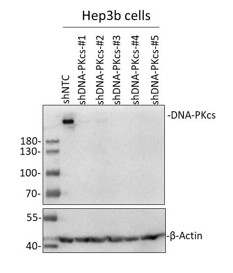
Western Blot: DNA-PKcs Antibody [NB100-658] - Hep3B human hepatocellular carcinoma cell line. 50 ug cell lysate per lane. 10% SDS-PAGE. DNA-PKcs antibody at 1:1000 dilution O/N. Secondary antibody: HRP-conjugated Donkey anti-rabbit IgG polyclonal antibody at 1:2000 dilution. WB image submitted by a verified customer review.![Knockdown Validated: DNA-PKcs Antibody [NB100-658]](https://resources.rndsystems.com/images/products/DNA-PKcs-Antibody-Knockdown-Validated-NB100-658-img0026.jpg "Western Blot: DNA-PKcs Antibody [NB100-658]")

Western Blot: DNA-PKcs Antibody [NB100-658] -
Western Blot: DNA-PKcs Antibody [NB100-658] - Molecular Characterization of G1 Resection(A) Schematic of the NHEJ reporter assay. The repair of two I-SceI-induced DSBs can result in loss of the intervening fragment, which is detected by a CD4+ signal (Rass et al., 2009). CD4+ clones were amplified by PCR (green arrows) across the repair site & sequenced. Repair of the two DSBs can also occur without loss of the intervening fragment, which escapes detection.(B) gamma H2AX foci in GC92 WT & Artemis KO cells treated with siDNA-PKcs or siCtIP. Cells were transfected with I-SceI, & foci were scored in I-SceI+ & I-SceI− cells (identified by immunofluorescence [IF] against I-SceI). Data are mean ± SEM.(C) End joining events in GC92 WT & Artemis KO cells containing the NHEJ reporter substrate. Cells were transfected with RFP or cMyc-Artemis constructs. Events were quantified by the fraction of CD4+ & RFP/cMyc+cells relative to all RFP/cMyc+cells, & results were normalized to WT cells. Data are mean ± SEM.(D) End joining events in GC92 WT & Artemis KO cells treated with siCtIP. Data are mean ± SEM.(E) Distribution of deletion sizes obtained from the sequence analysis of GC92 WT & siLig1/3-treated cells. nt, nucleotide.(F) End joining events in GC92 cells treated with siKu70/80, siLig4, siLig1/3, or siDNA-PKcs. Data are mean ± SEM.See also Table S1. Image collected & cropped by CiteAb from the following publication (https://pubmed.ncbi.nlm.nih.gov/28132842), licensed under a CC-BY license. Not internally tested by Novus Biologicals.Applications for DNA-PKcs Antibody
Application
Recommended Usage
Immunoprecipitation
2 - 5 ug/mg lysate
Western Blot
1:1000 - 1:10000
Application Notes
Ku-80 co-immunoprecipitates with DNA-PKcs when using this antibody. DNA-PKcs antibody is validated for WB from a verified customer review.
Reviewed Applications
Read 1 review rated 5 using NB100-658 in the following applications:
Formulation, Preparation, and Storage
Purification
Immunogen affinity purified
Formulation
TBS, 0.1% BSA
Preservative
0.09% Sodium Azide
Concentration
0.2 mg/ml
Shipping
The product is shipped with polar packs. Upon receipt, store it immediately at the temperature recommended below.
Stability & Storage
Store at 4C. Do not freeze.
Background: DNA-PKcs
Long Name
DNA-dependent Protein Kinase Catalytic Subunit
Alternate Names
DNAPK, DNAPKcs, DNPK1, HYRC1, p350, PRKDC, XRCC7
Entrez Gene IDs
5591 (Human)
Gene Symbol
PRKDC
UniProt
Additional DNA-PKcs Products
Product Documents for DNA-PKcs Antibody
Certificate of Analysis
To download a Certificate of Analysis, please enter a lot or batch number in the search box below.
Product Specific Notices for DNA-PKcs Antibody
This product is for research use only and is not approved for use in humans or in clinical diagnosis. Primary Antibodies are guaranteed for 1 year from date of receipt.
Related Research Areas
Citations for DNA-PKcs Antibody
 Powered by Bioz
Customer Reviews for DNA-PKcs Antibody (1)
5 out of 5
1 Customer Rating
Have you used DNA-PKcs Antibody?
Submit a review and receive an Amazon gift card!
$25/€18/£15/$25CAN/¥2500 Yen for a review with an image
$10/€7/£6/$10CAN/¥1110 Yen for a review without an image
Submit a review
Customer Images
Showing
1
-
1 of
1 review
Showing All
Filter By:
-
Application: Western BlotSample Tested: Hep3B human hepatocellular carcinoma cell lineSpecies: HumanVerified Customer | Posted 08/16/201950ug Hep3b cells lysate/lane, 10% SDS-PAGE; Primary antibody (DNA-PKcs Antibody (NB100-658), Lot: A1 ): 1:1000 diluted in 5% milk incubate at 4℃ for overnight; Secondary antibody: NA934V, Donkey Anti-Rabbit IgG Polyclonal Antibody (HRP (Horseradish Peroxidase)- GE Healthcare (Lot: 16908227) (1:2000 diluted in 5% milk). Incubate at room temperature for 1 hour.

There are no reviews that match your criteria.
Protocols
Find general support by application which include: protocols, troubleshooting, illustrated assays, videos and webinars.
- Cellular Response to Hypoxia Protocols
- Immunoprecipitation Protocol
- R&D Systems Quality Control Western Blot Protocol
- Troubleshooting Guide: Western Blot Figures
- Western Blot Conditions
- Western Blot Protocol
- Western Blot Protocol for Cell Lysates
- Western Blot Troubleshooting
- Western Blot Troubleshooting Guide
- View all Protocols, Troubleshooting, Illustrated assays and Webinars
Loading...