![Western Blot: DPY30 Antibody [NBP1-91848]](https://resources.rndsystems.com/images/products/DPY30-Antibody-Western-Blot-NBP1-91848-img0019.jpg "Western Blot: DPY30 Antibody [NBP1-91848]")
Key Product Details
Species Reactivity
Validated:
Predicted:
Applications
Label
Antibody Source
Format
Product Specifications
Immunogen
Clonality
Host
Isotype
Scientific Data Images for DPY30 Antibody - BSA Free
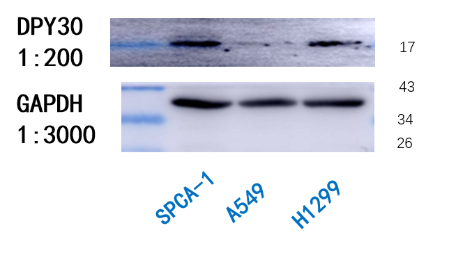
Western Blot: DPY30 Antibody [NBP1-91848]
Western Blot: DPY30 Antibody [NBP1-91848] - 40 ug total protein per lane, separated by 12% SDS-PAGE and blotted onto a PVDF membrane. DPY30 antibody at 1:200, GAPDH antibody at 1:3000. Secondary antibody at 1:5000. Detection by ECL. Image submitted by a verified customer review.![Immunocytochemistry/ Immunofluorescence: DPY30 Antibody [NBP1-91848]](https://resources.rndsystems.com/images/products/DPY30-Antibody-Immunocytochemistry-Immunofluorescence-NBP1-91848-img0012.jpg "Immunocytochemistry/ Immunofluorescence: DPY30 Antibody [NBP1-91848]")
Immunocytochemistry/ Immunofluorescence: DPY30 Antibody [NBP1-91848]
Immunocytochemistry/Immunofluorescence: DPY30 Antibody [NBP1-91848] - Staining of human cell line A-431 shows localization to nucleoplasm & the Golgi apparatus. Antibody staining is shown in green.![Immunohistochemistry-Paraffin: DPY30 Antibody [NBP1-91848]](https://resources.rndsystems.com/images/products/DPY30-Antibody-Immunohistochemistry-Paraffin-NBP1-91848-img0017.jpg "Immunohistochemistry-Paraffin: DPY30 Antibody [NBP1-91848]")
Immunohistochemistry-Paraffin: DPY30 Antibody [NBP1-91848]
Immunohistochemistry-Paraffin: DPY30 Antibody [NBP1-91848] - Staining of human small intestine shows moderate nuclear positivity in glandular cells.![Immunohistochemistry-Paraffin: DPY30 Antibody [NBP1-91848]](https://resources.rndsystems.com/images/products/DPY30-Antibody-Immunohistochemistry-Paraffin-NBP1-91848-img0013.jpg "Immunohistochemistry-Paraffin: DPY30 Antibody [NBP1-91848]")
Immunohistochemistry-Paraffin: DPY30 Antibody [NBP1-91848]
Immunohistochemistry-Paraffin: DPY30 Antibody [NBP1-91848] - Staining of human fallopian tube shows moderate to strong nuclear positivity in glandular cells.![Immunohistochemistry-Paraffin: DPY30 Antibody [NBP1-91848]](https://resources.rndsystems.com/images/products/DPY30-Antibody-Immunohistochemistry-Paraffin-NBP1-91848-img0014.jpg "Immunohistochemistry-Paraffin: DPY30 Antibody [NBP1-91848]")
Immunohistochemistry-Paraffin: DPY30 Antibody [NBP1-91848]
Immunohistochemistry-Paraffin: DPY30 Antibody [NBP1-91848] - Staining of human kidney shows moderate to strong nuclear positivity in cells in tubules.![Immunohistochemistry-Paraffin: DPY30 Antibody [NBP1-91848]](https://resources.rndsystems.com/images/products/DPY30-Antibody-Immunohistochemistry-Paraffin-NBP1-91848-img0015.jpg "Immunohistochemistry-Paraffin: DPY30 Antibody [NBP1-91848]")
Immunohistochemistry-Paraffin: DPY30 Antibody [NBP1-91848]
Immunohistochemistry-Paraffin: DPY30 Antibody [NBP1-91848] - Staining of human pancreas shows nuclear positivity.![Immunohistochemistry-Paraffin: DPY30 Antibody [NBP1-91848]](https://resources.rndsystems.com/images/products/DPY30-Antibody-Immunohistochemistry-Paraffin-NBP1-91848-img0016.jpg "Immunohistochemistry-Paraffin: DPY30 Antibody [NBP1-91848]")
Immunohistochemistry-Paraffin: DPY30 Antibody [NBP1-91848]
Immunohistochemistry-Paraffin: DPY30 Antibody [NBP1-91848] - Staining of human prostate shows moderate to strong nuclear positivity in glandular cells.Applications for DPY30 Antibody - BSA Free
Immunocytochemistry/ Immunofluorescence
Immunohistochemistry
Immunohistochemistry-Paraffin
Reviewed Applications
Read 1 review rated 3 using NBP1-91848 in the following applications:
Formulation, Preparation, and Storage
Purification
Formulation
Format
Preservative
Concentration
Shipping
Stability & Storage
Background: DPY30
Alternate Names
Gene Symbol
Additional DPY30 Products
Product Documents for DPY30 Antibody - BSA Free
Certificate of Analysis
To download a Certificate of Analysis, please enter a lot or batch number in the search box below.
Product Specific Notices for DPY30 Antibody - BSA Free
This product is for research use only and is not approved for use in humans or in clinical diagnosis. Primary Antibodies are guaranteed for 1 year from date of receipt.
Customer Reviews for DPY30 Antibody - BSA Free (1)
Have you used DPY30 Antibody - BSA Free?
Submit a review and receive an Amazon gift card!
$25/€18/£15/$25CAN/¥2500 Yen for a review with an image
$10/€7/£6/$10CAN/¥1110 Yen for a review without an image
Submit a review
Customer Images
-
Application: Western BlotSample Tested: SPCA-1 cells, H1299 whole cell lysate, SPCA-1 whole cell lysate and A549 whole cell lysateSpecies: HumanVerified Customer | Posted 12/05/2018The figure above can be used as a reference for Western blot.These are the main steps of our western blot. The protein concentration was assayed using the BCA protein assay(Thermo REF:23227). For this,40 μg of protein per lanewas separated by 12% SDS-PAGE and electroblotted onto a PVDF membrane. Then, non-specific binding was blocked by incubating with5% non-fat milk in TBST buffer at room temperature for 2 h. PVDF membranes were incubated overnight at 4 °C with primary antibodies which were diluted in TBS-T with 5% non-fat milk as follows: DPY30 (1:200), GAPDH (1:3000). This was followed by incubation with the corresponding secondary antibody(1:5000) at room temperature for 1 h. Finally, antigens were detected using the standard chemical luminescence method (ECL).

There are no reviews that match your criteria.
Protocols
Find general support by application which include: protocols, troubleshooting, illustrated assays, videos and webinars.
- Antigen Retrieval Protocol (PIER)
- Antigen Retrieval for Frozen Sections Protocol
- Appropriate Fixation of IHC/ICC Samples
- Cellular Response to Hypoxia Protocols
- Chromogenic IHC Staining of Formalin-Fixed Paraffin-Embedded (FFPE) Tissue Protocol
- Chromogenic Immunohistochemistry Staining of Frozen Tissue
- ClariTSA™ Fluorophore Kits
- Detection & Visualization of Antibody Binding
- Fluorescent IHC Staining of Frozen Tissue Protocol
- Graphic Protocol for Heat-induced Epitope Retrieval
- Graphic Protocol for the Preparation and Fluorescent IHC Staining of Frozen Tissue Sections
- Graphic Protocol for the Preparation and Fluorescent IHC Staining of Paraffin-embedded Tissue Sections
- Graphic Protocol for the Preparation of Gelatin-coated Slides for Histological Tissue Sections
- ICC Cell Smear Protocol for Suspension Cells
- ICC Immunocytochemistry Protocol Videos
- ICC for Adherent Cells
- IHC Sample Preparation (Frozen sections vs Paraffin)
- Immunocytochemistry (ICC) Protocol
- Immunocytochemistry Troubleshooting
- Immunofluorescence of Organoids Embedded in Cultrex Basement Membrane Extract
- Immunofluorescent IHC Staining of Formalin-Fixed Paraffin-Embedded (FFPE) Tissue Protocol
- Immunohistochemistry (IHC) and Immunocytochemistry (ICC) Protocols
- Immunohistochemistry Frozen Troubleshooting
- Immunohistochemistry Paraffin Troubleshooting
- Preparing Samples for IHC/ICC Experiments
- Preventing Non-Specific Staining (Non-Specific Binding)
- Primary Antibody Selection & Optimization
- Protocol for Heat-Induced Epitope Retrieval (HIER)
- Protocol for Making a 4% Formaldehyde Solution in PBS
- Protocol for VisUCyte™ HRP Polymer Detection Reagent
- Protocol for the Fluorescent ICC Staining of Cell Smears - Graphic
- Protocol for the Fluorescent ICC Staining of Cultured Cells on Coverslips - Graphic
- Protocol for the Preparation & Fixation of Cells on Coverslips
- Protocol for the Preparation and Chromogenic IHC Staining of Frozen Tissue Sections
- Protocol for the Preparation and Chromogenic IHC Staining of Frozen Tissue Sections - Graphic
- Protocol for the Preparation and Chromogenic IHC Staining of Paraffin-embedded Tissue Sections
- Protocol for the Preparation and Chromogenic IHC Staining of Paraffin-embedded Tissue Sections - Graphic
- Protocol for the Preparation and Fluorescent ICC Staining of Cells on Coverslips
- Protocol for the Preparation and Fluorescent ICC Staining of Non-adherent Cells
- Protocol for the Preparation and Fluorescent ICC Staining of Stem Cells on Coverslips
- Protocol for the Preparation and Fluorescent IHC Staining of Frozen Tissue Sections
- Protocol for the Preparation and Fluorescent IHC Staining of Paraffin-embedded Tissue Sections
- Protocol for the Preparation of Gelatin-coated Slides for Histological Tissue Sections
- Protocol for the Preparation of a Cell Smear for Non-adherent Cell ICC - Graphic
- TUNEL and Active Caspase-3 Detection by IHC/ICC Protocol
- The Importance of IHC/ICC Controls
- Troubleshooting Guide: Immunohistochemistry
- View all Protocols, Troubleshooting, Illustrated assays and Webinars
FAQs for DPY30 Antibody - BSA Free
-
Q: I had a question regarding your Anti-DPY-30 antibody (NBP1-91848). Online it states that the species reactivity is with human cell lines. When running a BLAST with the antigen sequence you supply, I noticed that it is 99% homologous with mouse and hamster cell lines. I was wondering if you happened to have any support/references of this antibody reacting with these cell lines, particularly in immunocytochemistry/immunofluorescence experiments?
A:
We have unfortunately only tested this antibody in human samples. As the sequences between mouse, hamster and our product are similar, the chance for detection using this antibody are good. If you would like to use this product in your experiment, we can offer you our Innovators Reward Program. In exchange for a review of your experiment on our product page, we will issue you a credit for the purchase price of the antibody.