EMILIN1 Antibody - BSA Free
Novus Biologicals | Catalog # NBP1-84127
![Immunohistochemistry-Paraffin: EMILIN1 Antibody [NBP1-84127]](https://resources.rndsystems.com/images/products/EMILIN1-Antibody-Immunohistochemistry-Paraffin-NBP1-84127-img0010.jpg "Immunohistochemistry-Paraffin: EMILIN1 Antibody [NBP1-84127]")
Loading...
Key Product Details
Species Reactivity
Validated:
Human
Cited:
Human, Mouse
Applications
Validated:
Immunohistochemistry, Immunohistochemistry-Paraffin, Western Blot
Cited:
Immunocytochemistry/ Immunofluorescence, IF/IHC
Label
Unconjugated
Antibody Source
Polyclonal Rabbit IgG
Format
BSA Free
Loading...
Product Specifications
Immunogen
This antibody was developed against Recombinant Protein corresponding to amino acids: PQSIMYRRFLRPRYRVAYKTVTDMEWRCCQGYGGDDCAESPAPALGPASSTPRPLARPARPNLSGSSAGSPLSGLGGEGPGESEKVQQLEEQVQSLTKELQGLRGVLQGLSGRLAEDVQRAVETAFNGRQQPADAAARPGVHETLNE
Clonality
Polyclonal
Host
Rabbit
Isotype
IgG
Scientific Data Images for EMILIN1 Antibody - BSA Free
Immunohistochemistry-Paraffin: EMILIN1 Antibody [NBP1-84127]
Immunohistochemistry-Paraffin: EMILIN1 Antibody [NBP1-84127] - Staining of human prostate shows strong membranous positivity in smooth muscle cells.![Immunohistochemistry-Paraffin: EMILIN1 Antibody [NBP1-84127]](https://resources.rndsystems.com/images/products/EMILIN1-Antibody-Immunohistochemistry-Paraffin-NBP1-84127-img0011.jpg "Immunohistochemistry-Paraffin: EMILIN1 Antibody [NBP1-84127]")
Immunohistochemistry-Paraffin: EMILIN1 Antibody [NBP1-84127]
Immunohistochemistry-Paraffin: EMILIN1 Antibody [NBP1-84127] - Staining of human skin shows strong positivity in connective tissue.![Immunohistochemistry-Paraffin: EMILIN1 Antibody [NBP1-84127]](https://resources.rndsystems.com/images/products/EMILIN1-Antibody-Immunohistochemistry-Paraffin-NBP1-84127-img0012.jpg "Immunohistochemistry-Paraffin: EMILIN1 Antibody [NBP1-84127]")
Immunohistochemistry-Paraffin: EMILIN1 Antibody [NBP1-84127]
Immunohistochemistry-Paraffin: EMILIN1 Antibody [NBP1-84127] - Staining of human testis shows moderate membranous positivity in peritubular myoid cells.![Immunohistochemistry-Paraffin: EMILIN1 Antibody [NBP1-84127]](https://resources.rndsystems.com/images/products/EMILIN1-Antibody-Immunohistochemistry-Paraffin-NBP1-84127-img0013.jpg "Immunohistochemistry-Paraffin: EMILIN1 Antibody [NBP1-84127]")
Immunohistochemistry-Paraffin: EMILIN1 Antibody [NBP1-84127]
Immunohistochemistry-Paraffin: EMILIN1 Antibody [NBP1-84127] - Staining of human cerebral cortex shows no positivity in neurons as expected.![EMILIN1 Antibody - BSA Free Western Blot: EMILIN1 Antibody - BSA Free [NBP1-84127]](https://resources.rndsystems.com/images/products/nbp1-84127_rabbit-polyclonal-emilin1-antibody-84202517195626.jpg "Western Blot: EMILIN1 Antibody - BSA Free [NBP1-84127]")
Western Blot: EMILIN1 Antibody - BSA Free [NBP1-84127]
Analysis in human cell line SH-SY5Y.Applications for EMILIN1 Antibody - BSA Free
Application
Recommended Usage
Immunohistochemistry
1:50 - 1:200
Immunohistochemistry-Paraffin
1:50 - 1:200
Western Blot
0.04 - 0.4 ug/mL
Application Notes
For IHC-Paraffin, HIER pH 6 retrieval is recommended.
Reviewed Applications
Read 2 reviews rated 4 using NBP1-84127 in the following applications:
Formulation, Preparation, and Storage
Purification
Affinity purified
Formulation
PBS (pH 7.2) and 40% Glycerol
Format
BSA Free
Preservative
0.02% Sodium Azide
Concentration
Concentrations vary lot to lot. See vial label for concentration. If unlisted please contact technical services.
Shipping
The product is shipped with polar packs. Upon receipt, store it immediately at the temperature recommended below.
Stability & Storage
Store at 4C short term. Aliquot and store at -20C long term. Avoid freeze-thaw cycles.
Background: EMILIN1
Alternate Names
DKFZp586M121, elastin microfibril interface-located protein 1, elastin microfibril interfacer 1, EMI, EMILIN, EMILIN-1, gp115
Gene Symbol
EMILIN1
Additional EMILIN1 Products
Product Documents for EMILIN1 Antibody - BSA Free
Certificate of Analysis
To download a Certificate of Analysis, please enter a lot or batch number in the search box below.
Product Specific Notices for EMILIN1 Antibody - BSA Free
This product is for research use only and is not approved for use in humans or in clinical diagnosis. Primary Antibodies are guaranteed for 1 year from date of receipt.
Citations for EMILIN1 Antibody - BSA Free
 Powered by Bioz
Customer Reviews for EMILIN1 Antibody - BSA Free (2)
4 out of 5
2 Customer Ratings
Have you used EMILIN1 Antibody - BSA Free?
Submit a review and receive an Amazon gift card!
$25/€18/£15/$25CAN/¥2500 Yen for a review with an image
$10/€7/£6/$10CAN/¥1110 Yen for a review without an image
Submit a review
Customer Images
Showing
1
-
2 of
2 reviews
Showing All
Filter By:
-
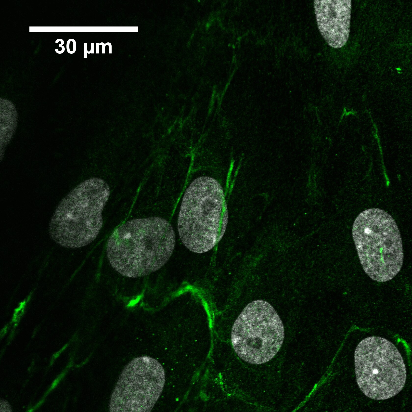
Application: ImmunocytochemistrySample Tested: Cell cultureSpecies: HumanVerified Customer | Posted 08/03/2021Human periodontal ligament cells stained with anti-emilin1 1:300 and goat anti-rabbit Alexa Fluor 488 1:500. Nuclei costained with 300nM DAPI.
 Bio-Techne ResponseThis review was submitted through the legacy Novus Innovators Program, reflecting a new species or application tested on a primary antibody.
 -
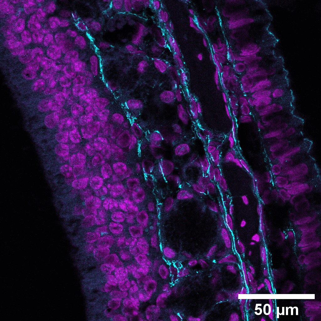
Application: Immunohistochemistry-ParaffinSample Tested: FFPE and IHC-P Sample TestedSpecies: RatVerified Customer | Posted 08/03/20215um section of formalin-fixed paraffin-embedded rat nasal concha stained with anti-emilin1 (1:300) and goat anti-rabbit Alexa Fluor 488 (1:500) and DAPI (300nM).Citrate buffer antigen retrieval 60C overnight.
Bio-Techne ResponseThis review was submitted through the legacy Novus Innovators Program, reflecting a new species or application tested on a primary antibody.
There are no reviews that match your criteria.
Protocols
Find general support by application which include: protocols, troubleshooting, illustrated assays, videos and webinars.
- Antigen Retrieval Protocol (PIER)
- Antigen Retrieval for Frozen Sections Protocol
- Appropriate Fixation of IHC/ICC Samples
- Cellular Response to Hypoxia Protocols
- Chromogenic IHC Staining of Formalin-Fixed Paraffin-Embedded (FFPE) Tissue Protocol
- Chromogenic Immunohistochemistry Staining of Frozen Tissue
- ClariTSA™ Fluorophore Kits
- Detection & Visualization of Antibody Binding
- Fluorescent IHC Staining of Frozen Tissue Protocol
- Graphic Protocol for Heat-induced Epitope Retrieval
- Graphic Protocol for the Preparation and Fluorescent IHC Staining of Frozen Tissue Sections
- Graphic Protocol for the Preparation and Fluorescent IHC Staining of Paraffin-embedded Tissue Sections
- Graphic Protocol for the Preparation of Gelatin-coated Slides for Histological Tissue Sections
- IHC Sample Preparation (Frozen sections vs Paraffin)
- Immunofluorescent IHC Staining of Formalin-Fixed Paraffin-Embedded (FFPE) Tissue Protocol
- Immunohistochemistry (IHC) and Immunocytochemistry (ICC) Protocols
- Immunohistochemistry Frozen Troubleshooting
- Immunohistochemistry Paraffin Troubleshooting
- Preparing Samples for IHC/ICC Experiments
- Preventing Non-Specific Staining (Non-Specific Binding)
- Primary Antibody Selection & Optimization
- Protocol for Heat-Induced Epitope Retrieval (HIER)
- Protocol for Making a 4% Formaldehyde Solution in PBS
- Protocol for VisUCyte™ HRP Polymer Detection Reagent
- Protocol for the Preparation & Fixation of Cells on Coverslips
- Protocol for the Preparation and Chromogenic IHC Staining of Frozen Tissue Sections
- Protocol for the Preparation and Chromogenic IHC Staining of Frozen Tissue Sections - Graphic
- Protocol for the Preparation and Chromogenic IHC Staining of Paraffin-embedded Tissue Sections
- Protocol for the Preparation and Chromogenic IHC Staining of Paraffin-embedded Tissue Sections - Graphic
- Protocol for the Preparation and Fluorescent IHC Staining of Frozen Tissue Sections
- Protocol for the Preparation and Fluorescent IHC Staining of Paraffin-embedded Tissue Sections
- Protocol for the Preparation of Gelatin-coated Slides for Histological Tissue Sections
- R&D Systems Quality Control Western Blot Protocol
- TUNEL and Active Caspase-3 Detection by IHC/ICC Protocol
- The Importance of IHC/ICC Controls
- Troubleshooting Guide: Immunohistochemistry
- Troubleshooting Guide: Western Blot Figures
- Western Blot Conditions
- Western Blot Protocol
- Western Blot Protocol for Cell Lysates
- Western Blot Troubleshooting
- Western Blot Troubleshooting Guide
- View all Protocols, Troubleshooting, Illustrated assays and Webinars
Loading...