FAM134B Antibody - BSA Free
Novus Biologicals | Catalog # NBP2-55248

Loading...
Key Product Details
Species Reactivity
Human
Applications
Immunocytochemistry/ Immunofluorescence
Label
Unconjugated
Antibody Source
Polyclonal Rabbit IgG
Format
BSA Free
Loading...
Product Specifications
Immunogen
This antibody was developed against a recombinant protein corresponding to the following amino acid sequence: ENGMGTNDEDELSLGLPTELKRKKEQLDSGHRPSKETQSAAGLTLPLNSDQTFHLMSNLAGDVITAAVTAAIKDQLEGVQQALSQAAPIPEEDTDTEEGDDFELLDQSELDQIESELGLTQDQEAEAQQNKKSSG
Reactivity Notes
Mouse 86%, Rat 84%
Clonality
Polyclonal
Host
Rabbit
Isotype
IgG
Scientific Data Images for FAM134B Antibody - BSA Free
Western Blot: FAM134B Antibody [NBP2-55248] -
Western Blot: FAM134B Antibody [NBP2-55248] - Palmitate inhibits EBSS-induced ER-phagy. a–c Cells transiently expressing mRFP-LC3 were starved in EBSS for 3 h in the presence or absence of palmitate (0.1 mM) & were co-treated for 3 h with Baf A1. Treated cells were stained for the ER marker KDEL. Representative micrographs (a) & quantification (b) of colocalization between mRFP-LC3 & KDEL (n = 10 per each group). Scale bar, 10 μm. Data are mean ± SEM; ****p < 0.0001 vs. control, ###p = 0.0002, ####p < 0.0001, ††††p < 0.0001. c The ratio of the average number of KDEL-positive mRFP-LC3 puncta to the total number of mRFP-LC3 puncta per cell (n = 10 per each group). Data are mean ± SEM; ****p < 0.0001 vs. control, ####p < 0.0001, ††††p < 0.0001. d & e Immunoblotting analysis (d) & quantification (e) of ER-phagy receptors & LC3-II in cells starved in EBSS for 3 h in the presence or absence of palmitate (0.1 mM) & were co-treated for 3 h with Baf A1 (FAM134B; n = 8 for control, PA, EBSS, PA + EBSS, n = 5 for control + Baf A1, PA + Baf A1, EBSS + Baf A1, PA + EBSS + Baf A1, RTN3; n = 4 per each group, CCPG1; n = 3 per each group, LC3-II; n = 9 for control, PA, EBSS, PA + EBSS, n = 4 for control + Baf A1, PA + Baf A1, EBSS + Baf A1, PA + EBSS + Baf A1). Data are mean ± SEM; FAM134B; ****p < 0.0001 vs. control, ##p = 0.0016, ††p = 0.0094, CCPG1; ****p < 0.0001 vs. control, LC3-II; *p = 0.0331, ****p < 0.0001 vs. control, ###p = 0.0002 at PA vs. PA + Baf A1, ###p = 0.0005 at PA + EBSS vs. PA + EBSS + Baf A1, ####p < 0.0001, †p = 0.0203. n.s., no significant difference Image collected & cropped by CiteAb from the following publication (https://pubmed.ncbi.nlm.nih.gov/33823883), licensed under a CC-BY license. Not internally tested by Novus Biologicals.
Western Blot: FAM134B Antibody [NBP2-55248] -
Western Blot: FAM134B Antibody [NBP2-55248] - Palmitate impairs ER-phagy. a & b Immunoblotting analysis (a) & quantification (b) of ER-phagy receptors (FAM134B, RTN3, & CCPG1) in cells treated with 0.1 mM palmitate for the indicated times (FAM134B & RTN3; n = 4 per each group, CCPG1; n = 3 per each group). Data are mean ± SEM; FAM134B; **p = 0.0053, ***p = 0.0005 vs. control, CCPG1; *p = 0.0388, ***p = 0.0004 vs. control. c & d Cells transiently expressing mRFP-LC3 were treated with 0.1 mM palmitate for the indicated times in the presence or absence of Baf A1 (200 nM). Treated cells were stained for the ER marker KDEL. Representative micrographs (c) & quantification (d) of colocalization between mRFP-LC3 & KDEL (n = 13 per each group). Scale bar, 10 μm. Data are mean ± SEM; **p = 0.0029, ***p = 0.0008 vs. control, ##p = 0.0025. n.s., no significant difference Image collected & cropped by CiteAb from the following publication (https://pubmed.ncbi.nlm.nih.gov/33823883), licensed under a CC-BY license. Not internally tested by Novus Biologicals.Applications for FAM134B Antibody - BSA Free
Application
Recommended Usage
Immunocytochemistry/ Immunofluorescence
0.25-2 ug/ml
Application Notes
ICC/IF Fixation Permeabilization: Use PFA/Triton X-100.
Reviewed Applications
Read 1 review rated 5 using NBP2-55248 in the following applications:
Formulation, Preparation, and Storage
Purification
Affinity purified
Formulation
PBS (pH 7.2) and 40% Glycerol
Format
BSA Free
Preservative
0.02% Sodium Azide
Concentration
Concentrations vary lot to lot. See vial label for concentration. If unlisted please contact technical services.
Shipping
The product is shipped with polar packs. Upon receipt, store it immediately at the temperature recommended below.
Stability & Storage
Store at 4C short term. Aliquot and store at -20C long term. Avoid freeze-thaw cycles.
Background: FAM134B
Alternate Names
FAM134B protein, family with sequence similarity 134, member B, FLJ20152, FLJ22155, FLJ22179, HSAN2B, JK1
Gene Symbol
RETREG1
Additional FAM134B Products
Product Documents for FAM134B Antibody - BSA Free
Certificate of Analysis
To download a Certificate of Analysis, please enter a lot or batch number in the search box below.
Product Specific Notices for FAM134B Antibody - BSA Free
This product is for research use only and is not approved for use in humans or in clinical diagnosis. Primary Antibodies are guaranteed for 1 year from date of receipt.
Citations for FAM134B Antibody - BSA Free
 Powered by Bioz
Customer Reviews for FAM134B Antibody - BSA Free (1)
5 out of 5
1 Customer Rating
Have you used FAM134B Antibody - BSA Free?
Submit a review and receive an Amazon gift card!
$25/€18/£15/$25CAN/¥2500 Yen for a review with an image
$10/€7/£6/$10CAN/¥1110 Yen for a review without an image
Submit a review
Customer Images
Showing
1
-
1 of
1 review
Showing All
Filter By:
-
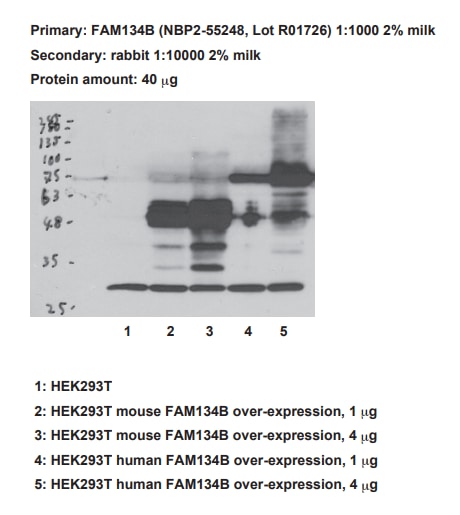
Application: Western BlotSample Tested: HEK293 cell lysateSpecies: Mouse and HumanVerified Customer | Posted 05/12/20171: HEK293T 2: HEK293T mouse FAM134B over-expression, 1 μg 3: HEK293T mouse FAM134B over-expression, 4 μg 4: HEK293T human FAM134B over-expression, 1 μg 5: HEK293T human FAM134B over-expression, 4 μg

There are no reviews that match your criteria.
Protocols
Find general support by application which include: protocols, troubleshooting, illustrated assays, videos and webinars.
- Appropriate Fixation of IHC/ICC Samples
- Cellular Response to Hypoxia Protocols
- ClariTSA™ Fluorophore Kits
- Detection & Visualization of Antibody Binding
- ICC Cell Smear Protocol for Suspension Cells
- ICC Immunocytochemistry Protocol Videos
- ICC for Adherent Cells
- Immunocytochemistry (ICC) Protocol
- Immunocytochemistry Troubleshooting
- Immunofluorescence of Organoids Embedded in Cultrex Basement Membrane Extract
- Immunohistochemistry (IHC) and Immunocytochemistry (ICC) Protocols
- Preparing Samples for IHC/ICC Experiments
- Preventing Non-Specific Staining (Non-Specific Binding)
- Primary Antibody Selection & Optimization
- Protocol for VisUCyte™ HRP Polymer Detection Reagent
- Protocol for the Fluorescent ICC Staining of Cell Smears - Graphic
- Protocol for the Fluorescent ICC Staining of Cultured Cells on Coverslips - Graphic
- Protocol for the Preparation and Fluorescent ICC Staining of Cells on Coverslips
- Protocol for the Preparation and Fluorescent ICC Staining of Non-adherent Cells
- Protocol for the Preparation and Fluorescent ICC Staining of Stem Cells on Coverslips
- Protocol for the Preparation of a Cell Smear for Non-adherent Cell ICC - Graphic
- TUNEL and Active Caspase-3 Detection by IHC/ICC Protocol
- The Importance of IHC/ICC Controls
- View all Protocols, Troubleshooting, Illustrated assays and Webinars
Loading...