FCHO2 Antibody - BSA Free
Novus Biologicals | Catalog # NBP2-32694
![Western Blot: FCHO2 Antibody [NBP2-32694]](https://resources.rndsystems.com/images/products/FCHO2-Antibody-Western-Blot-NBP2-32694-img0018.jpg "Western Blot: FCHO2 Antibody [NBP2-32694]")
Loading...
Key Product Details
Validated by
Knockout/Knockdown, Orthogonal Validation, Independent Antibodies
Species Reactivity
Validated:
Human
Predicted:
Mouse (97%), Rat (99%). Backed by our 100% Guarantee.
Applications
Validated:
Knockout Validated, Western Blot, Simple Western
Cited:
Western Blot
Label
Unconjugated
Antibody Source
Polyclonal Rabbit IgG
Format
BSA Free
Loading...
Product Specifications
Immunogen
This antibody was developed against a recombinant protein corresponding to amino acids: KAAVKSKKATDTYKLYVEKYALAKADFEQKMTETAQKFQDIEETHLIHIKEIIGSLSNAIKEIHLQIG
Clonality
Polyclonal
Host
Rabbit
Isotype
IgG
Scientific Data Images for FCHO2 Antibody - BSA Free
Western Blot: FCHO2 Antibody [NBP2-32694]
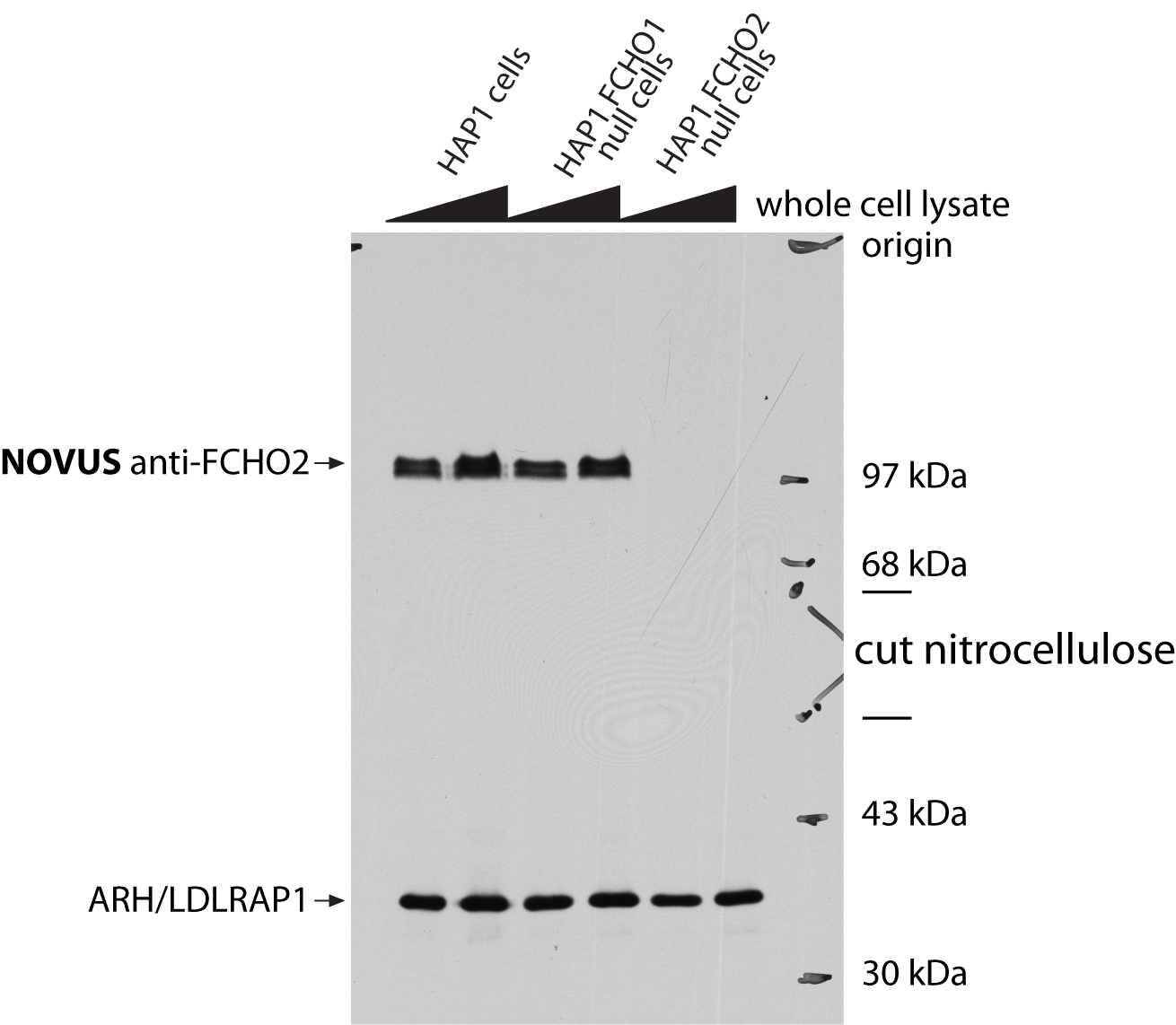
Western Blot: FCHO2 Antibody [NBP2-32694] - HAP1 cells probed with anti-FCHO2 antibody (upper panel) or anti-ARH (LDLRAP1) antibody (lower panel). Image from a verified customer review.![Western Blot: FCHO2 Antibody [NBP2-32694]](https://resources.rndsystems.com/images/products/FCHO2-Antibody-Western-Blot-NBP2-32694-img0006.jpg "Western Blot: FCHO2 Antibody [NBP2-32694]")
Western Blot: FCHO2 Antibody [NBP2-32694]
Western Blot: FCHO2 Antibody [NBP2-32694] - Lane 1: Marker [kDa] 250, 130, 95, 72, 55, 36, 28, 17, 10. Lane 2: Human cell line RT-4![Simple Western: FCHO2 Antibody [NBP2-32694]](https://resources.rndsystems.com/images/products/FCHO2-Antibody-Simple-Western-NBP2-32694-img0009.jpg "Simple Western: FCHO2 Antibody [NBP2-32694]")
Simple Western: FCHO2 Antibody [NBP2-32694]
Simple Western: FCHO2 Antibody [NBP2-32694] - Simple Western lane view shows a specific band for FCHO2 in 0.2 mg/ml of RT-4 (left), A431 (right) lysate. This experiment was performed under reducing conditions using the 12-230 kDa separation system.![Simple Western: FCHO2 Antibody [NBP2-32694]](https://resources.rndsystems.com/images/products/FCHO2-Antibody-Simple-Western-NBP2-32694-img0010.jpg "Simple Western: FCHO2 Antibody [NBP2-32694]")
Simple Western: FCHO2 Antibody [NBP2-32694]
Simple Western: FCHO2 Antibody [NBP2-32694] - Electropherogram image(s) of corresponding Simple Western lane view. FCHO2 antibody was used at 1:20 dilution on RT-4 and A431 lysate(s).![Knockout Validated: FCHO2 Antibody [NBP2-32694]](https://resources.rndsystems.com/images/products/FCHO2-Antibody-Knockout-Validated-NBP2-32694-img0020.jpg "Western Blot: FCHO2 Antibody [NBP2-32694]")
![FCHO2 Antibody Immunohistochemistry-Paraffin: FCHO2 Antibody Antibody [NBP2-32694]](https://resources.rndsystems.com/images/products/nbp2-32694_rabbit-polyclonal-fcho2-antibody-104202413384416.jpg "Immunohistochemistry-Paraffin: FCHO2 Antibody Antibody [NBP2-32694]")
![FCHO2 Antibody - BSA Free Immunohistochemistry-Paraffin: FCHO2 Antibody [NBP2-32694]](https://resources.rndsystems.com/images/products/nbp2-32694_-immunohistochemistry-paraffin-639173302846935235.jpg "Immunohistochemistry-Paraffin: FCHO2 Antibody [NBP2-32694]")
Applications for FCHO2 Antibody - BSA Free
Application
Recommended Usage
Simple Western
1:20
Western Blot
0.04-0.4 ug/ml
Application Notes
For IHC-Paraffin, HIER pH 6 retrieval is recommended. In Simple Western only 10 - 15 uL of the recommended dilution is used per data point.
See Simple Western Antibody Database for Simple Western validation: Tested in RT-4, A431, separated by Size, antibody dilution of 1:20, apparent MW was 117 kDa.
See Simple Western Antibody Database for Simple Western validation: Tested in RT-4, A431, separated by Size, antibody dilution of 1:20, apparent MW was 117 kDa.
Reviewed Applications
Read 1 review rated 5 using NBP2-32694 in the following applications:
Formulation, Preparation, and Storage
Purification
Affinity purified
Formulation
PBS (pH 7.2) and 40% Glycerol
Format
BSA Free
Preservative
0.02% Sodium Azide
Concentration
Concentrations vary lot to lot. See vial label for concentration. If unlisted please contact technical services.
Shipping
The product is shipped with polar packs. Upon receipt, store it immediately at the temperature recommended below.
Stability & Storage
Store at 4C short term. Aliquot and store at -20C long term. Avoid freeze-thaw cycles.
Background: FCHO2
Alternate Names
FCH domain only 2
Gene Symbol
FCHO2
Additional FCHO2 Products
Product Documents for FCHO2 Antibody - BSA Free
Certificate of Analysis
To download a Certificate of Analysis, please enter a lot or batch number in the search box below.
Product Specific Notices for FCHO2 Antibody - BSA Free
This product is for research use only and is not approved for use in humans or in clinical diagnosis. Primary Antibodies are guaranteed for 1 year from date of receipt.
Citations for FCHO2 Antibody - BSA Free
 Powered by Bioz
Customer Reviews for FCHO2 Antibody - BSA Free (1)
5 out of 5
1 Customer Rating
Have you used FCHO2 Antibody - BSA Free?
Submit a review and receive an Amazon gift card!
$25/€18/£15/$25CAN/¥2500 Yen for a review with an image
$10/€7/£6/$10CAN/¥1110 Yen for a review without an image
Submit a review
Customer Images
Showing
1
-
1 of
1 review
Showing All
Filter By:
-
Application: Western BlotSample Tested: human haploid HAP1 cell whole cell lysatesSpecies: HumanVerified Customer | Posted 01/16/2015HAP1 cells HAP1 cells probed with Novus anti-FCHO2 antibody (upper panel) or anti-ARH (LDLRAP1) antibody (lower panel)

There are no reviews that match your criteria.
Protocols
Find general support by application which include: protocols, troubleshooting, illustrated assays, videos and webinars.
- Cellular Response to Hypoxia Protocols
- R&D Systems Quality Control Western Blot Protocol
- Troubleshooting Guide: Western Blot Figures
- Western Blot Conditions
- Western Blot Protocol
- Western Blot Protocol for Cell Lysates
- Western Blot Troubleshooting
- Western Blot Troubleshooting Guide
- View all Protocols, Troubleshooting, Illustrated assays and Webinars
Loading...