FLNC Antibody - BSA Free
Novus Biologicals | Catalog # NBP1-89300
![Immunohistochemistry-Paraffin: FLNC Antibody [NBP1-89300]](https://resources.rndsystems.com/images/products/FLNC-Antibody-Immunohistochemistry-Paraffin-NBP1-89300-img0010.jpg "Immunohistochemistry-Paraffin: FLNC Antibody [NBP1-89300]")
Loading...
Key Product Details
Validated by
Orthogonal Validation
Species Reactivity
Validated:
Human
Cited:
Human, Mouse
Predicted:
Mouse (99%), Rat (99%). Backed by our 100% Guarantee.
Applications
Validated:
Immunohistochemistry, Immunohistochemistry-Paraffin, Immunocytochemistry/ Immunofluorescence
Cited:
Immunohistochemistry-Paraffin, Western Blot
Label
Unconjugated
Antibody Source
Polyclonal Rabbit IgG
Format
BSA Free
Loading...
Product Specifications
Immunogen
This antibody was developed against Recombinant Protein corresponding to amino acids: HSLHETSTVLVETVTKSSSSRGSSYSSIPKFSSDASKVVTRGPGLSQAFVGQKNSFTVDCSKAGTNMMMVGVHGPKTPCEEVYVKHMGNRVYNVTYTVKEKGDY
Clonality
Polyclonal
Host
Rabbit
Isotype
IgG
Scientific Data Images for FLNC Antibody - BSA Free
![Immunocytochemistry/ Immunofluorescence: FLNC Antibody [NBP1-89300]](https://resources.rndsystems.com/images/products/FLNC-Antibody-Immunocytochemistry-Immunofluorescence-NBP1-89300-img0009.jpg "Immunocytochemistry/ Immunofluorescence: FLNC Antibody [NBP1-89300]")
Immunocytochemistry/ Immunofluorescence: FLNC Antibody [NBP1-89300]
Immunocytochemistry/Immunofluorescence: FLNC Antibody [NBP1-89300] - Staining of human cell line U-2 OS shows localization to plasma membrane and cytosol. Antibody staining shown in green.![Immunohistochemistry-Paraffin: FLNC Antibody [NBP1-89300]](https://resources.rndsystems.com/images/products/FLNC-Antibody-Immunohistochemistry-Paraffin-NBP1-89300-img0015.jpg "Immunohistochemistry-Paraffin: FLNC Antibody [NBP1-89300]")
Immunohistochemistry-Paraffin: FLNC Antibody [NBP1-89300]
Immunohistochemistry-Paraffin: FLNC Antibody [NBP1-89300] - Staining of human liver shows no positivity in hepatocytes as expected.![Immunohistochemistry-Paraffin: FLNC Antibody [NBP1-89300]](https://resources.rndsystems.com/images/products/FLNC-Antibody-Immunohistochemistry-Paraffin-NBP1-89300-img0006.jpg "Immunohistochemistry-Paraffin: FLNC Antibody [NBP1-89300]")
Immunohistochemistry-Paraffin: FLNC Antibody [NBP1-89300]
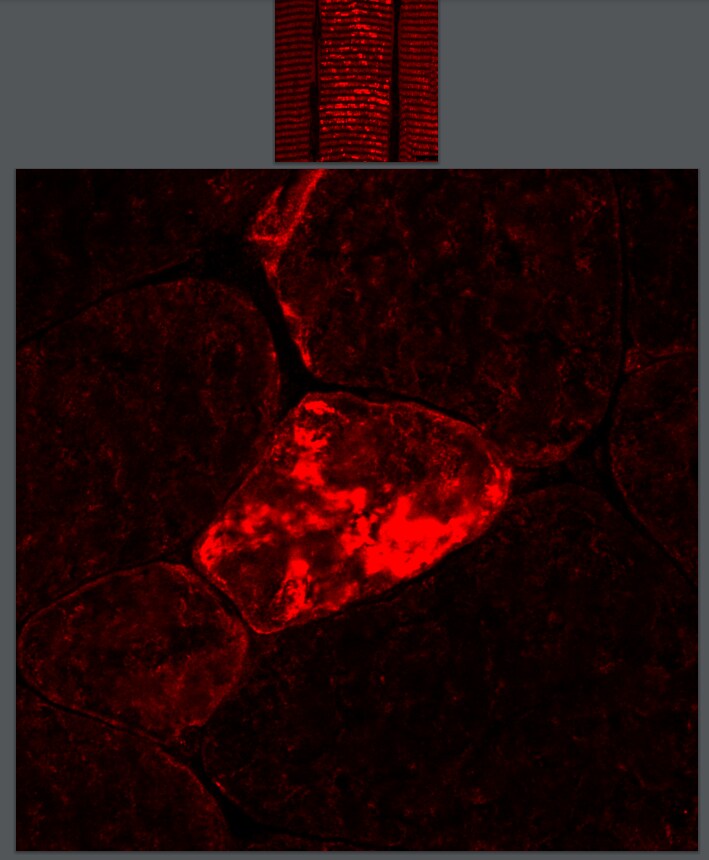
Immunohistochemistry-Paraffin: FLNC Antibody [NBP1-89300] - Staining of human skeletal muscle shows high expression.![Immunohistochemistry-Paraffin: FLNC Antibody [NBP1-89300]](https://resources.rndsystems.com/images/products/FLNC-Antibody-Immunohistochemistry-Paraffin-NBP1-89300-img0013.jpg "Immunohistochemistry-Paraffin: FLNC Antibody [NBP1-89300]")
Immunohistochemistry-Paraffin: FLNC Antibody [NBP1-89300]
Immunohistochemistry-Paraffin: FLNC Antibody [NBP1-89300] - Staning of human tonsil shows no positivity in non-germinal center cells as expected.![Immunohistochemistry-Paraffin: FLNC Antibody [NBP1-89300]](https://resources.rndsystems.com/images/products/FLNC-Antibody-Immunohistochemistry-Paraffin-NBP1-89300-img0014.jpg "Immunohistochemistry-Paraffin: FLNC Antibody [NBP1-89300]")
Immunohistochemistry-Paraffin: FLNC Antibody [NBP1-89300]
Immunohistochemistry-Paraffin: FLNC Antibody [NBP1-89300] - Staining of human prostate shows moderate cytoplasmic positivity in smooth muscle cells.Applications for FLNC Antibody - BSA Free
Application
Recommended Usage
Immunocytochemistry/ Immunofluorescence
0.25 - 2 ug/mL
Immunohistochemistry
1:200 - 1:500
Immunohistochemistry-Paraffin
1:200 - 1:500
Application Notes
IHC-Paraffin, HIER pH 6 retrieval is recommended. ICC/IF, Fixation Permeabilization: Use PFA/Triton X-100.
Reviewed Applications
Read 1 review rated 4 using NBP1-89300 in the following applications:
Formulation, Preparation, and Storage
Purification
Affinity purified
Formulation
PBS (pH 7.2) and 40% Glycerol
Format
BSA Free
Preservative
0.02% Sodium Azide
Concentration
Concentrations vary lot to lot. See vial label for concentration. If unlisted please contact technical services.
Shipping
The product is shipped with polar packs. Upon receipt, store it immediately at the temperature recommended below.
Stability & Storage
Store at 4C short term. Aliquot and store at -20C long term. Avoid freeze-thaw cycles.
Background: FLNC
Three isoforms of Filamin are expressed in mammals, identified as FLNA, FLNB and FLNC. FLNC is considered a muscle specific Filamin isoform for its high expression in cardiac and skeletal muscles. In muscle tissue, FLNC localizes to the Z-line, sarcolemma, myotendinous and intercalated disks (2). FLNC interacts with various muscle proteins via a non-conserved sequence within its Ig domain 20. Some muscle binding partners for FLNC include gamma and delta-sarcoglycans in the sarcolemma, and FATZ, myozenins, myotilin and myopodin in the Z-discs (2, 4). FLNC variants are associated with various inherited pathological conditions including myofibrillar myopathy 5, distal myopathy 4, dilated cardiomyopathy, hypertrophic cardiomyopathy, and restrictive cardiomyopathy (2, 5).
References
1. Chiang, W., Greaser, M. L., & Lyons, G. E. (2000). Filamin isogene expression during mouse myogenesis. Developmental Dynamics. https://doi.org/10.1002/(sici)1097-0177(200001)217:199::aid-dvdy9>3.3.co;2-x
2. Leber, Y., Ruparelia, A. A., Kirfel, G., van der Ven, P. F. M., Hoffmann, B., Merkel, R.,... Furst, D. O. (2016). Filamin C is a highly dynamic protein associated with fast repair of myofibrillar microdamage. Human Molecular Genetics. https://doi.org/10.1093/hmg/ddw135
3. Nakamura, F., Stossel, T. P., & Hartwig, J. H. (2011). The filamins: Organizers of cell structure and function. Cell Adhesion and Migration. https://doi.org/10.4161/cam.5.2.14401
4. Thompson, T. G., Chan, Y. M., Hack, A. A., Brosius, M., Rajala, M., Lidov, H. G. W.,... Kunkel, L. M. (2000). Filamin 2 (FLN2): A muscle-specific sarcoglycan interacting protein. Journal of Cell Biology. https://doi.org/10.1083/jcb.148.1.115
5. Brodehl, A., Ferrier, R. A., Hamilton, S. J., Greenway, S. C., Brundler, M. A., Yu, W.,... Scherer, S. (2016). Mutations in FLNC are Associated with Familial Restrictive Cardiomyopathy. Human Mutation. https://doi.org/10.1002/humu.22942
Alternate Names
ABP-280, ABP280A, ABP-280-like protein, ABPA, ABP-L, ABPLABP-L, gamma filamin, Actin-binding-like protein, filamin 2, filamin C, gamma, filamin C, gamma (actin binding protein 280), Filamin-2, filamin-C, FLJ10186, FLN2actin binding protein 280, FLNc, FLN-C, Gamma-filamin
Gene Symbol
FLNC
Additional FLNC Products
Product Documents for FLNC Antibody - BSA Free
Certificate of Analysis
To download a Certificate of Analysis, please enter a lot or batch number in the search box below.
Product Specific Notices for FLNC Antibody - BSA Free
This product is for research use only and is not approved for use in humans or in clinical diagnosis. Primary Antibodies are guaranteed for 1 year from date of receipt.
Citations for FLNC Antibody - BSA Free
 Powered by Bioz
Customer Reviews for FLNC Antibody - BSA Free (1)
4 out of 5
1 Customer Rating
Have you used FLNC Antibody - BSA Free?
Submit a review and receive an Amazon gift card!
$25/€18/£15/$25CAN/¥2500 Yen for a review with an image
$10/€7/£6/$10CAN/¥1110 Yen for a review without an image
Submit a review
Customer Images
Showing
1
-
1 of
1 review
Showing All
Filter By:
-
Application: Immunohistochemistry-FrozenSample Tested: Skeletal muscle tissue and Mouse skeletal muscleSpecies: MouseVerified Customer | Posted 06/28/20211. TA mouse muscle longitudinal section and (2.) Vasus transverse section stained with Filamin C antibody (NBP1-89300)Pathak, P., Blech-Hermoni, Y., Subedi, K. et al. Myopathy associated LDB3 mutation causes Z-disc disassembly and protein aggregation through PKC alpha and TSC2-mTOR downregulation. Commun Biol 4, 355 (2021). https://doi.org/10.1038/s42003-021-01864-1
 Bio-Techne ResponseThis review was submitted through the legacy Novus Innovators Program, reflecting a new species or application tested on a primary antibody.
There are no reviews that match your criteria.
Protocols
Find general support by application which include: protocols, troubleshooting, illustrated assays, videos and webinars.
- Antigen Retrieval Protocol (PIER)
- Antigen Retrieval for Frozen Sections Protocol
- Appropriate Fixation of IHC/ICC Samples
- Cellular Response to Hypoxia Protocols
- Chromogenic IHC Staining of Formalin-Fixed Paraffin-Embedded (FFPE) Tissue Protocol
- Chromogenic Immunohistochemistry Staining of Frozen Tissue
- ClariTSA™ Fluorophore Kits
- Detection & Visualization of Antibody Binding
- Fluorescent IHC Staining of Frozen Tissue Protocol
- Graphic Protocol for Heat-induced Epitope Retrieval
- Graphic Protocol for the Preparation and Fluorescent IHC Staining of Frozen Tissue Sections
- Graphic Protocol for the Preparation and Fluorescent IHC Staining of Paraffin-embedded Tissue Sections
- Graphic Protocol for the Preparation of Gelatin-coated Slides for Histological Tissue Sections
- ICC Cell Smear Protocol for Suspension Cells
- ICC Immunocytochemistry Protocol Videos
- ICC for Adherent Cells
- IHC Sample Preparation (Frozen sections vs Paraffin)
- Immunocytochemistry (ICC) Protocol
- Immunocytochemistry Troubleshooting
- Immunofluorescence of Organoids Embedded in Cultrex Basement Membrane Extract
- Immunofluorescent IHC Staining of Formalin-Fixed Paraffin-Embedded (FFPE) Tissue Protocol
- Immunohistochemistry (IHC) and Immunocytochemistry (ICC) Protocols
- Immunohistochemistry Frozen Troubleshooting
- Immunohistochemistry Paraffin Troubleshooting
- Preparing Samples for IHC/ICC Experiments
- Preventing Non-Specific Staining (Non-Specific Binding)
- Primary Antibody Selection & Optimization
- Protocol for Heat-Induced Epitope Retrieval (HIER)
- Protocol for Making a 4% Formaldehyde Solution in PBS
- Protocol for VisUCyte™ HRP Polymer Detection Reagent
- Protocol for the Fluorescent ICC Staining of Cell Smears - Graphic
- Protocol for the Fluorescent ICC Staining of Cultured Cells on Coverslips - Graphic
- Protocol for the Preparation & Fixation of Cells on Coverslips
- Protocol for the Preparation and Chromogenic IHC Staining of Frozen Tissue Sections
- Protocol for the Preparation and Chromogenic IHC Staining of Frozen Tissue Sections - Graphic
- Protocol for the Preparation and Chromogenic IHC Staining of Paraffin-embedded Tissue Sections
- Protocol for the Preparation and Chromogenic IHC Staining of Paraffin-embedded Tissue Sections - Graphic
- Protocol for the Preparation and Fluorescent ICC Staining of Cells on Coverslips
- Protocol for the Preparation and Fluorescent ICC Staining of Non-adherent Cells
- Protocol for the Preparation and Fluorescent ICC Staining of Stem Cells on Coverslips
- Protocol for the Preparation and Fluorescent IHC Staining of Frozen Tissue Sections
- Protocol for the Preparation and Fluorescent IHC Staining of Paraffin-embedded Tissue Sections
- Protocol for the Preparation of Gelatin-coated Slides for Histological Tissue Sections
- Protocol for the Preparation of a Cell Smear for Non-adherent Cell ICC - Graphic
- TUNEL and Active Caspase-3 Detection by IHC/ICC Protocol
- The Importance of IHC/ICC Controls
- Troubleshooting Guide: Immunohistochemistry
- View all Protocols, Troubleshooting, Illustrated assays and Webinars
Loading...