![Western Blot: G3BP1 Antibody [NBP1-18922]](https://resources.rndsystems.com/images/products/G3BP1-Antibody-Western-Blot-NBP1-18922-img0008.jpg "Western Blot: G3BP1 Antibody [NBP1-18922]")
Loading...
Key Product Details
Validated by
Independent Antibodies, Biological Validation
Species Reactivity
Validated:
Human, Mouse, Mammal
Cited:
Human, Mammal
Predicted:
Orangutan (100%). Backed by our 100% Guarantee.
Applications
Validated:
Western Blot, Immunocytochemistry/ Immunofluorescence, Immunoprecipitation
Cited:
Immunocytochemistry/ Immunofluorescence, IF/IHC
Label
Unconjugated
Antibody Source
Polyclonal Rabbit IgG
Loading...
Product Specifications
Immunogen
The immunogen recognized by this antibody maps to a region between residue 275 and 325 of human GTPase activating protein (SH3 domain) binding protein 1 using the numbering given in entry NP_005745.1 (GeneID 10146).
Reactivity Notes
Mammal reactivity reported in scientific literature (PMID: 28794026).
Clonality
Polyclonal
Host
Rabbit
Isotype
IgG
Scientific Data Images for G3BP1 Antibody
Western Blot: G3BP1 Antibody [NBP1-18922]
Western Blot: G3BP1 Antibody [NBP1-18922] - Whole cell lysate (50 ug) from HeLa, MCF-7, U2OS, TCMK-1, and NIH 3T3 cells prepared using NETN lysis buffer. Antibody: Affinity purified rabbit anti-G3BP1antibody used for WB at 0.04 ug/ml. Detection: Chemiluminescence with anexposure time of 1 second.![Immunocytochemistry/ Immunofluorescence: G3BP1 Antibody [NBP1-18922]](https://resources.rndsystems.com/images/products/G3BP1-Antibody-Immunocytochemistry-Immunofluorescence-NBP1-18922-img0007.jpg "Immunocytochemistry/ Immunofluorescence: G3BP1 Antibody [NBP1-18922]")
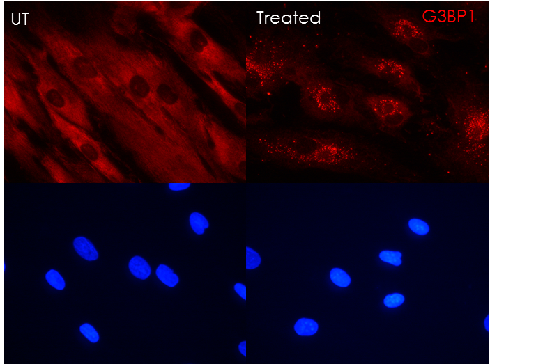
Immunocytochemistry/ Immunofluorescence: G3BP1 Antibody [NBP1-18922]
Immunocytochemistry/Immunofluorescence: G3BP1 Antibody [NBP1-18922] - G3BP1 in red (Alexa fluor 555) and nucleus in blue (DAPI). Human primary fibroblasts untreated in the left and treated in the right with G3BP1 stress granules labeling. Alexa fluor 555 1s exposure and DAPI 1s exposure. Fixation: PFA 4% 10min RT, permeabilization: Triton 0.1% TBS, blocking: Donkey Serum 10% TBS-T, Ab dilution: 1:1000, Secondary Ab: Alexa fluor 555 Donkey anti-rabbit 1:600, Mounting medium with DAPI. Image submitted by a verified customer review.![Western Blot: G3BP1 Antibody [NBP1-18922]](https://resources.rndsystems.com/images/products/G3BP1-Antibody-Western-Blot-NBP1-18922-img0006.jpg "Western Blot: G3BP1 Antibody [NBP1-18922]")
Western Blot: G3BP1 Antibody [NBP1-18922]
Western Blot: G3BP1 Antibody [NBP1-18922] - Whole cell lysate from HeLa (5, 15 and 50 mcg for WB; 1 mg for IP, 20% of IP loaded), 293T (T; 50 mcg) and mouse NIH3T3 (M; 50mcg) cells. Affinity purified rabbit anti-G3BP1 antibody used for WB at 0.04 mcg/ml (A) and 1 mcg/ml (B) and used for IP at 3 mcg/mg lysate. G3BP1 was also immunoprecipitated by rabbit anti-G3BP1 antibody NBP1-18923, which recognizes a downstream epitope.Applications for G3BP1 Antibody
Application
Recommended Usage
Immunocytochemistry/ Immunofluorescence
Validated from a verified customer review.
Immunoprecipitation
2-10 ug/mg lysate
Western Blot
1:2000-1:10000
Reviewed Applications
Read 1 review rated 5 using NBP1-18922 in the following applications:
Formulation, Preparation, and Storage
Purification
Immunogen affinity purified
Formulation
TBS and 0.1% BSA
Preservative
0.09% Sodium Azide
Concentration
0.2 mg/ml
Shipping
The product is shipped with polar packs. Upon receipt, store it immediately at the temperature recommended below.
Stability & Storage
Store at 4C. Do not freeze.
Background: G3BP1
Alternate Names
ATP-dependent DNA helicase VIII, EC 3.6.1, EC 3.6.4.12, EC 3.6.4.13, G3BP-1, G3BPRas-GTPase-activating protein SH3-domain-binding protein, GAP binding protein, GAP SH3 domain-binding protein 1, GTPase activating protein (SH3 domain) binding protein 1, hDH VIII, HDH-VIII, MGC111040, ras GTPase-activating protein-binding protein 1, RasGAP-associated endoribonuclease G3BP
Gene Symbol
G3BP1
UniProt
Additional G3BP1 Products
Product Documents for G3BP1 Antibody
Certificate of Analysis
To download a Certificate of Analysis, please enter a lot or batch number in the search box below.
Product Specific Notices for G3BP1 Antibody
This product is for research use only and is not approved for use in humans or in clinical diagnosis. Primary Antibodies are guaranteed for 1 year from date of receipt.
Citations for G3BP1 Antibody
 Powered by Bioz
Customer Reviews for G3BP1 Antibody (1)
5 out of 5
1 Customer Rating
Have you used G3BP1 Antibody?
Submit a review and receive an Amazon gift card!
$25/€18/£15/$25CAN/¥2500 Yen for a review with an image
$10/€7/£6/$10CAN/¥1110 Yen for a review without an image
Submit a review
Customer Images
Showing
1
-
1 of
1 review
Showing All
Filter By:
-
Application: ImmunocytochemistrySample Tested: Human primary fibroblastSpecies: HumanVerified Customer | Posted 02/12/2019G3BP1 in red (Alexa fluor 555) and nucleus in blue (DAPI). Human primary fibroblasts untreated in the left and treated in the right with G3BP1 stress granules labeling. Alexa fluor 555 1s exposure and DAPI 1s exposure.ICC-IF Fixation: PFA 4% 10min RT permeabilization: Triton 0.1% TBS blocking: Donkey Serum 10% TBS-T 1. Ab dilution: 1:1000 Secondary Ab: Alexa fluor 555 Donkey anti-rabbit 1:600 Mounting medium with DAPI

There are no reviews that match your criteria.
Protocols
Find general support by application which include: protocols, troubleshooting, illustrated assays, videos and webinars.
- Appropriate Fixation of IHC/ICC Samples
- Cellular Response to Hypoxia Protocols
- ClariTSA™ Fluorophore Kits
- Detection & Visualization of Antibody Binding
- ICC Cell Smear Protocol for Suspension Cells
- ICC Immunocytochemistry Protocol Videos
- ICC for Adherent Cells
- Immunocytochemistry (ICC) Protocol
- Immunocytochemistry Troubleshooting
- Immunofluorescence of Organoids Embedded in Cultrex Basement Membrane Extract
- Immunohistochemistry (IHC) and Immunocytochemistry (ICC) Protocols
- Immunoprecipitation Protocol
- Preparing Samples for IHC/ICC Experiments
- Preventing Non-Specific Staining (Non-Specific Binding)
- Primary Antibody Selection & Optimization
- Protocol for VisUCyte™ HRP Polymer Detection Reagent
- Protocol for the Fluorescent ICC Staining of Cell Smears - Graphic
- Protocol for the Fluorescent ICC Staining of Cultured Cells on Coverslips - Graphic
- Protocol for the Preparation and Fluorescent ICC Staining of Cells on Coverslips
- Protocol for the Preparation and Fluorescent ICC Staining of Non-adherent Cells
- Protocol for the Preparation and Fluorescent ICC Staining of Stem Cells on Coverslips
- Protocol for the Preparation of a Cell Smear for Non-adherent Cell ICC - Graphic
- R&D Systems Quality Control Western Blot Protocol
- TUNEL and Active Caspase-3 Detection by IHC/ICC Protocol
- The Importance of IHC/ICC Controls
- Troubleshooting Guide: Western Blot Figures
- Western Blot Conditions
- Western Blot Protocol
- Western Blot Protocol for Cell Lysates
- Western Blot Troubleshooting
- Western Blot Troubleshooting Guide
- View all Protocols, Troubleshooting, Illustrated assays and Webinars
Loading...