![Immunohistochemistry: GFAP Antibody [NB300-141]](https://resources.rndsystems.com/images/products/GFAP-Antibody-Immunocytochemistry-Immunofluorescence-NB300-141-img0011.jpg "Immunohistochemistry: GFAP Antibody [NB300-141]")
Loading...
Key Product Details
Species Reactivity
Validated:
Human, Mouse, Rat, Porcine, Bovine, Chicken, Equine, Guinea Pig, Rabbit
Cited:
Human, Mouse, Rat, Avian - Chicken, Guinea Pig, Rabbit
Applications
Validated:
Immunohistochemistry, Immunohistochemistry-Paraffin, Immunohistochemistry-Frozen, Western Blot, Immunocytochemistry/ Immunofluorescence, Simple Western
Cited:
Immunohistochemistry, Immunohistochemistry-Paraffin, Immunohistochemistry-Frozen, Western Blot, Immunocytochemistry/ Immunofluorescence, Simple Western, Chemotaxis, IF/IHC
Label
Unconjugated
Antibody Source
Polyclonal Rabbit
Loading...
Product Specifications
Immunogen
This GFAP Antibody was developed against recombinant full length human GFAP isotype 1 expressed in and purified from E. coli.
Reactivity Notes
Predicted to work with most mammals. Chicken reactivity reported in scientific literature (PMID: 20844134). Rabbit and Guinea Pig reactivity reported in scientific literature (PMID: 4559710).
Marker
Astrocyte Marker
Clonality
Polyclonal
Host
Rabbit
Theoretical MW
50 kDa.
Disclaimer note: The observed molecular weight of the protein may vary from the listed predicted molecular weight due to post translational modifications, post translation cleavages, relative charges, and other experimental factors.
Disclaimer note: The observed molecular weight of the protein may vary from the listed predicted molecular weight due to post translational modifications, post translation cleavages, relative charges, and other experimental factors.
Scientific Data Images for GFAP Antibody
Immunohistochemistry: GFAP Antibody [NB300-141]
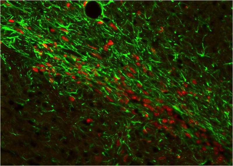
Immunohistochemistry: GFAP Antibody [NB300-141] - Analysis of a rat cerebellum section stained with rabbit polyclonal antibody to GFAP, NB300-141, dilution 1:5000 in green and mouse monoclonal antibody to MeCP2, dilution 1:500, in red. The blue is DAPI staining of nuclear DNA. Following transcardial perfusion of rat with 4% paraformaldehyde, brain was post fixed for 1 hour, cut to 45 uM, and free-floating sections were stained with above antibodies. The GFAP antibody stains the network of astrocytic cells and the processes of Bergmann glia in the molecular layer. The MeCP2 antibody specifically labels nuclei of certain neurons.![Immunocytochemistry/ Immunofluorescence: GFAP Antibody [NB300-141]](https://resources.rndsystems.com/images/products/GFAP-Antibody-Immunocytochemistry-Immunofluorescence-NB300-141-img0002.jpg "Immunocytochemistry/ Immunofluorescence: GFAP Antibody [NB300-141]")
Immunocytochemistry/ Immunofluorescence: GFAP Antibody [NB300-141]
Immunocytochemistry/Immunofluorescence: GFAP Antibody [NB300-141] - Rat neurons stained with Neurofilament Heavy antibody NB300-217 (red) and GFAP antibody NB300-141 (green).![Simple Western: GFAP Antibody [NB300-141]](https://resources.rndsystems.com/images/products/GFAP-Antibody-Simple-Western-NB300-141-img0004.jpg "Simple Western: GFAP Antibody [NB300-141]")
Simple Western: GFAP Antibody [NB300-141]
Simple Western: GFAP Antibody [NB300-141] - Simple Western lane view shows a specific band for GFAP in 0.05 mg/mL of Human Brain lysate. This experiment was performed under reducing conditions using the 12-230 kDa separation system.![Immunocytochemistry/ Immunofluorescence: GFAP Antibody [NB300-141]](https://resources.rndsystems.com/images/products/GFAP-Antibody-Immunocytochemistry-Immunofluorescence-NB300-141-img0014.jpg "Immunocytochemistry/ Immunofluorescence: GFAP Antibody [NB300-141]")
Immunocytochemistry/ Immunofluorescence: GFAP Antibody [NB300-141]
GFAP-Antibody-Immunocytochemistry-Immunofluorescence-NB300-141-img0014.jpg![Immunohistochemistry-Paraffin: GFAP Antibody [NB300-141]](https://resources.rndsystems.com/images/products/GFAP-Antibody-Immunohistochemistry-Paraffin-NB300-141-img0013.jpg "Immunohistochemistry-Paraffin: GFAP Antibody [NB300-141]")
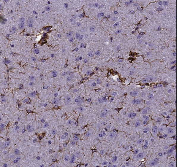
Immunohistochemistry-Paraffin: GFAP Antibody [NB300-141]
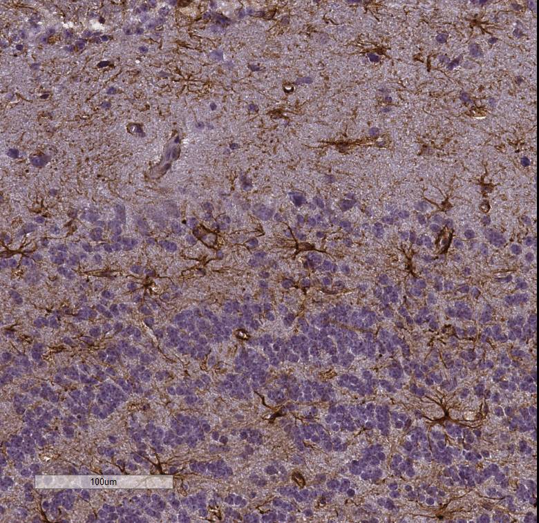
Immunohistochemistry-Paraffin: GFAP Antibody [NB300-141] - Mouse brain section, 20x magnification. Antibody at 1:1000. Detection with Polymer-HRP. IHC image submitted by a verified customer review.![Western Blot: GFAP Antibody [NB300-141]](https://resources.rndsystems.com/images/products/GFAP-Antibody-Western-Blot-NB300-141-img0006.jpg "Western Blot: GFAP Antibody [NB300-141]")
Western Blot: GFAP Antibody [NB300-141]
Western Blot: GFAP Antibody [NB300-141] - Analysis of Rat brain lysate. Antibody at 1:5000. Specific band running with an apparent SDS-PAGE molecular weight of ~50 kDa corresponds to rodent GFAP was observed.![Western Blot: GFAP Antibody [NB300-141]](https://resources.rndsystems.com/images/products/GFAP-Antibody-Western-Blot-NB300-141-img0009.jpg "Western Blot: GFAP Antibody [NB300-141]")
Western Blot: GFAP Antibody [NB300-141]
Western Blot: GFAP Antibody [NB300-141] - Analysis of GFAP expression in whole rat cerebellum homogenate.![Western Blot: GFAP Antibody [NB300-141]](https://resources.rndsystems.com/images/products/GFAP-Antibody-Western-Blot-NB300-141-img0012.jpg "Western Blot: GFAP Antibody [NB300-141]")
Western Blot: GFAP Antibody [NB300-141]
Western Blot: GFAP Antibody [NB300-141] - Analysis of different tissue lysates using rabbit polyclonal antibody to GFAP, NB300-141, dilution 1:5000 in green: [1] protein standard (red), [2] rat brain, [3] rat spinal cord, [4] mouse brain, [5] mouse spinal cord. Strong band at about 50 kDa corresponds to the major isotype of the GFAP protein. Smaller isotypes and proteolytic fragments of GFAP are also detected on the blot.![Immunocytochemistry/ Immunofluorescence: GFAP Antibody [NB300-141]](https://resources.rndsystems.com/images/products/GFAP-Antibody-Immunocytochemistry-Immunofluorescence-NB300-141-img0007.jpg "Immunocytochemistry/ Immunofluorescence: GFAP Antibody [NB300-141]")
Immunocytochemistry/ Immunofluorescence: GFAP Antibody [NB300-141]
Immunocytochemistry/Immunofluorescence: GFAP Antibody [NB300-141] - Analysis of mixed neuron-glial cultures using GFAP antibody NB300-141 (red) and Vimentin antibody NB300-223 (green). The fibroblastic cells contain only Vimentin and so are green. The astrocytes contain either Vimentin and GFAP (appearing golden) or predominantly GFAP (appearing red). Blue is nuclear DNA stain.![Immunocytochemistry/ Immunofluorescence: GFAP Antibody [NB300-141]](https://resources.rndsystems.com/images/products/GFAP-Antibody-Immunocytochemistry-Immunofluorescence-NB300-141-img0008.jpg "Immunocytochemistry/ Immunofluorescence: GFAP Antibody [NB300-141]")
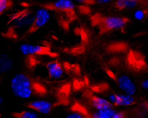
Immunocytochemistry/ Immunofluorescence: GFAP Antibody [NB300-141]
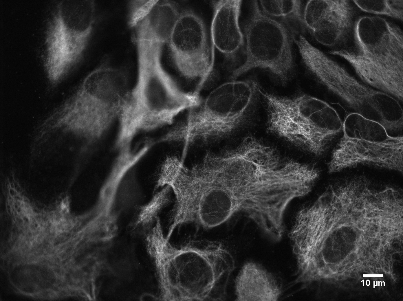
Immunocytochemistry/Immunofluorescence: GFAP Antibody [NB300-141] - Cultured Rat hippocampal neurons. ICC/IF image submitted by a verified customer review.![Immunohistochemistry: GFAP Antibody [NB300-141]](https://resources.rndsystems.com/images/products/GFAP-Antibody-Immunocytochemistry-Immunofluorescence-NB300-141-img0003.jpg "Immunohistochemistry: GFAP Antibody [NB300-141]")
Immunohistochemistry: GFAP Antibody [NB300-141]
Immunohistochemistry: GFAP Antibody [NB300-141] - Xenografted mouse brain section: astocyte and human nuclei. ICC/IF image submitted by a verified customer review.![Immunohistochemistry-Frozen: GFAP Antibody [NB300-141]](https://resources.rndsystems.com/images/products/GFAP-Antibody-Immunohistochemistry-Frozen-NB300-141-img0010.jpg "Immunohistochemistry-Frozen: GFAP Antibody [NB300-141]")
Immunohistochemistry-Frozen: GFAP Antibody [NB300-141]
Immunohistochemistry-Frozen: GFAP Antibody [NB300-141] - Imaging of mouse brain (cortex), 20x magnification. IHC image submitted by a verified customer review.
Immunocytochemistry/Immunofluorescence: GFAP Antibody [NB300-141] -
EGFP+ neurons are positive for RABV antigen.Brains were collected from Cre reporter mice fifteen days post-infection, cryosectioned, and EGFP+ regions compared to cell-specific labeling, A) NeuN (blue, neuronal nuclei antibody, 20× fluorescence imaging), B) GFAP (blue, astrocyte antibody, 40× confocal imaging), or C) RABV P antigen (purple) and DAPI nuclear stain (blue, 63× confocal imaging). White arrows in (C) indicate regions positive for RABV P.
Immunocytochemistry/ Immunofluorescence: GFAP Antibody [NB300-141] -
Immunocytochemistry/ Immunofluorescence: GFAP Antibody [NB300-141] - EGFP+ neurons are positive for RABV antigen.Brains were collected from Cre reporter mice fifteen days post-infection, cryosectioned, & EGFP+ regions compared to cell-specific labeling, A) NeuN (blue, neuronal nuclei antibody, 20× fluorescence imaging), B) GFAP (blue, astrocyte antibody, 40× confocal imaging), or C) RABV P antigen (purple) & DAPI nuclear stain (blue, 63× confocal imaging). White arrows in (C) indicate regions positive for RABV P. Image collected & cropped by CiteAb from the following publication (https://dx.plos.org/10.1371/journal.ppat.1002971), licensed under a CC-BY license. Not internally tested by Novus Biologicals.
Western Blot: GFAP Antibody [NB300-141] -
Western Blot: GFAP Antibody [NB300-141] - Effects of the repeated administration of NVP CXCR2 20 (NVP; 10 μg/5 μl; i.t.; 16 h & 1 h before CCI & then once a day for 7 days) on the protein levels of CXCR2, IBA1, GFAP, CXCL1, CXCL2, & CXCL3 proteins (A–I) in the spinal cord (A–F) & DRG (G–I) on the 7th day after CCI in rats. The data are presented as the mean fold changes relative to the control ± SEM (5–6 samples per group). Intergroup differences were analyzed using ANOVA with Bonferroni's multiple comparisons test. *p < 0.05, **p < 0.01, ***p < 0.001 indicate differences vs. naive rats. #p < 0.05, indicate differences between V-treated & NVP-treated rats. CCI, chronic constriction injury; N, naive; V, vehicle; NVP, NVP CXCR2 20. Image collected & cropped by CiteAb from the following publication (https://pubmed.ncbi.nlm.nih.gov/31616413), licensed under a CC-BY license. Not internally tested by Novus Biologicals.
Immunocytochemistry/ Immunofluorescence: GFAP Antibody [NB300-141] -
Immunocytochemistry/ Immunofluorescence: GFAP Antibody [NB300-141] - Increased astrocyte activation in IL-1 beta -stimulated two-week old offspring is reduced by maternal LB supplementation. Representative images of fluorescence microscopy of claudin-5+ (location of the brain capillaries, red), GFAP+ astrocyte (green), & DAPI (nuclei, blue). Three to five sections per mouse were examined & at least three mice were examined in each group. Stronger than control SPF GFAP staining was observed around the blood vessel (see arrow) after IL-1 beta insult. Maternal supplemented group (LB) with or without postnatal insult had GFAP levels similar to the control group indicating that LB supplementation prevented astrocyte activation around the BBB endothelium. Image collected & cropped by CiteAb from the following publication (https://pubmed.ncbi.nlm.nih.gov/32424168), licensed under a CC-BY license. Not internally tested by Novus Biologicals.
Immunocytochemistry/ Immunofluorescence: GFAP Antibody [NB300-141] -
Immunocytochemistry/ Immunofluorescence: GFAP Antibody [NB300-141] - Glial cells density is differently modulated during AD progression.a Retinal slices were immunolabeled with anti-GFAP antibody (green) & Hoechst for nuclei visualization (blue) at different ages of 3xTg-AD & non-Tg mice & density of GFAP signal was quantified as shown in b (**p < 0.01 pre vs early; n = 16 fields/four slices for each condition; two-way ANOVA, Holm-Sidak; ##p < 0.01 for comparison with age-matched non-Tg mice, two-way ANOVA, Holm-Sidak). c Representative multiarea image of retinal slice immunolabeled with anti-Iba1 antibody (green) & Hoechst for nuclei visualization (blue); density of Iba1+ cells was quantified as shown in d (**p < 0.01 vs 3xTg-AD pre; ##p < 0.01 vs age-matched non-Tg mice, n = 16 fields/four slices for each condition, two-way ANOVA, Holm-Sidak method for multiple comparison) Image collected & cropped by CiteAb from the following publication (https://pubmed.ncbi.nlm.nih.gov/29880901), licensed under a CC-BY license. Not internally tested by Novus Biologicals.Applications for GFAP Antibody
Application
Recommended Usage
Immunocytochemistry/ Immunofluorescence
1:1000 - 1:5000
Immunohistochemistry
1:1000 - 1:5000
Simple Western
1:10,000
Western Blot
1:5000
Application Notes
In WB a band can be seen at 50-55 kDa representing GFAP. A lower band may be seen around 45 kDa representing a proteolytic fragment derived from the GFAP molecule. GFAP antibody validated for IHC-P from a verified customer review. IHC-Fr has been reported in scientific literature (PMID: 28040732).
See Simple Western Antibody Database for Simple Western validation: tested in human brain lysate (0.05 mg/ml); separated by size, antibody dilution of 1:10,000; detects a band at 50 kDa; matrix was 12-230 kDa.
See Simple Western Antibody Database for Simple Western validation: tested in human brain lysate (0.05 mg/ml); separated by size, antibody dilution of 1:10,000; detects a band at 50 kDa; matrix was 12-230 kDa.
Reviewed Applications
Read 12 reviews rated 4.7 using NB300-141 in the following applications:
Formulation, Preparation, and Storage
Purification
Unpurified
Formulation
Supplied as serum
Preservative
5mM Sodium Azide
Concentration
This product is unpurified. The exact concentration of antibody is not quantifiable.
Shipping
The product is shipped with polar packs. Upon receipt, store it immediately at the temperature recommended below.
Stability & Storage
Store at 4C short term. Aliquot and store at -20C long term. Avoid freeze-thaw cycles.
Background: GFAP
An increase in GFAP levels is often associated with neuroinflammation which results in the activation and proliferation of astroglia cell population (1,2). GFAP expression is also observed in brains of patients with neurodegenerative diseases including Alzheimer's and Parkinson's, epilepsy disorders, and brain injuries (1-4). Lesion sites associated with neurodegeneration can exhibit an array of gliosis characteristics from glial scarring with reduced astrocyte proliferation to activated, GFAP-positive astrocytes surrounding amyloid plaques (2). Furthermore, the GFAP gene is a target of single nucleotide polymorphisms in the coding region, considered a gain-of-function mutation, characterized by astrocytic inclusions, termed Rosenthal fibers, resulting in Alexander Disease (1-4). GFAP is also a center of many post-translational modifications, such as phosphorylation, which can alter various aspects of filament assembly (1,4).
References
1. Yang, Z., & Wang, K. K. (2015). Glial fibrillary acidic protein: from intermediate filament assembly and gliosis to neurobiomarker. Trends in Neurosciences. https://doi.org/10.1016/j.tins.2015.04.003
2. Hol, E. M., & Capetanaki, Y. (2017). Type III Intermediate Filaments Desmin, Glial Fibrillary Acidic Protein (GFAP), Vimentin, and Peripherin. Cold Spring Harbor Perspectives in Biology. https://doi.org/10.1101/cshperspect.a021642
3. Potokar, M., Morita, M., Wiche, G., & Jorgacevski, J. (2020). The Diversity of Intermediate Filaments in Astrocytes. Cells. https://doi.org/10.3390/cells9071604
4. Viedma-Poyatos, a., Pajares, M. A., & Perez-Sala, D. (2020). Type III intermediate filaments as targets and effectors of electrophiles and oxidants. Redox Biology. https://doi.org/10.1016/j.redox.2020.101582
Long Name
Glial Fibrillary Acidic Protein
Alternate Names
ALXDRD, FLJ45472, GFAP, GFAP astrocytes, glial fibrillary acidic protein, GFAP immunohistochemistry, GFAP mouse, GFAP rabbit, GFAP stain
Gene Symbol
GFAP
UniProt
Additional GFAP Products
Product Documents for GFAP Antibody
Certificate of Analysis
To download a Certificate of Analysis, please enter a lot or batch number in the search box below.
Product Specific Notices for GFAP Antibody
This product is for research use only and is not approved for use in humans or in clinical diagnosis. Primary Antibodies are guaranteed for 1 year from date of receipt.
Related Research Areas
Citations for GFAP Antibody
 Powered by Bioz
Customer Reviews for GFAP Antibody (12)
4.7 out of 5
12 Customer Ratings
Have you used GFAP Antibody?
Submit a review and receive an Amazon gift card!
$25/€18/£15/$25CAN/¥2500 Yen for a review with an image
$10/€7/£6/$10CAN/¥1110 Yen for a review without an image
Submit a review
Customer Images
Showing
1
-
5 of
12 reviews
Showing All
Filter By:
-
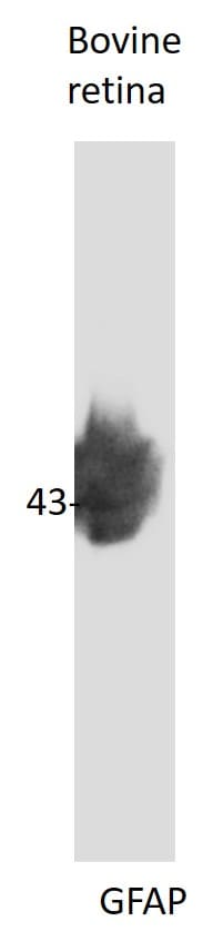
Application: Western BlotSample Tested: retinaSpecies: BovineVerified Customer | Posted 07/28/2020good reactivity

-
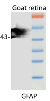
Application: Western BlotSample Tested: retina and 30ugSpecies: GoatVerified Customer | Posted 07/28/2020Very specific bands observed around 42 kDa and use at a concentration of 1:500
-
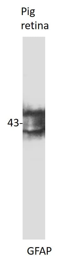
Application: Western BlotSample Tested: retinaSpecies: PigVerified Customer | Posted 07/28/2020good reactivity. use at 1:500.use at 1:500
-
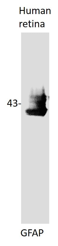
Application: Western BlotSample Tested: retinaSpecies: HumanVerified Customer | Posted 07/28/2020good reaction
-
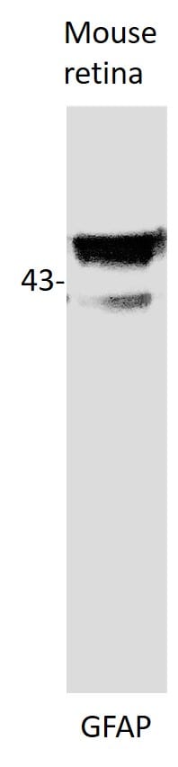
Application: Western BlotSample Tested: retinaSpecies: MouseVerified Customer | Posted 07/28/2020very good reaction
-
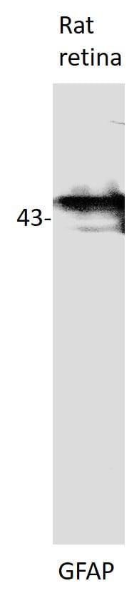
Application: Western BlotSample Tested: retinaSpecies: RatVerified Customer | Posted 07/28/2020good reaction
-
Application: ImmunocytochemistrySample Tested: mixed neuronal primary cultureSpecies: HumanVerified Customer | Posted 06/23/2020
-
Application: Immunohistochemistry-ParaffinSample Tested: Mouse brainSpecies: MouseVerified Customer | Posted 06/07/2019Mouse brain, 20x magnification.Proteinasi K treatment; Primary Ab 1:1000. Detection with Polymer-HRP.
-
Application: ImmunohistochemistrySample Tested: PFA Fixed Frozen Section of Mouse BrainSpecies: MouseVerified Customer | Posted 06/29/2017IHC GFAP NB300-141 on mouse brain (cortex), 20xWD [1:K]; Secondary ab RTU Polymer-HRP
-
Application: ImmunocytochemistrySample Tested: Primary mouse cortical astrocytesSpecies: MouseVerified Customer | Posted 04/18/2017
-
Application: ImmunocytochemistrySample Tested: Primary rat cortical neuronsSpecies: RatVerified Customer | Posted 03/27/2017Fixation: 4% paraformaldehyde (TAAB) in PBS, 15 minutes RT Blocking: 10% donkey serum and 1% BSA in PBS with 0.1% saponin, 1 hour RT Permeabilization: 0.1% saponin Primary Antibody: 1:1000 in PBS with 0.1% saponin, overnight, 4°C Wash: three times wash with PBS with 0.1% saponin, 10 min each Secondary Antibody: Invitrogen goat anti-rabbit Alexa647, 1:500 in PBS with 0.1% saponin, 2 hours, RT Wash: two washes with PBS with 0.1% saponin, 10 min each and three washes with PBS, 10 min each Microscope: Burker Vutara 350 The antibody labels intermediate filaments in cultured astrocytes.
-
Application: ImmunofluorescenceSample Tested:Species: MouseVerified Customer | Posted 01/17/2014xenografted mouse brain section : astocyte and human nuclei
There are no reviews that match your criteria.
Protocols
Find general support by application which include: protocols, troubleshooting, illustrated assays, videos and webinars.
- Antigen Retrieval Protocol (PIER)
- Antigen Retrieval for Frozen Sections Protocol
- Appropriate Fixation of IHC/ICC Samples
- Cellular Response to Hypoxia Protocols
- Chromogenic IHC Staining of Formalin-Fixed Paraffin-Embedded (FFPE) Tissue Protocol
- Chromogenic Immunohistochemistry Staining of Frozen Tissue
- ClariTSA™ Fluorophore Kits
- Detection & Visualization of Antibody Binding
- Fluorescent IHC Staining of Frozen Tissue Protocol
- Graphic Protocol for Heat-induced Epitope Retrieval
- Graphic Protocol for the Preparation and Fluorescent IHC Staining of Frozen Tissue Sections
- Graphic Protocol for the Preparation and Fluorescent IHC Staining of Paraffin-embedded Tissue Sections
- Graphic Protocol for the Preparation of Gelatin-coated Slides for Histological Tissue Sections
- ICC Cell Smear Protocol for Suspension Cells
- ICC Immunocytochemistry Protocol Videos
- ICC for Adherent Cells
- IHC Sample Preparation (Frozen sections vs Paraffin)
- Immunocytochemistry (ICC) Protocol
- Immunocytochemistry Troubleshooting
- Immunofluorescence of Organoids Embedded in Cultrex Basement Membrane Extract
- Immunofluorescent IHC Staining of Formalin-Fixed Paraffin-Embedded (FFPE) Tissue Protocol
- Immunohistochemistry (IHC) and Immunocytochemistry (ICC) Protocols
- Immunohistochemistry Frozen Troubleshooting
- Immunohistochemistry Paraffin Troubleshooting
- Preparing Samples for IHC/ICC Experiments
- Preventing Non-Specific Staining (Non-Specific Binding)
- Primary Antibody Selection & Optimization
- Protocol for Heat-Induced Epitope Retrieval (HIER)
- Protocol for Making a 4% Formaldehyde Solution in PBS
- Protocol for VisUCyte™ HRP Polymer Detection Reagent
- Protocol for the Fluorescent ICC Staining of Cell Smears - Graphic
- Protocol for the Fluorescent ICC Staining of Cultured Cells on Coverslips - Graphic
- Protocol for the Preparation & Fixation of Cells on Coverslips
- Protocol for the Preparation and Chromogenic IHC Staining of Frozen Tissue Sections
- Protocol for the Preparation and Chromogenic IHC Staining of Frozen Tissue Sections - Graphic
- Protocol for the Preparation and Chromogenic IHC Staining of Paraffin-embedded Tissue Sections
- Protocol for the Preparation and Chromogenic IHC Staining of Paraffin-embedded Tissue Sections - Graphic
- Protocol for the Preparation and Fluorescent ICC Staining of Cells on Coverslips
- Protocol for the Preparation and Fluorescent ICC Staining of Non-adherent Cells
- Protocol for the Preparation and Fluorescent ICC Staining of Stem Cells on Coverslips
- Protocol for the Preparation and Fluorescent IHC Staining of Frozen Tissue Sections
- Protocol for the Preparation and Fluorescent IHC Staining of Paraffin-embedded Tissue Sections
- Protocol for the Preparation of Gelatin-coated Slides for Histological Tissue Sections
- Protocol for the Preparation of a Cell Smear for Non-adherent Cell ICC - Graphic
- R&D Systems Quality Control Western Blot Protocol
- TUNEL and Active Caspase-3 Detection by IHC/ICC Protocol
- The Importance of IHC/ICC Controls
- Troubleshooting Guide: Immunohistochemistry
- Troubleshooting Guide: Western Blot Figures
- Western Blot Conditions
- Western Blot Protocol
- Western Blot Protocol for Cell Lysates
- Western Blot Troubleshooting
- Western Blot Troubleshooting Guide
- View all Protocols, Troubleshooting, Illustrated assays and Webinars
FAQs for GFAP Antibody
Showing
1
-
2 of
2 FAQs
Showing All
-
Q: I would like to use your GFAP-antibody (cat # NB300-141) in a Western Blot experiment. Could you tell me the dilution of this antibody?
A: The dilution recommended for Western blot using NB300-141 is 1:50,000.
-
Q: Our customer would like to know if your antibody is suitable for frozen sample by IF.
A: NB300-141 has been tested and validated in IHC-Frozen and IF; this is covered by our guarantee.
-
Q: I would like to use your GFAP-antibody (cat # NB300-141) in a Western Blot experiment. Could you tell me the dilution of this antibody?
A: The dilution recommended for Western blot using NB300-141 is 1:50,000.
-
Q: Our customer would like to know if your antibody is suitable for frozen sample by IF.
A: NB300-141 has been tested and validated in IHC-Frozen and IF; this is covered by our guarantee.
Loading...
Associated Pathways
