Best Seller
GFP Antibody - BSA Free
Novus Biologicals | Catalog # NB600-308

Key Product Details
Species Reactivity
Validated:
Non-species specific
Cited:
Human, Mouse, Rat, Nematode - Caenorhabditis elegans, Non-species specific, Rabbit
Applications
Validated:
Immunohistochemistry, Immunohistochemistry-Paraffin, Immunohistochemistry-Frozen, Immunohistochemistry Free-Floating, Immunohistochemistry Whole-Mount, Western Blot, ELISA, Flow Cytometry, Immunocytochemistry/ Immunofluorescence, Immunoprecipitation, Dot Blot, Electron Microscopy, Knockdown Validated
Cited:
Immunohistochemistry, Immunohistochemistry-Paraffin, Immunohistochemistry-Frozen, Immunohistochemistry Free-Floating, Immunohistochemistry Whole-Mount, Western Blot, Immunoblotting, ELISA, Flow Cytometry, Immunocytochemistry/ Immunofluorescence, Immunoprecipitation, Chromatin Immunoprecipitation, Cytometric Bead Assay Standard, IF/IHC, In Situ Hybridization, Electron Microscopy, Knockdown Validated
Label
Unconjugated
Antibody Source
Polyclonal Rabbit IgG
Format
BSA Free
Loading...
Product Specifications
Immunogen
The immunogen is a Green Fluorescent Protein (GFP) fusion protein corresponding to the full length amino acid sequence (246aa) derived from the jellyfish Aequorea victoria.
Reactivity Notes
No reaction was observed against Human, Mouse serum proteins. Suitable for detecting fusion proteins containing the GFP sequence expressed in Human, Mouse, Rat, C. elegans, Drosophila and in vitro transcription/translation systems and transgenic animals. Known cross reactivity with wt and all variants such as rGFP, eGFP, S65T-GFP, RS-GFP, YFP and EGFP.
GFP Transgenic Rat reactivity reported in scientific literature (PMID:25724725).
Mouse reactivity reported in multiple pieces of scientific literature.
Transgenic C. elegans reactivity reported in scientific literature (PMID: 27110099).
Use in Mouse reported in secitific publication (PMID: 32765228).
Plant reactivity reported in scientific literature (PMID:32896843)
Specificity
No reaction was observed against Human, Mouse or Rat serum proteins.
Clonality
Polyclonal
Host
Rabbit
Isotype
IgG
Description
This antibody was prepared from monospecific antiserum by immunoaffinity chromatography using Green Fluorescent Protein (Aequorea victoria) coupled to agarose beads followed by solid phase adsorption(s) to remove any unwanted reactivities. Assay by immunoelectrophoresis resulted in a single precipitin arc against anti-Rabbit Serum and purified and partially purified Green Fluorescent Protein (Aequorea victoria)
GFP antibody is stable for several weeks at 4C as an undiluted liquid.
GFP antibody is stable for several weeks at 4C as an undiluted liquid.
Scientific Data Images for GFP Antibody - BSA Free
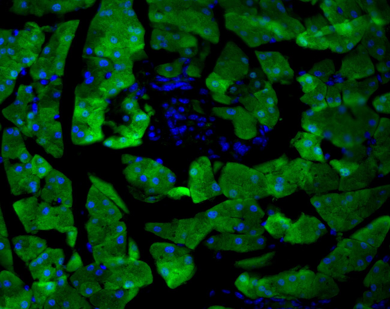
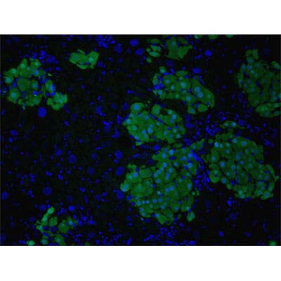
Immunocytochemistry/Immunofluorescence Detection of GFP in Transgenic Mouse Pancreas Cells
Analysis of GFP in transgenic mouse pancreas in OCT. The blue region in the center is an islet which does not express GFP and is negative. Image from verified customer review.
Western Blot Detection of GFP Tagged PKA Subunits in HEK293T Cells
Analysis of GFP tagged PKA subunits over-expressed in HEK293T cells using GFP antibody. Image from verified customer review.
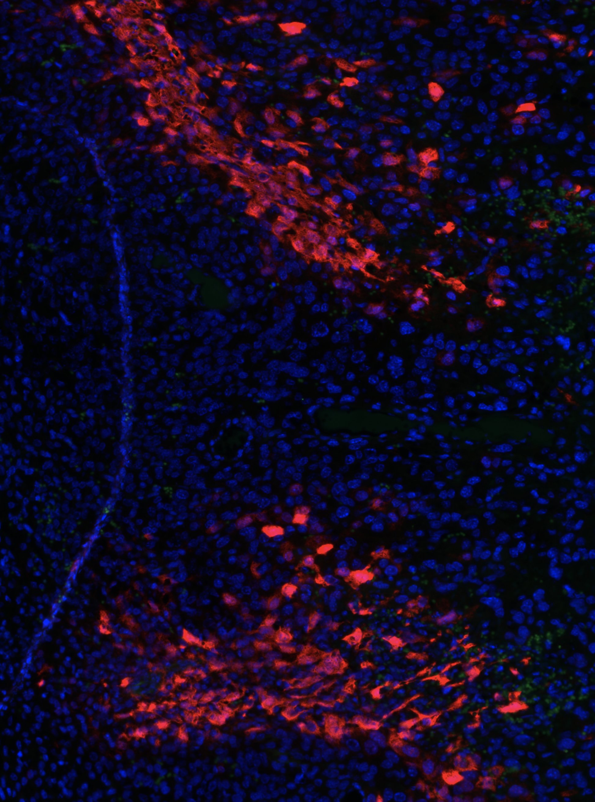
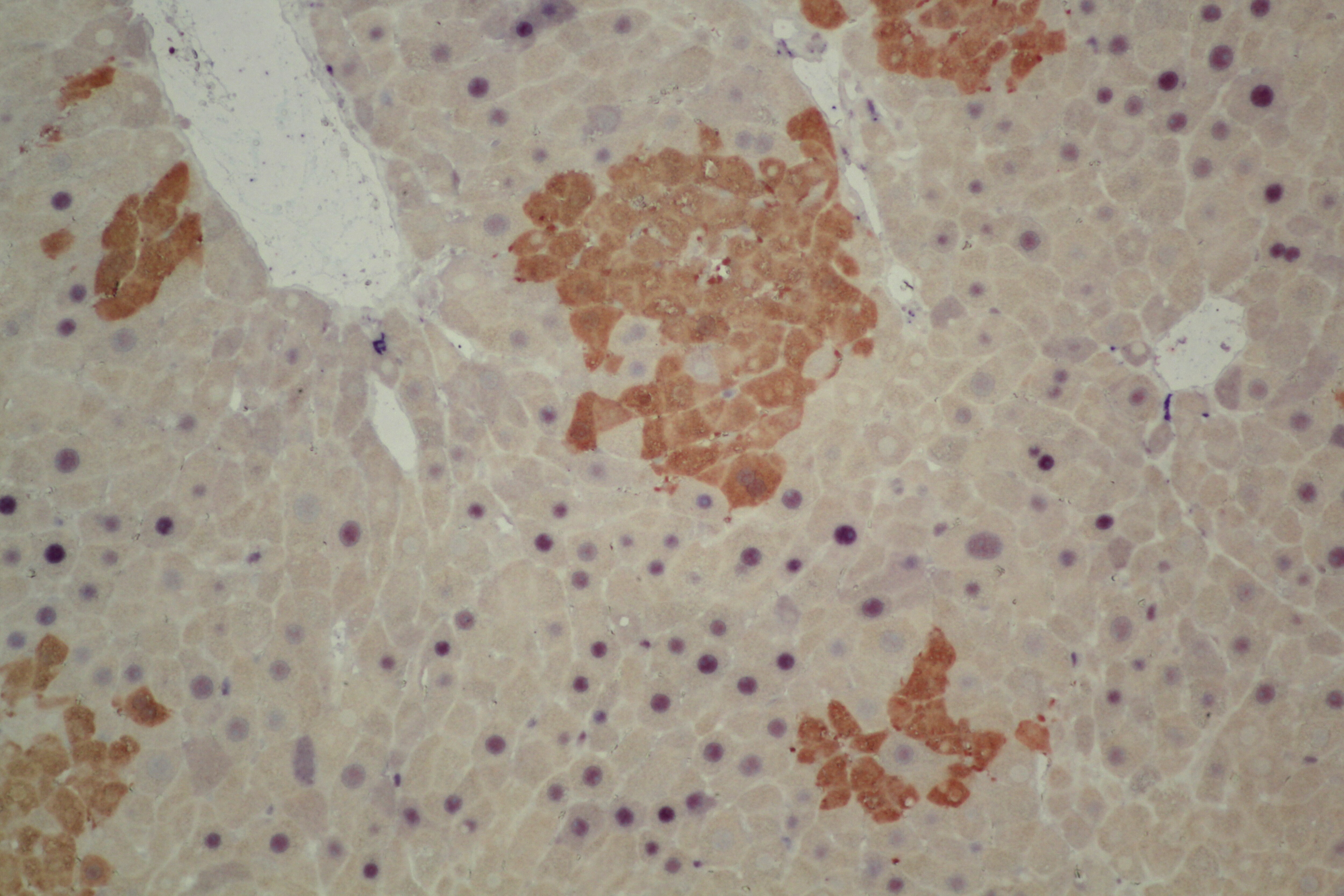
Immunohistochemical Staining of GFP+ Hepatocytes in Paraffin Embedded Liver
Staining of GFP+ hepatocytes (brown) transplanted into the liver of a recipient animal using anti-GFP antibody. Cyclin D1 double staining in blue. Image from verified customer review.
Detection of GFP Tagged Protein in Western Blot Using HRP Conjugated Antibody
Total cell lysates (20ug) with protein of interest tagged with GFP was loaded. Blotted with GFP antibody [HRP] NB600-308H, without any secondary antibody. Image from verified customer review.
Immunohistochemical Staining of GFP in WT and Mutant Drosophila Ovary
GFP Antibody-Immunohistochemistry-NB600-308-img0037.jpg
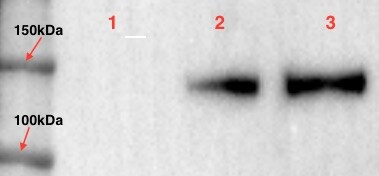
GFP Detection in Spiked HeLa Whole Cell Lysate by Western Blot
Western Blot of GFP antibody. Lane 1: Wild type GFP (0.1 ug) was used to spike HeLa whole cell lysate. Lane 2: none. Load: 30 ug per lane.Primary antibody: GFP antibody at 1:1000 for overnight at 4C.Secondary antibody: IRDye800(TM) Goat-a-Rabbit IgG [H&L] MX10 at 1:10,000 for 45 min at RT.Block: 5% BLOTTO in PBS overnight at 4C.Predicted/Observed size: 27 kDa for epitope tag GFP. Other band(s): none.
Western Blot Analysis of GFP in Transfected and Positive Control 293FT Cells
Western Blot of GFP antibody. Lane 1: 293FT cells transfected with CDK4 dominant negative (C-).Lane 2: 293FT cells positive control (C+).Load: 25 ug per lane.Primary antibody: GFP antibody at 1:400 for overnight at 4C.Secondary antibody: IRDye800(TM) rabbit secondary antibody at 1:10,000 for 45 min at RT.Block: 5% BLOTTO overnight at 4C.Predicted/Observed size: 27 kDa for GFP.
Western Blot Analysis of GFP in Multiple Cell Lysates and Proteins
Western Blot of GFP antibody. Marker: Opal Pre-stained ladder. Lane 1: HEK293 lysate. Lane 2: HeLa Lysate. Lane 3: CHO/K1 Lysate. Lane 4: MDA-MB-231. Lane 5: A431 Lysate. Lane 6: Jurkat Lysate. Lane 7: NIH/3T3 Lysate. Lane 8: E-coli HCP Control. Lane 9: FLAG Positive Control Lysate. Lane 10: Red Fluorescent Protein. Lane 11: Green Fluorescent Protein. Lane 12: Glutathione-S-Transferase Protein. Lane 13: Maltose Binding Protein. Load: 10 ug of lysate or 50ng of purified protein per lane. Primary antibody: GFP antibody at 1ug/mL overnight at 4C. Secondary antibody: Peroxidase rabbit secondary antibody at 1:30,000 for 60 min at RT. Blocking Buffer: 1% Casein-TTBS for 30 min at RT.Predicted/Observed size: 30 kDa for GFP.
Immunohistochemical Analysis of GFP in Frozen Mouse Brain
Adult mouse brain tissue. This image was submitted via customer review.
Immunocytochemistry/Immunofluorescence Imaging of GFP Labeled Dengue Virus in Dendritic Cells and Dermal Macrophages
Immuno-microscopy of GFP antibody. Monocyte derived dendritic cells and dermal macrophages were challenged and directly visualized with eGFP labeled Dengue virus to localize sequestration of virus particles in the different cells (upper). The location of the GFP was confirmed by TEM (lower magnified view) using rabbit anti GFP Primary antibody (1:200) and a gold labeled secondary antibody.
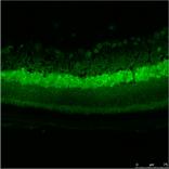
Immunohistochemical Staining of GFP in Mouse Retinal Cells
Analysis of GFP in mouse retinal cells. Image courtesy of anonymous customer review.
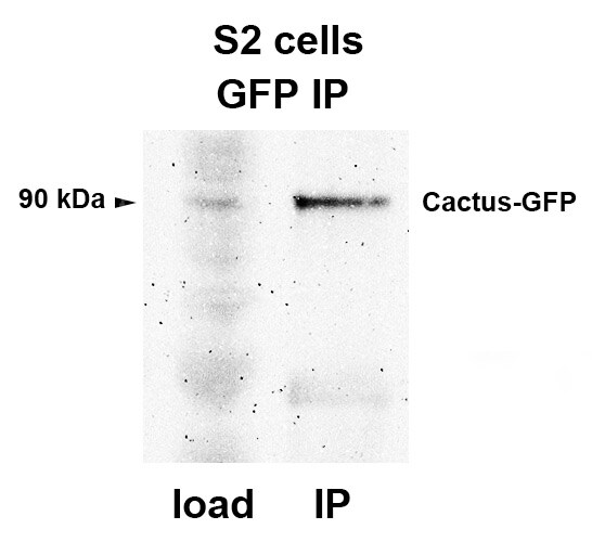
Immunoprecipitation Analysis in S2 Cells Using GFP Antibody
IP analysis of GFP in S2 Cells (Drosophila). Image from verified customer review.
Immunoprecipitation Analysis in HeLa Cell Lysate Using GFP Antibody
GFP-Antibody-Immunoprecipitation-NB600-308-img0035.jpg
Immunocytochemistry/ Immunofluorescence: GFP Antibody [NB600-308] -
Immunocytochemistry/ Immunofluorescence: GFP Antibody [NB600-308] - Myc is post-transcriptionally regulated by the IIS-GSK3 cascade.(A) Germaria from ovaries of ctrl, chicoRNAi, & bskRNAi driven by nos-Gal4 expressing LacZ under the control of myc endogenous promoter stained with anti-beta -galactosidase & anti-1B1. Scale bars, 10 μm. (B) A representative live imaging of germaria from flies expressing GFP-tagged Sgg under its endogenous promoter. Scale bar, 10 μm. (C) A germarium from wt flies endogenously expressing Myc-GFP stained with anti-GFP (green) & anti-p-GSK3 (red). Germarium regions are indicated. Myc protein highly corresponds to inhibited form of GSK3. Scale bar, 10 μm. Image collected & cropped by CiteAb from the following publication (https://pubmed.ncbi.nlm.nih.gov/31612862), licensed under a CC0-1.0 license. Not internally tested by Novus Biologicals.
Immunocytochemistry/ Immunofluorescence: GFP Antibody [NB600-308] -
Immunocytochemistry/ Immunofluorescence: GFP Antibody [NB600-308] - Myc protein pattern in the ovary.(A) A representative low-magnification image of ovarioles from flies endogenously expressing Myc-GFP stained with anti-GFP, anti-1B1, & DAPI. Note that Myc level is low in early germarium stages, becomes high from germarium region 2B, & reduces from the stage-10 egg chamber. Scale bar, 50 μm. Image collected & cropped by CiteAb from the following publication (https://pubmed.ncbi.nlm.nih.gov/31612862), licensed under a CC0-1.0 license. Not internally tested by Novus Biologicals.
Western Blot: GFP Antibody [NB600-308] -
Western Blot: GFP Antibody [NB600-308] - MISP interacts & co-localizes with IQGAP1. (a) HEK293T cells transiently overexpressing GFP-IQGAP1 & FLAG-MISP were used for co-immunoprecipitation experiments from both sides (GFP trap or FLAG M2 beads). (b) Endogenous immunoprecipitation using IQGAP1 antibody or control immunoglobulin (IgG) was carried out in HeLa cell lysates & MISP was detected in the eluate. (c) In vitro interaction between purified MBP-MISP & GST-IQGAP1 was detected by GST & MBP pull-down experiments. (d) Immunoprecipiation experiments showing that GFP-MISP binds endogenous IQGAP1 (upper panel) & MISP binds to IQGAP1 endogenously using specific antibodies (lower panel) in mitotically arrested (taxol & nocodazole, respectively) HeLa cell lysates. (a–d) Images were gained from the same Western blot membrane after cutting/cropping & presented with different exposure times or contrast enhancement for better presentation purposes. The dividing lane marks the grouping of images of the same (or different) membrane. Uncropped blots, where applicable, are included in Supplementary Fig. 4. (e) HeLa cells were immunostained for MISP & IQGAP1 & co-localization was visualized in mitosis in single-plane confocal images, scale bar: 5 μm. Lower pictures: After manual thresholding white spots mark the co-localizing areas in the merge images. Image collected & cropped by CiteAb from the following publication (https://pubmed.ncbi.nlm.nih.gov/29679050), licensed under a CC-BY license. Not internally tested by Novus Biologicals.
Immunocytochemistry/ Immunofluorescence: GFP Antibody [NB600-308] -
Immunocytochemistry/ Immunofluorescence: GFP Antibody [NB600-308] - BRC-1 & BRD-1 are inter-dependent for localization.A) Co-localization between BRD-1::GFP (green) & TagRFP-T::BRC-1 (red) at late pachytene in live worms. Scale bar = 10 μm. B) Stills of germline nuclei from live worms expressing GFP::BRC-1 & mCherry::Histone H2B (mCherry::his-58; gfp::brc-1); merge & GFP channel; top two panels, respectively. GFP::BRC-1 expression in brd-1(ok1623) mutant at indicated meiotic stages. Bottom two panels show BRD-1::GFP localization in wild type & the brc-1(xoe4) mutant. Images are projections through half of the gonad. TZ = transition zone; EP = early pachytene; MP = mid pachytene; LP = late pachytene; DP = diplotene; DK = diakinesis. Scale bar = 5 μm. C) Immunoblot of whole worm extracts from indicated worms probed with anti-GFP & alpha -tubulin antibodies. Lane 1 = N2: wild type; Lane 2 = JEL515: gfp::brc-1; Lane 3 = JEL520: gfp::brc-1 brd-1(ok1623); Lane 4 = JEL744: brc-1(xoe4) brd-1::gfp; Lane 5 = JEL657: brd-1::gfp; Lane 6 = JEL678: brc-1(tm1145) brd-1::gfp. Image collected & cropped by CiteAb from the following publication (https://pubmed.ncbi.nlm.nih.gov/30383767), licensed under a CC-BY license. Not internally tested by Novus Biologicals.
Western Blot: GFP Antibody [NB600-308] -
Western Blot: GFP Antibody [NB600-308] - BRC-1 & BRD-1 are inter-dependent for localization.A) Co-localization between BRD-1::GFP (green) & TagRFP-T::BRC-1 (red) at late pachytene in live worms. Scale bar = 10 μm. B) Stills of germline nuclei from live worms expressing GFP::BRC-1 & mCherry::Histone H2B (mCherry::his-58; gfp::brc-1); merge & GFP channel; top two panels, respectively. GFP::BRC-1 expression in brd-1(ok1623) mutant at indicated meiotic stages. Bottom two panels show BRD-1::GFP localization in wild type & the brc-1(xoe4) mutant. Images are projections through half of the gonad. TZ = transition zone; EP = early pachytene; MP = mid pachytene; LP = late pachytene; DP = diplotene; DK = diakinesis. Scale bar = 5 μm. C) Immunoblot of whole worm extracts from indicated worms probed with anti-GFP & alpha -tubulin antibodies. Lane 1 = N2: wild type; Lane 2 = JEL515: gfp::brc-1; Lane 3 = JEL520: gfp::brc-1 brd-1(ok1623); Lane 4 = JEL744: brc-1(xoe4) brd-1::gfp; Lane 5 = JEL657: brd-1::gfp; Lane 6 = JEL678: brc-1(tm1145) brd-1::gfp. Image collected & cropped by CiteAb from the following publication (https://pubmed.ncbi.nlm.nih.gov/30383767), licensed under a CC-BY license. Not internally tested by Novus Biologicals.
Immunocytochemistry/ Immunofluorescence: GFP Antibody [NB600-308] -
Immunocytochemistry/ Immunofluorescence: GFP Antibody [NB600-308] - Other IIS components are not altered by bsk RNAi.(A) Confocal images for germaria expressing Chico-GFP, myc-Dp110 or PDK1-GFP in the background of ctrl or bskRNAi from ovaries stained with anti-GFP or anti-myc. Scale bars, 10 μm. (B) Ovaries expressing LacZ driven by the puc promoter stained with anti-beta -galactosidase, anti-1B1, & DAPI. Note that JNK activity is moderately induced in late germarium & decreased in the following stages, while JNK activity is much stronger in follicle cells of a maturing egg. Scale bars, 20 μm. (C) Germaria from ovaries of ctrl & bskRNAi driven by nos-Gal4 stained with anti-AKT & anti-1B1. Scale bars, 10 μm. (D) Quantification of AKT intensity from cysts in germarium region 2A & 2B of ovaries with ctrl & bskRNAi driven by nos-Gal4. Intensities are normalized to the value of ctrl at region 2B. n = 7 & 8 germaria for ctrl & bskRNAi, respectively. Error bars represent SEM. *p<0.005. (E) Visualization of the InR mRNA by FISH in germaria from ovaries of ctrl & bskRNAi driven by nos-Gal4. Note that InR mRNA level is decreased by bsk RNAi. Germaria are outlined with dotted lines. Scale bars, 10 μm. (F) Quantification of InR mRNA density in region 2A & 2B germ cells from ovaries with indicated genotypes. n = 9 germaria for each genotype. Error bars represent SEM. *p<0.005. (G) Quantification of relative Myc-GFP intensity in region 2B cysts of ovaries illustrated in Figure 5H. n = 10 germaria for each genotype. Error bars represent SEM. *p<0.005.10.7554/eLife.49309.017Figure 5—figure supplement 1—source data 1.Relative AKT intensity, InR mRNA density, & Myc-GFP intensity in the germarium.Relative AKT intensity, InR mRNA density, & Myc-GFP intensity in the germarium. Image collected & cropped by CiteAb from the following publication (https://pubmed.ncbi.nlm.nih.gov/31612862), licensed under a CC0-1.0 license. Not internally tested by Novus Biologicals.
Western Blot: GFP Antibody [NB600-308] -
Western Blot: GFP Antibody [NB600-308] - GFP Expression within A. thaliana upon infiltration with SWNT-PM-CytKH9/pDNA complexes.a Reporter construct design of transient GFP expression for the nucleus & mitochondria. b Representative confocal laser scanning microscopy images of A. thaliana root cells from a minimum of five seedlings for each condition (n = 5) 18 h post infiltration with SWNT-PM-CytKH9/pDNA complexes containing pDONR-35S-GFP or pDONR-Cox2-GFP reporter constructs for nuclear or mitochondrial expression, respectively. Mitochondria were stained with MitoTracker Red (CMXRos), & colocalization analysis was performed on GFP expression & MitoTracker signals. Scale bars represent 20 μm. c, d Representative western blots & their quantification from cytosolic (c) & mitochondrial (d) fractions isolated from approximately 60 seedlings 18 h post infiltration with SWNT NCs probed for GFP expression. Actin & cytochrome c were used as loading controls for cytosolic & mitochondrial proteins, respectively. Data points from five biological replicates (n = 5) are represented as the mean ± standard deviation. Statistical significance was determined by Brown-Forsythe & Welch one-way ANOVA test. For the cytosolic samples, P-values are 0.0027, & 0.0020 between DNA only & SWNT-PM-CytKH9, SWNT-PM-Cytcox, respectively, & 0.0253 between SWNT-PM-CytKH9 & SWNT-PM-Cytcox. For the mitochondrial samples, P-values are 0.0019, & 0.0333 between DNA only & SWNT-PM-CytKH9, & SWNT-PM-Cytcox, respectively, & 0.0359 between SWNT-PM-CytKH9 & SWNT-PM-Cytcox. ns – not statistically significant, *P < 0.05, **P < 0.01. Source data are provided as a Source Data file. Image collected & cropped by CiteAb from the following publication (https://pubmed.ncbi.nlm.nih.gov/35577779), licensed under a CC-BY license. Not internally tested by Novus Biologicals.
Western Blot: GFP Antibody [NB600-308] -
Western Blot: GFP Antibody [NB600-308] - VGLL1 regulates MMP9 transcription in gastric cancer cells. (a,b) Viability (a) & invasion (b) assay of NUGC3 cells. Cells were treated with 20 nM siScramble (SC) or siMMP9 for 48 h, & then stained with sulforhodamine B. Data are presented as mean ±SD. n = 3; * p < 0.05 (Student’s t-test). (c) MMP9 mRNA expression regulation by VGLL1 in gastric cancer cells treated with siVGLL1 or siSC (control) was measured by qPCR. (d) Changes in MMP9 expression upon knockdown or overexpression of VGLL1 assayed by western blotting in NUGC3 cells. (e) IHC of VGLL1 & MMP9 expression in the liver & lungs of an in vivo metastasis mouse model using surgical resection of tumors. Scale bar, 200 µm. (f) Construction of various luciferase reporter systems under control of the MMP9 promoter. (g) MMP9 promoter activities of the reporter systems containing modified TEA-binding sites were measured in NUGC3 cells. n = 3; * p < 0.05 (Student’s t-test). (h) Effect of TEAD4 on VGLL1-regulated MMP9 transcriptional activity. NUGC3 cells treated with siSC or siTEAD4 for 24 h were transfected with MMP9-luc, Renilla-luc, pCDNA3.1, & pcDNA3.1-myc-VGLL1 vectors for 48 h. n = 3; * p < 0.05 (Student’s t-test). (i) Interaction between VGLL1 & TEAD4. Lysates of NUGC3 cells that were transfected with pcDNA3.1-myc-VGLL1 & pEGFP-N1-TEAD4 were immunoprecipitated using anti-IgG, anti-GFP, & anti-Myc antibodies. Protein expression was analyzed by immunoblotting. (j) ChIP assays while using nuclear extracts of NUGC3 cells treated with siTEAD4. The ChIP-enriched DNA was subjected to PCR. (k) Target genes of VGLL1 & YAP. NUGC3 cells were transfected with pcDNA3, pcDNA3-myc-VGLL1, or pcDNA3-myc-YAP, & the mRNA expression levels were analyzed by RT-PCR. Image collected & cropped by CiteAb from the following publication (https://pubmed.ncbi.nlm.nih.gov/31816819), licensed under a CC-BY license. Not internally tested by Novus Biologicals.
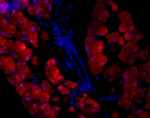
Immunocytochemistry/Immunofluorescence: Rabbit GFP pAb [NB600-308]
Immunocytochemistry/Immunofluorescence: Rabbit GFP pAb [NB600-308] - Analysis of GFP in human Hs746T gastric cancer cell line. Image from a verified customer review.Applications for GFP Antibody - BSA Free
Application
Recommended Usage
ELISA
1:20000-1:120000
Electron Microscopy
1:10-1:500
Flow Cytometry
1:10-1:1000
Immunocytochemistry/ Immunofluorescence
1:500-1:5000
Immunohistochemistry
1:200-1:3000
Immunohistochemistry-Frozen
1:50-1:250
Immunohistochemistry-Paraffin
1:10-1:500
Immunoprecipitation
1:10-1:500
Western Blot
1:500-1:5000
Application Notes
This product is designed to detect GFP and its variants. GFP antibody has been tested by western blot and ELISA. This product can be used to detect GFP by ELISA (sandwich or capture) for the direct binding of antigen and recognizes wild type, recombinant and enhanced forms of GFP. Biotin conjugated polyclonal anti-GFP used in a sandwich ELISA is well suited to titrate GFP in solution using this antibody in combination with monoclonal anti-GFP using either form of the antibody as the capture or detection antibodies. However, use the monoclonal form only for the detection of wild type or recombinant GFP as this form does not sufficiently detect 'enhanced' GFP. The detection antibody is typically conjugated to biotin and subsequently reacted with streptavidin conjugated HRP. Fluorochrome conjugated polyclonal anti-GFP can be used to detect GFP by immunofluorescence microscopy in prokaryotic (E.coli) and eukaryotic (CHO cells) expression systems and can detect GFP containing inserts. Significant amplification of signal is achieved using fluorochrome conjugated polyclonal anti-GFP relative to the fluorescence of GFP alone. For immunoblotting use either alkaline phosphatase or peroxidase conjugated polyclonal anti-GFP to detect GFP or GFP containing proteins on western blots. Optimal titers for applications should be determined by the researcher.
Use in Immunoprecipitation reported in scientific literature (PMID:34887587).
Use in DB reported in scientific literature (PMID:34242364).
Use in Knockdown Validated reported in scientific literature
(PMID: 32905777).
Use in Immunoprecipitation reported in scientific literature (PMID:34887587).
Use in DB reported in scientific literature (PMID:34242364).
Use in Knockdown Validated reported in scientific literature
(PMID: 32905777).
Reviewed Applications
Read 19 reviews rated 4.5 using NB600-308 in the following applications:
- Immunocytochemistry (1 Review)
- Immunofluorescence (5 Reviews)
- Immunofluorescence in fixed cells (1 Review)
- Immunofluorescence on tissues. (1 Review)
- Immunohistochemistry (1 Review)
- Immunohistochemistry-Frozen (2 Reviews)
- Immunohistochemistry-Paraffin (1 Review)
- Immunoprecipitation (2 Reviews)
- Western Blot (5 Reviews)
Flow Cytometry Panel Builder
Bio-Techne Knows Flow Cytometry
Save time and reduce costly mistakes by quickly finding compatible reagents using the Panel Builder Tool.
Advanced Features
- Spectra Viewer - Custom analysis of spectra from multiple fluorochromes
- Spillover Popups - Visualize the spectra of individual fluorochromes
- Antigen Density Selector - Match fluorochrome brightness with antigen density

Formulation, Preparation, and Storage
Purification
Immunogen affinity purified
Formulation
0.02 M Potassium Phosphate, 0.15 M Sodium Chloride, pH 7.2
Format
BSA Free
Preservative
0.01% Sodium Azide
Concentration
Please see the vial label for concentration. If unlisted please contact technical services.
Shipping
The product is shipped with polar packs. Upon receipt, store it immediately at the temperature recommended below.
Stability & Storage
Store at -20C. Avoid freeze-thaw cycles.
Background: GFP
References
1. Shi, C., Pan, F. C., Kim, J. N., Washington, M. K., Padmanabhan, C., Meyer, C. T.,... Means, A. L. (2019). Differential Cell Susceptibilities to Kras(G12D) in the Setting of Obstructive Chronic Pancreatitis. Cell Mol Gastroenterol Hepatol. doi:10.1016/j.jcmgh.2019.07.001
2. Zhao, S., Fortier, T. M., & Baehrecke, E. H. (2018). Autophagy Promotes Tumor-like Stem Cell Niche Occupancy. Curr Biol, 28(19), 3056-3064.e3053. doi:10.1016/j.cub.2018.07.075
3. Zusso, M., Lunardi, V., Franceschini, D., Pagetta, A., Lo, R., Stifani, S.,... Moro, S. (2019). Ciprofloxacin and levofloxacin attenuate microglia inflammatory response via TLR4/NF-kB pathway. J Neuroinflammation, 16(1), 148. doi:10.1186/s12974-019-1538-9
Long Name
Green Fluorescent Protein
Alternate Names
eGFP, GFPuv, anti-gfp (green fluorescent protein) pab, GFP immunofluorescence, GFP immunoprecipitation, GFP mouse, GFP staining, GFP western blot
UniProt
Additional GFP Products
Product Documents for GFP Antibody - BSA Free
Certificate of Analysis
To download a Certificate of Analysis, please enter a lot or batch number in the search box below.
Product Specific Notices for GFP Antibody - BSA Free
This product is for research use only and is not approved for use in humans or in clinical diagnosis. Primary Antibodies are guaranteed for 1 year from date of receipt.
Citations for GFP Antibody - BSA Free
 Powered by Bioz
Customer Reviews for GFP Antibody - BSA Free (19)
4.5 out of 5
19 Customer Ratings
Have you used GFP Antibody - BSA Free?
Submit a review and receive an Amazon gift card!
$25/€18/£15/$25CAN/¥2500 Yen for a review with an image
$10/€7/£6/$10CAN/¥1110 Yen for a review without an image
Submit a review
Customer Images
Showing
1
-
5 of
19 reviews
Showing All
Filter By:
-
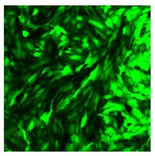
Application: Immunofluorescence in fixed cellsSample Tested: Hs746T gastric cancer cell lineSpecies: HumanVerified Customer | Posted 03/13/2025This is a HS746T gastric cancer cell line expressing GFP. The fluorescence is amazing with this antibody.

-
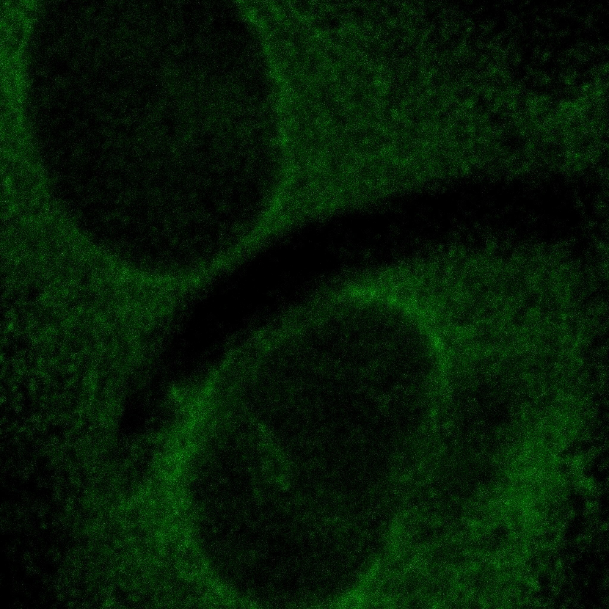
Application: Immunohistochemistry-FrozenSample Tested: Adult mouse brain tissueSpecies: MouseVerified Customer | Posted 04/12/2018Enhanced endogenous eGFP signalDilutions 1:400
-
Application: ImmunocytochemistrySample Tested: FFPESpecies: MouseVerified Customer | Posted 03/22/2017Trypsin retrieval
-
Application: Western BlotSample Tested: whole cellular protein and fractionated (soluble and membrane protein)Species: HumanVerified Customer | Posted 03/10/2017HEK293T cells expressing Aedes aegypti leucine-rich repeat-containing GPCR (lgr1) fused C-terminally tail with EGFP (139kDa). Lane 1 (untransfected), lane 2 (transient expression) and lane 3 (stable expression). 50ug protein per well.Human embryonic kidney 293T HEK293T cells
-
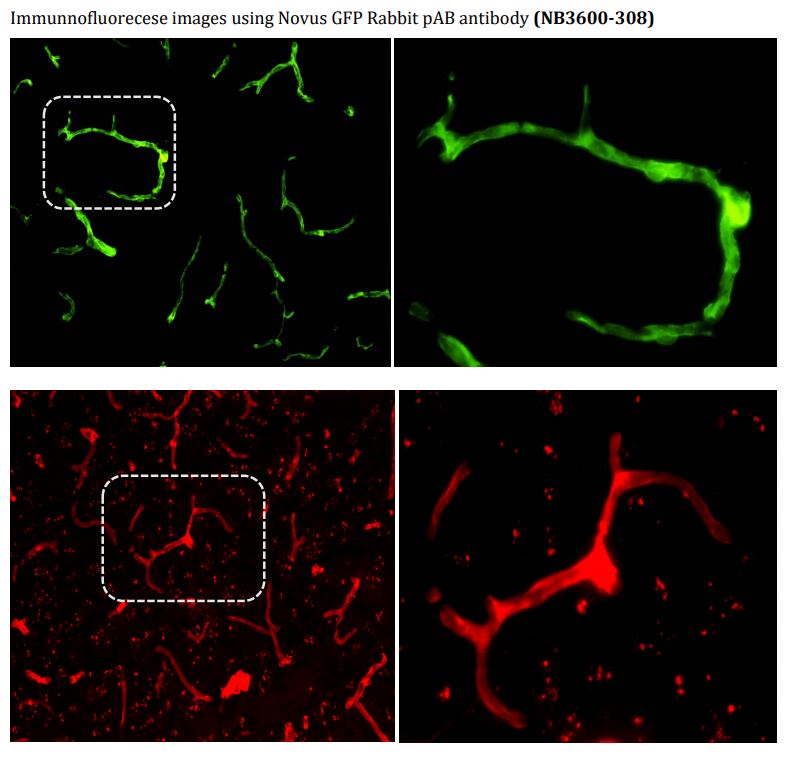
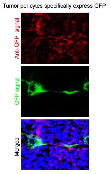
Application: Immunofluorescence on tissues.Sample Tested: Human Xenograft TumorSpecies: HumanVerified Customer | Posted 03/02/2017GFP is specifically expressed in vascular pericytes in human glioma stem cell-derived xenografts in mouse brains. The frozen sections were stained with anti-GFP (NB600-308, 1:100). The antibody worked but with strong noise.
-
Application: ImmunofluorescenceSample Tested: Pig TM cellsSpecies: OtherVerified Customer | Posted 07/27/2016
-
Application: ImmunofluorescenceSample Tested: Mouse, 3T3 cellsSpecies: MouseVerified Customer | Posted 10/10/2015GRK2-GFP over-expressed in 3T3 cells
-
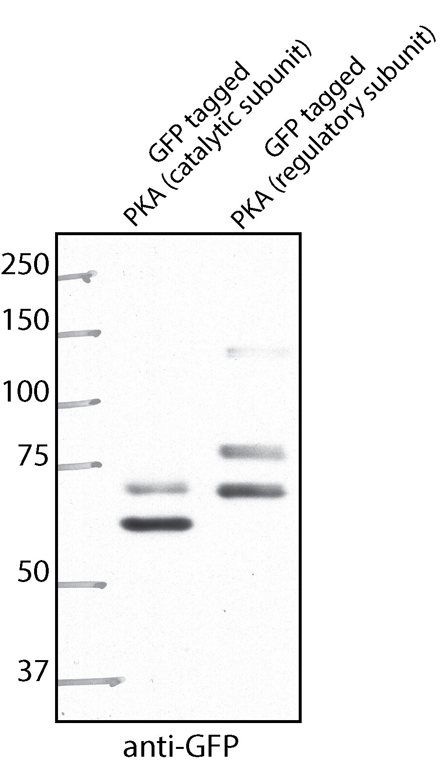
Application: Western BlotSample Tested: HEK293T whole cell extractSpecies: HumanVerified Customer | Posted 10/10/2015GFP tagged PKA subunits over-expressed in HEK293T cells
-
Application: ImmunoprecipitationSample Tested: S2 Cells (Drosophila)Species: OtherVerified Customer | Posted 04/02/2015GFP immunoprecipitation
-
Application: ImmunofluorescenceSample Tested: transgenic mouse pancreasSpecies: MouseVerified Customer | Posted 11/25/2014transgenic mouse pancreas in O.C.T.
-
Application: Immunohistochemistry-ParaffinVerified Customer | Posted 10/05/2014IHC staining of GPF+ hepatocytes (brown) transplantand into the liver of a recipient animal. Cyclin D1 double staining in blue
-
Application: Immunohistochemistry-FrozenSample Tested: Transgenic mouse pancreas in OCTSpecies: MouseVerified Customer | Posted 11/06/2013GFP detection in transgenic mouse pancreas in OCT
-
Application: ImmunofluorescenceSample Tested: Rat liverSpecies: RatVerified Customer | Posted 07/13/2012
-
Application: ImmunoprecipitationSample Tested: Hela whole cell lysateSpecies: HumanVerified Customer | Posted 04/24/2012Ago2 Immunoprecipitation from HeLa cells stably expressing Ago2-GFP using the Novus rabbit polyclonal GFP antibody
-
Application: Western BlotSample Tested: YeastSpecies: OtherVerified Customer | Posted 11/15/2011
-
Application: ImmunohistochemistrySample Tested: IHC on parrafin sections (mouse)Species: MouseVerified Customer | Posted 09/28/2011
-
Application: ImmunofluorescenceSample Tested: Mouse Retinal CellsSpecies: MouseVerified Customer | Posted 07/20/2011
-
Application: Western BlotSample Tested: Pig ear fibroblasts, Sample Amount: 10 ulSpecies: OtherVerified Customer | Posted 11/23/2010
-
Application: Western BlotSample Tested: P19 cells transfectedwith a plasmid DNA expressing a GFP fusion protein, Sample Amount: 20 microgramsSpecies: MouseVerified Customer | Posted 09/23/2010
There are no reviews that match your criteria.
Protocols
Find general support by application which include: protocols, troubleshooting, illustrated assays, videos and webinars.
- 7-Amino Actinomycin D (7-AAD) Cell Viability Flow Cytometry Protocol
- Antigen Retrieval Protocol (PIER)
- Antigen Retrieval for Frozen Sections Protocol
- Appropriate Fixation of IHC/ICC Samples
- Cellular Response to Hypoxia Protocols
- Chromogenic IHC Staining of Formalin-Fixed Paraffin-Embedded (FFPE) Tissue Protocol
- Chromogenic Immunohistochemistry Staining of Frozen Tissue
- ClariTSA™ Fluorophore Kits
- Detection & Visualization of Antibody Binding
- ELISA Sample Preparation & Collection Guide
- ELISA Troubleshooting Guide
- Extracellular Membrane Flow Cytometry Protocol
- Flow Cytometry Protocol for Cell Surface Markers
- Flow Cytometry Protocol for Staining Membrane Associated Proteins
- Flow Cytometry Staining Protocols
- Flow Cytometry Troubleshooting Guide
- Fluorescent IHC Staining of Frozen Tissue Protocol
- Graphic Protocol for Heat-induced Epitope Retrieval
- Graphic Protocol for the Preparation and Fluorescent IHC Staining of Frozen Tissue Sections
- Graphic Protocol for the Preparation and Fluorescent IHC Staining of Paraffin-embedded Tissue Sections
- Graphic Protocol for the Preparation of Gelatin-coated Slides for Histological Tissue Sections
- How to Run an R&D Systems DuoSet ELISA
- How to Run an R&D Systems Quantikine ELISA
- How to Run an R&D Systems Quantikine™ QuicKit™ ELISA
- ICC Cell Smear Protocol for Suspension Cells
- ICC Immunocytochemistry Protocol Videos
- ICC for Adherent Cells
- IHC Sample Preparation (Frozen sections vs Paraffin)
- Immunocytochemistry (ICC) Protocol
- Immunocytochemistry Troubleshooting
- Immunofluorescence of Organoids Embedded in Cultrex Basement Membrane Extract
- Immunofluorescent IHC Staining of Formalin-Fixed Paraffin-Embedded (FFPE) Tissue Protocol
- Immunohistochemistry (IHC) and Immunocytochemistry (ICC) Protocols
- Immunohistochemistry Frozen Troubleshooting
- Immunohistochemistry Paraffin Troubleshooting
- Immunoprecipitation Protocol
- Intracellular Flow Cytometry Protocol Using Alcohol (Methanol)
- Intracellular Flow Cytometry Protocol Using Detergents
- Intracellular Nuclear Staining Flow Cytometry Protocol Using Detergents
- Intracellular Staining Flow Cytometry Protocol Using Alcohol Permeabilization
- Intracellular Staining Flow Cytometry Protocol Using Detergents to Permeabilize Cells
- Preparing Samples for IHC/ICC Experiments
- Preventing Non-Specific Staining (Non-Specific Binding)
- Primary Antibody Selection & Optimization
- Propidium Iodide Cell Viability Flow Cytometry Protocol
- Protocol for Heat-Induced Epitope Retrieval (HIER)
- Protocol for Liperfluo
- Protocol for Making a 4% Formaldehyde Solution in PBS
- Protocol for VisUCyte™ HRP Polymer Detection Reagent
- Protocol for the Characterization of Human Th22 Cells
- Protocol for the Characterization of Human Th9 Cells
- Protocol for the Fluorescent ICC Staining of Cell Smears - Graphic
- Protocol for the Fluorescent ICC Staining of Cultured Cells on Coverslips - Graphic
- Protocol for the Preparation & Fixation of Cells on Coverslips
- Protocol for the Preparation and Chromogenic IHC Staining of Frozen Tissue Sections
- Protocol for the Preparation and Chromogenic IHC Staining of Frozen Tissue Sections - Graphic
- Protocol for the Preparation and Chromogenic IHC Staining of Paraffin-embedded Tissue Sections
- Protocol for the Preparation and Chromogenic IHC Staining of Paraffin-embedded Tissue Sections - Graphic
- Protocol for the Preparation and Fluorescent ICC Staining of Cells on Coverslips
- Protocol for the Preparation and Fluorescent ICC Staining of Non-adherent Cells
- Protocol for the Preparation and Fluorescent ICC Staining of Stem Cells on Coverslips
- Protocol for the Preparation and Fluorescent IHC Staining of Frozen Tissue Sections
- Protocol for the Preparation and Fluorescent IHC Staining of Paraffin-embedded Tissue Sections
- Protocol for the Preparation of Gelatin-coated Slides for Histological Tissue Sections
- Protocol for the Preparation of a Cell Smear for Non-adherent Cell ICC - Graphic
- Protocol: Annexin V and PI Staining by Flow Cytometry
- Protocol: Annexin V and PI Staining for Apoptosis by Flow Cytometry
- Quantikine HS ELISA Kit Assay Principle, Alkaline Phosphatase
- Quantikine HS ELISA Kit Principle, Streptavidin-HRP Polymer
- R&D Systems Quality Control Western Blot Protocol
- Sandwich ELISA (Colorimetric) – Biotin/Streptavidin Detection Protocol
- Sandwich ELISA (Colorimetric) – Direct Detection Protocol
- TUNEL and Active Caspase-3 Detection by IHC/ICC Protocol
- The Importance of IHC/ICC Controls
- Troubleshooting Guide: ELISA
- Troubleshooting Guide: Fluorokine Flow Cytometry Kits
- Troubleshooting Guide: Immunohistochemistry
- Troubleshooting Guide: Western Blot Figures
- Western Blot Conditions
- Western Blot Protocol
- Western Blot Protocol for Cell Lysates
- Western Blot Troubleshooting
- Western Blot Troubleshooting Guide
- View all Protocols, Troubleshooting, Illustrated assays and Webinars
FAQs for GFP Antibody - BSA Free
Showing
1
-
3 of
3 FAQs
Showing All
-
Q: Does the GFP antibody NB600-308 also work against AcGFP (93% identical to EGFP) in IP and Western blot?
A: For our product NB600-308, we have unfortunately not yet tested the product for cross reactivity with AcGFP. However, since the antibody is polyclonal and made to the full length AvGFP sequence, there is a good chance for cross reactivity.
-
Q: Hello, my lab purchased a GFP antibody from Novus in the past and I am now in need of an antibody to detect YFP and CFP as well. The GFP antibody we order has a part number of NB600-308. Would it possibly recognize YPF and CFP as well or will I need to purchase additional antibodies?
A: NB600-308 is polyclonal to full length GFP, hence it will recognize most of its mutants. It will react with YFP and CFP.
-
Q: We are looking for a rabbit anti-GFP antibody as a primary antibody for immunofluorescence and WB. I've seen that you carry several anti-GFP antibodies and I would like to ask which one you would recommend. The one with Cat.No NB600-308 has been used in a lot of publications and I reckon it is a solid antibody. Is this our best choice, or is there a better alternative?
A: NB600-308 is an excellent GFP antibody and I think it would work very well for you. It has been reviewed and published with which usually makes our customer feel much more confident, seeing as it worked in other people's hand.
-
Q: Does the GFP antibody NB600-308 also work against AcGFP (93% identical to EGFP) in IP and Western blot?
A: For our product NB600-308, we have unfortunately not yet tested the product for cross reactivity with AcGFP. However, since the antibody is polyclonal and made to the full length AvGFP sequence, there is a good chance for cross reactivity.
-
Q: Hello, my lab purchased a GFP antibody from Novus in the past and I am now in need of an antibody to detect YFP and CFP as well. The GFP antibody we order has a part number of NB600-308. Would it possibly recognize YPF and CFP as well or will I need to purchase additional antibodies?
A: NB600-308 is polyclonal to full length GFP, hence it will recognize most of its mutants. It will react with YFP and CFP.
-
Q: We are looking for a rabbit anti-GFP antibody as a primary antibody for immunofluorescence and WB. I've seen that you carry several anti-GFP antibodies and I would like to ask which one you would recommend. The one with Cat.No NB600-308 has been used in a lot of publications and I reckon it is a solid antibody. Is this our best choice, or is there a better alternative?
A: NB600-308 is an excellent GFP antibody and I think it would work very well for you. It has been reviewed and published with which usually makes our customer feel much more confident, seeing as it worked in other people's hand.
Loading...