
SSEA-4 is expressed on the surface of human embryonal carcinoma (EC) cells (the pluripotent stem cells of teratocarcinomas), human embryonic germ cells (EG), and human embryonic stem cells (ES). Expression of SSEA-4 is down-regulated following differentiation of human EC cells. In contrast, the differentiation of murine EC and ES cells may be accompanied by an increase in SSEA-4 expression (1-4).
GloLIVE Human/Mouse SSEA-4 NL557 Live Cell Antibody
R&D Systems | Catalog # NLLC1435R
50X Concentration, Azide Free

Loading...
Key Product Details
Species
Human, Mouse
Applications
Immunocytochemistry/ Immunofluorescence
Conjugate
NL557
Product Summary for GloLIVE Human/Mouse SSEA-4 NL557 Live Cell Antibody
Kit Summary
To detect SSEA-4 expression in live cells by immunocytochemical staining.
Why confirm marker expression in live cells before colony selection?
Maintaining pluripotent human embryonic stem cells and deriving induced pluripotent stem cells require colony selection and cell expansion. Picking colonies that contain pluripotent stem cells is typically achieved by manually analyzing colony morphology. Unfortunately, this critical process is time-consuming and labor-intensive.
In addition, morphology-based colony selection does not consistently guarantee pluripotent starting populations, since cells can fail to be fully reprogrammed or can naturally differentiate within a colony.
To address this need, R&D Systems offers GloLIVE™ antibodies that allow researchers to confirm stem cell marker expression in live cells before colony selection.
Live pluripotent stem cell imaging:
- Promotes the selection of high quality, undifferentiated stem cell colonies to reduce experimental variation.
- Verifies stem cell pluripotency in 30 minutes, ensuring efficient use of time and reagents.
- Allows for the continued culture of pluripotent cells with no adverse effects on cell proliferation or stemness following staining.
Antibody Components
- GloLIVE NL557-conjugated Mouse Anti-Human/Mouse SSEA-4 Monoclonal Antibody.
- Supplied as 50X concentration of antibody in 0.5 mL PBS
Azide free and endotoxin tested (= 5 EU/mL), this antibody can be used for 12 tests, if a staining volume of 2 mL is used.
Loading...
Product Specifications
Specificity
Recognizes a carbohydrate epitope of SSEA-4 (1, 2).
Clone
MC-813-70
Scientific Data Images for GloLIVE Human/Mouse SSEA-4 NL557 Live Cell Antibody
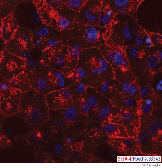
Detection of SSEA-4 in Live BG01V Human Embryonic Stem Cells.
The stem cell marker SSEA-4 was visualized in live BG01V human embryonic stem cells using a GloLIVE NL557-conjugated Mouse Anti-Human/Mouse SSEA-4 Monoclonal Antibody (Catalog # NLLC1435R; red). The cells were counterstained with Hoechst 33342 (blue). Positive staining for SSEA-4 in human stem cells is an indicator of pluripotency.Formulation, Preparation, and Storage
Formulation
Supplied in a saline solution containing BSA and Sodium Azide.
Shipping
The product is shipped with polar packs. Upon receipt, store it immediately at the temperature recommended below.
Background: SSEA-4
References
- Shevinsky, L.H. et al. (1982) Cell 30:697.
- Kannagi, R. et al. (1983) EMBO J. 2:2355.
- Thomson, J.A. and J.S. Odorico (2000) Trends Biotechnol. 18:53.
- Draper, J.S. et al. (2002) J. Anat. 200:249.
Long Name
Stage-specific Embryonic Antigen-4
Alternate Names
SSEA4
Additional SSEA-4 Products
Product Documents for GloLIVE Human/Mouse SSEA-4 NL557 Live Cell Antibody
Certificate of Analysis
To download a Certificate of Analysis, please enter a lot or batch number in the search box below.
Note: Certificate of Analysis not available for kit components.
Product Specific Notices for GloLIVE Human/Mouse SSEA-4 NL557 Live Cell Antibody
For research use only
Related Research Areas
Customer Reviews for GloLIVE Human/Mouse SSEA-4 NL557 Live Cell Antibody
There are currently no reviews for this product. Be the first to review GloLIVE Human/Mouse SSEA-4 NL557 Live Cell Antibody and earn rewards!
Have you used GloLIVE Human/Mouse SSEA-4 NL557 Live Cell Antibody?
Submit a review and receive an Amazon gift card!
$25/€18/£15/$25CAN/¥2500 for a review with an image
$10/€7/£6/$10CAN/¥1110 for a review without an image
Submit a review
Protocols
View specific protocols for GloLIVE Human/Mouse SSEA-4 NL557 Live Cell Antibody (NLLC1435R):
Refer to the product datasheet for complete product details.

Precautions
Use aseptic technique and sterile culturing conditions to prevent contamination.
Reagents Provided
GloLIVE NL557-conjugated Mouse Anti-Human/Mouse SSEA-4 Monoclonal Antibody. (Catalog # NLLC1435R):
Other Supplies Required
Reagents
- Stem cell culture media
Materials
- Human or mouse pluripotent stem cells
- Pipettes and pipette tips
- Serological pipette
Equipment
- 37 °C and 5% CO2 incubator
- Fluorescence microscope
Live Cell Immunocytochemistry Procedure Overview
To ensure sterility of cultures, all steps should be performed under sterile conditions.

- Add fluorochrome-conjugated primary antibody in appropriate culture media to cells.
- Incubate for 30 minutes.
- Replace with fresh culture media.

- Visualize using a fluorescence microscope.
Note: Culture with GloLIVE antibodies does not appear to affect cell proliferation or stemness as assayed by proliferation curves for 3 days post-antibody incubation and expression levels of SSEA-4 and Oct-3/4.
Find general support by application which include: protocols, troubleshooting, illustrated assays, videos and webinars.
- Appropriate Fixation of IHC/ICC Samples
- Cellular Response to Hypoxia Protocols
- ClariTSA™ Fluorophore Kits
- Detection & Visualization of Antibody Binding
- ICC Cell Smear Protocol for Suspension Cells
- ICC Immunocytochemistry Protocol Videos
- ICC for Adherent Cells
- Immunocytochemistry (ICC) Protocol
- Immunocytochemistry Troubleshooting
- Immunofluorescence of Organoids Embedded in Cultrex Basement Membrane Extract
- Immunohistochemistry (IHC) and Immunocytochemistry (ICC) Protocols
- Preparing Samples for IHC/ICC Experiments
- Preventing Non-Specific Staining (Non-Specific Binding)
- Primary Antibody Selection & Optimization
- Protocol for VisUCyte™ HRP Polymer Detection Reagent
- Protocol for the Fluorescent ICC Staining of Cell Smears - Graphic
- Protocol for the Fluorescent ICC Staining of Cultured Cells on Coverslips - Graphic
- Protocol for the Preparation and Fluorescent ICC Staining of Cells on Coverslips
- Protocol for the Preparation and Fluorescent ICC Staining of Non-adherent Cells
- Protocol for the Preparation and Fluorescent ICC Staining of Stem Cells on Coverslips
- Protocol for the Preparation of a Cell Smear for Non-adherent Cell ICC - Graphic
- TUNEL and Active Caspase-3 Detection by IHC/ICC Protocol
- The Importance of IHC/ICC Controls
- View all Protocols, Troubleshooting, Illustrated assays and Webinars
FAQs for GloLIVE Human/Mouse SSEA-4 NL557 Live Cell Antibody
Showing
1
-
1 of
1 FAQ
Showing All
-
Q: What is the recommended starting confluence to observe staining?
A: The optimal confluence would depend on the purpose of the staining. If the plan is to use all the cells in the flask after they have proliferated, then one can wait for the cells to become confluent or almost confluent (80-90% confluent) and then check the staining. If the goal is to to check on the cells mid-way through their growth and there are sufficient cells in the flask, one could check the staining then as well. The staining is not dependent on how many cells are in the flask, as long as there are sufficient cells.
Loading...