GR/NR3C1 [p Ser226] Antibody (S02-1H0)
Novus Biologicals | Catalog # NBP3-14939
Recombinant Monoclonal Antibody
Loading...
Key Product Details
Species Reactivity
Human
Applications
Immunohistochemistry, Immunohistochemistry-Paraffin, Western Blot
Label
Unconjugated
Antibody Source
Recombinant Monoclonal Rabbit IgG Clone # S02-1H0 expressed in HEK293
Loading...
Product Specifications
Immunogen
A synthetic phosphopeptide corresponding to residues surrounding Ser226 of human GR/NR3C1 (Uniprot#: P04150)
Modification
p Ser 226
Clonality
Monoclonal
Host
Rabbit
Isotype
IgG
Theoretical MW
94 kDa.
Disclaimer note: The observed molecular weight of the protein may vary from the listed predicted molecular weight due to post translational modifications, post translation cleavages, relative charges, and other experimental factors.
Disclaimer note: The observed molecular weight of the protein may vary from the listed predicted molecular weight due to post translational modifications, post translation cleavages, relative charges, and other experimental factors.
Scientific Data Images for GR/NR3C1 [p Ser226] Antibody (S02-1H0)
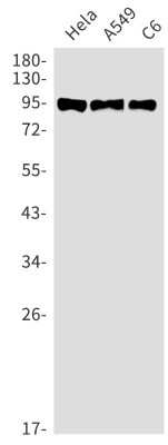
Western Blot: GR/NR3C1 [p Ser226] Antibody (S02-1H0) [NBP3-14939] - Western blot detection of GR/NR3C1 [p Ser226] in Hela, A549, C6 cell lysates using GR/NR3C1 [p Ser226] Rabbit mAb (1:1000 diluted). Predicted band size: 86kDa. Observed band size: 94kDa.
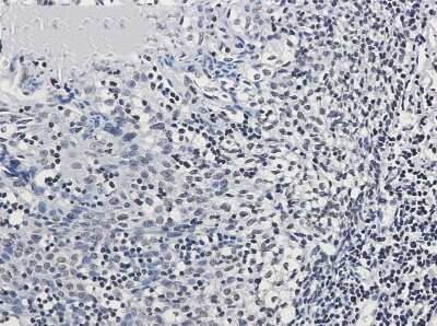
Immunohistochemistry-Paraffin: GR/NR3C1 [p Ser226] Antibody (S02-1H0) [NBP3-14939] - Immunohistochemistry of GR/NR3C1 [p Ser226] in paraffin-embedded Human tonsil using GR/NR3C1 [p Ser226] Rabbit mAb at dilution 1/50
Applications for GR/NR3C1 [p Ser226] Antibody (S02-1H0)
Application
Recommended Usage
Immunohistochemistry
1/100
Western Blot
1/1000
Formulation, Preparation, and Storage
Purification
Affinity purified
Formulation
50mM Tris-Glycine (pH 7.4), 0.15M NaCl, 40% Glycerol, 0.05% BSA
Preservative
0.01% Sodium Azide
Concentration
Please see the vial label for concentration. If unlisted please contact technical services.
Shipping
The product is shipped with polar packs. Upon receipt, store it immediately at the temperature recommended below.
Stability & Storage
Store at 4C short term. Aliquot and store at -20C long term. Avoid freeze-thaw cycles.
Background: GR/NR3C1
Long Name
Glucocorticoid Receptor
Alternate Names
NR3C1
Gene Symbol
NR3C1
Additional GR/NR3C1 Products
Product Documents for GR/NR3C1 [p Ser226] Antibody (S02-1H0)
Certificate of Analysis
To download a Certificate of Analysis, please enter a lot or batch number in the search box below.
Product Specific Notices for GR/NR3C1 [p Ser226] Antibody (S02-1H0)
This product is for research use only and is not approved for use in humans or in clinical diagnosis. Primary Antibodies are guaranteed for 1 year from date of receipt.
Related Research Areas
Customer Reviews for GR/NR3C1 [p Ser226] Antibody (S02-1H0)
There are currently no reviews for this product. Be the first to review GR/NR3C1 [p Ser226] Antibody (S02-1H0) and earn rewards!
Have you used GR/NR3C1 [p Ser226] Antibody (S02-1H0)?
Submit a review and receive an Amazon gift card!
$25/€18/£15/$25CAN/¥2500 Yen for a review with an image
$10/€7/£6/$10CAN/¥1110 Yen for a review without an image
Submit a review
Protocols
Find general support by application which include: protocols, troubleshooting, illustrated assays, videos and webinars.
- Antigen Retrieval Protocol (PIER)
- Antigen Retrieval for Frozen Sections Protocol
- Appropriate Fixation of IHC/ICC Samples
- Cellular Response to Hypoxia Protocols
- Chromogenic IHC Staining of Formalin-Fixed Paraffin-Embedded (FFPE) Tissue Protocol
- Chromogenic Immunohistochemistry Staining of Frozen Tissue
- ClariTSA™ Fluorophore Kits
- Detection & Visualization of Antibody Binding
- Fluorescent IHC Staining of Frozen Tissue Protocol
- Graphic Protocol for Heat-induced Epitope Retrieval
- Graphic Protocol for the Preparation and Fluorescent IHC Staining of Frozen Tissue Sections
- Graphic Protocol for the Preparation and Fluorescent IHC Staining of Paraffin-embedded Tissue Sections
- Graphic Protocol for the Preparation of Gelatin-coated Slides for Histological Tissue Sections
- IHC Sample Preparation (Frozen sections vs Paraffin)
- Immunofluorescent IHC Staining of Formalin-Fixed Paraffin-Embedded (FFPE) Tissue Protocol
- Immunohistochemistry (IHC) and Immunocytochemistry (ICC) Protocols
- Immunohistochemistry Frozen Troubleshooting
- Immunohistochemistry Paraffin Troubleshooting
- Preparing Samples for IHC/ICC Experiments
- Preventing Non-Specific Staining (Non-Specific Binding)
- Primary Antibody Selection & Optimization
- Protocol for Heat-Induced Epitope Retrieval (HIER)
- Protocol for Making a 4% Formaldehyde Solution in PBS
- Protocol for VisUCyte™ HRP Polymer Detection Reagent
- Protocol for the Preparation & Fixation of Cells on Coverslips
- Protocol for the Preparation and Chromogenic IHC Staining of Frozen Tissue Sections
- Protocol for the Preparation and Chromogenic IHC Staining of Frozen Tissue Sections - Graphic
- Protocol for the Preparation and Chromogenic IHC Staining of Paraffin-embedded Tissue Sections
- Protocol for the Preparation and Chromogenic IHC Staining of Paraffin-embedded Tissue Sections - Graphic
- Protocol for the Preparation and Fluorescent IHC Staining of Frozen Tissue Sections
- Protocol for the Preparation and Fluorescent IHC Staining of Paraffin-embedded Tissue Sections
- Protocol for the Preparation of Gelatin-coated Slides for Histological Tissue Sections
- R&D Systems Quality Control Western Blot Protocol
- TUNEL and Active Caspase-3 Detection by IHC/ICC Protocol
- The Importance of IHC/ICC Controls
- Troubleshooting Guide: Immunohistochemistry
- Troubleshooting Guide: Western Blot Figures
- Western Blot Conditions
- Western Blot Protocol
- Western Blot Protocol for Cell Lysates
- Western Blot Troubleshooting
- Western Blot Troubleshooting Guide
- View all Protocols, Troubleshooting, Illustrated assays and Webinars
Loading...
Associated Pathways
