HDAC11 Antibody - BSA Free
Novus Biologicals | Catalog # NBP2-16789
![Simple Western: HDAC11 Antibody [NBP2-16789]](https://resources.rndsystems.com/images/products/HDAC11-Antibody-Simple-Western-NBP2-16789-img0006.jpg "Simple Western: HDAC11 Antibody [NBP2-16789]")
Loading...
Key Product Details
Validated by
Knockout/Knockdown
Species Reactivity
Validated:
Human, Mouse
Cited:
Human
Predicted:
Rhesus Macaque (98%). Backed by our 100% Guarantee.
Applications
Validated:
Western Blot, Simple Western, Knockdown Validated
Cited:
IF/IHC, Microarray
Label
Unconjugated
Antibody Source
Polyclonal Rabbit IgG
Format
BSA Free
Loading...
Product Specifications
Immunogen
Recombinant protein encompassing a sequence within the center region of human HDAC11. The exact sequence is proprietary.
Reactivity Notes
Rat (89%), Bovine (88%)
Localization
Nucleus
Clonality
Polyclonal
Host
Rabbit
Isotype
IgG
Theoretical MW
39 kDa.
Disclaimer note: The observed molecular weight of the protein may vary from the listed predicted molecular weight due to post translational modifications, post translation cleavages, relative charges, and other experimental factors.
Disclaimer note: The observed molecular weight of the protein may vary from the listed predicted molecular weight due to post translational modifications, post translation cleavages, relative charges, and other experimental factors.
Scientific Data Images for HDAC11 Antibody - BSA Free
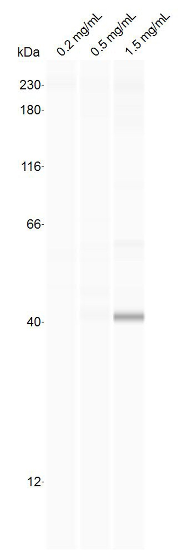
Simple Western: HDAC11 Antibody [NBP2-16789]
Simple Western: HDAC11 Antibody [NBP2-16789] - 1:25 dilution against the indicated concentration of protein lysate of human SVG-A cells. Image submitted by a verified customer review.![Knockdown Validated: HDAC11 Antibody [NBP2-16789]](https://resources.rndsystems.com/images/products/HDAC11-Antibody-Knockdown-Validated-NBP2-16789-img0011.jpg "Western Blot: HDAC11 Antibody [NBP2-16789]")

Western Blot: HDAC11 Antibody [NBP2-16789] -
Western Blot: HDAC11 Antibody [NBP2-16789] - HDAC11 silencing decreases expression of downstream targets & abrogates self-renewal of CSCs. (A) Depletion of HDAC11 using two IPTG inducible shRNA shows a significant decrease in Sox2, YAP1 & Gli1 protein expression as compared to IPTG treated control shRNA. The glycolysis pathway targets HK2 & PDK2 also show reduced protein expression in the absence of HDAC11. No change is observed in PDK1 expression; the full images of the western blots are provided as Supplementary Fig. 4. (B) Quantitation of the western blot band intensities using ImageJ analysis. (C) Sphere formation assay with SP cells from shHDAC11 H1650 cells treated with IPTG show abrogation of self-renewal ability in the absence of HDAC11. (D) Quantitation of sphere formation assay reveal that IPTG treated SP cells from two different clones of HDAC11 shRNA containing H1650 form fewer spheres as compared to IPTG untreated or control shRNA containing cells. (E,F) Real time PCR analysis of H1650 cells with two different HDAC11 shRNA show a decrease in mRNA expression of Sox2, HK2, PDK1, PDK2 with IPTG treatment. No significant change was observed in Oct4 & Nanog expression. The depletion of HDAC11 was confirmed using RT-PCR. Image collected & cropped by CiteAb from the following publication (https://pubmed.ncbi.nlm.nih.gov/32170113), licensed under a CC-BY license. Not internally tested by Novus Biologicals.Applications for HDAC11 Antibody - BSA Free
Application
Recommended Usage
Simple Western
1:25
Western Blot
1:500-1:3000
Application Notes
See Simple Western Antibody Database for Simple Western validation: Tested in Human SVG-A cell protein lysate, separated by Size, antibody dilution of 1:25
Reviewed Applications
Read 1 review rated 4 using NBP2-16789 in the following applications:
Formulation, Preparation, and Storage
Purification
Antigen Affinity-purified
Formulation
PBS, 20% Glycerol
Format
BSA Free
Preservative
0.025% Proclin 300
Concentration
Concentrations vary lot to lot. See vial label for concentration. If unlisted please contact technical services.
Shipping
The product is shipped with polar packs. Upon receipt, store it immediately at the temperature recommended below.
Stability & Storage
Aliquot and store at -20C or -80C. Avoid freeze-thaw cycles.
Background: HDAC11
Long Name
Histone deacetylase 11
Alternate Names
EC 3.5.1.98, HD11
Gene Symbol
HDAC11
UniProt
Additional HDAC11 Products
Product Documents for HDAC11 Antibody - BSA Free
Certificate of Analysis
To download a Certificate of Analysis, please enter a lot or batch number in the search box below.
Product Specific Notices for HDAC11 Antibody - BSA Free
This product is for research use only and is not approved for use in humans or in clinical diagnosis. Primary Antibodies are guaranteed for 1 year from date of receipt.
⚠ WARNING: This product can expose you to chemicals including mercury, which is known to the State of California to cause reproductive toxicity with developmental effects. For more information go to www.P65Warnings.ca.gov.Citations for HDAC11 Antibody - BSA Free
 Powered by Bioz
Customer Reviews for HDAC11 Antibody - BSA Free (1)
4 out of 5
1 Customer Rating
Have you used HDAC11 Antibody - BSA Free?
Submit a review and receive an Amazon gift card!
$25/€18/£15/$25CAN/¥2500 Yen for a review with an image
$10/€7/£6/$10CAN/¥1110 Yen for a review without an image
Submit a review
Customer Images
Showing
1
-
1 of
1 review
Showing All
Filter By:
-
Application: Simple WesternSample Tested: SVG-A whole cell lysateSpecies: HumanVerified Customer | Posted 03/08/2019Antibody tested by Simple Western at 1:25 dilution against the indicated concentration of protein lysate of human SVG-A cells.

There are no reviews that match your criteria.
Protocols
Find general support by application which include: protocols, troubleshooting, illustrated assays, videos and webinars.
- Cellular Response to Hypoxia Protocols
- R&D Systems Quality Control Western Blot Protocol
- Troubleshooting Guide: Western Blot Figures
- Western Blot Conditions
- Western Blot Protocol
- Western Blot Protocol for Cell Lysates
- Western Blot Troubleshooting
- Western Blot Troubleshooting Guide
- View all Protocols, Troubleshooting, Illustrated assays and Webinars
Loading...