HOXC11 Antibody (OTI3E10)
Novus Biologicals | Catalog # NBP2-00499
Key Product Details
Species Reactivity
Validated:
Cited:
Applications
Validated:
Cited:
Label
Antibody Source
Product Specifications
Immunogen
Reactivity Notes
Clonality
Host
Isotype
Theoretical MW
Disclaimer note: The observed molecular weight of the protein may vary from the listed predicted molecular weight due to post translational modifications, post translation cleavages, relative charges, and other experimental factors.
Scientific Data Images for HOXC11 Antibody (OTI3E10)
![Western Blot: HOXC11 Antibody (OTI3E10) [NBP2-00499]](https://resources.rndsystems.com/images/products/HOXC11-Antibody-3E10-Western-Blot-NBP2-00499-img0011.jpg "Western Blot: HOXC11 Antibody (OTI3E10) [NBP2-00499]")
Western Blot: HOXC11 Antibody (OTI3E10) [NBP2-00499]
Western Blot: HOXC11 Antibody (3E10) [NBP2-00499] - Analysis of extracts (10ug) from 9 Human tissue by using anti-HOXC11 monoclonal antibody at 1:200 (1: Testis; 2: Omentum; 3: Uterus; 4: Breast; 5: Brain; 6: Liver; 7: Ovary; 8: Thyroid gland; 9: colon)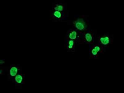
![Immunohistochemistry-Paraffin: HOXC11 Antibody (OTI3E10) [NBP2-00499]](https://resources.rndsystems.com/images/products/HOXC11-Antibody-3E10-Immunohistochemistry-Paraffin-NBP2-00499-img0007.jpg "Immunohistochemistry-Paraffin: HOXC11 Antibody (OTI3E10) [NBP2-00499]")
Immunohistochemistry-Paraffin: HOXC11 Antibody (OTI3E10) [NBP2-00499]
Immunohistochemistry-Paraffin: HOXC11 Antibody (3E10) [NBP2-00499] - Staining of paraffin-embedded Human tonsil using anti-HOXC11 mouse monoclonal antibody.![Flow Cytometry: HOXC11 Antibody (OTI3E10) [NBP2-00499]](https://resources.rndsystems.com/images/products/HOXC11-Antibody-3E10-Flow-Cytometry-NBP2-00499-img0001.jpg "Flow Cytometry: HOXC11 Antibody (OTI3E10) [NBP2-00499]")
Flow Cytometry: HOXC11 Antibody (OTI3E10) [NBP2-00499]
Flow Cytometry: HOXC11 Antibody (3E10) [NBP2-00499] - HEK293T cells transfected with either overexpression plasmid (Red) or empty vector control plasmid (Blue) were immunostained by anti-HOXC11 antibody, and then analyzed by flow cytometry.![Western Blot: HOXC11 Antibody (OTI3E10) [NBP2-00499]](https://resources.rndsystems.com/images/products/HOXC11-Antibody-3E10-Western-Blot-NBP2-00499-img0009.jpg "Western Blot: HOXC11 Antibody (OTI3E10) [NBP2-00499]")
Western Blot: HOXC11 Antibody (OTI3E10) [NBP2-00499]
Western Blot: HOXC11 Antibody (3E10) [NBP2-00499] - HEK293T cells were transfected with the pCMV6-ENTRY control (Left lane) or pCMV6-ENTRY HOXC11 (Right lane) cDNA for 48 hrs and lysed. Equivalent amounts of cell lysates (5 ug per lane) were separated by SDS-PAGE and immunoblotted with anti-HOXC11.![Western Blot: HOXC11 Antibody (OTI3E10) [NBP2-00499]](https://resources.rndsystems.com/images/products/HOXC11-Antibody-3E10-Western-Blot-NBP2-00499-img0010.jpg "Western Blot: HOXC11 Antibody (OTI3E10) [NBP2-00499]")
Western Blot: HOXC11 Antibody (OTI3E10) [NBP2-00499]
Western Blot: HOXC11 Antibody (3E10) [NBP2-00499] - Analysis of extracts (10ug) from a mouse cell line and 3 different mouse tissues by using antiHOXC11 monoclonal antibody.(1:200)![Immunohistochemistry-Paraffin: HOXC11 Antibody (OTI3E10) [NBP2-00499]](https://resources.rndsystems.com/images/products/HOXC11-Antibody-3E10-Immunohistochemistry-Paraffin-NBP2-00499-img0003.jpg "Immunohistochemistry-Paraffin: HOXC11 Antibody (OTI3E10) [NBP2-00499]")
Immunohistochemistry-Paraffin: HOXC11 Antibody (OTI3E10) [NBP2-00499]
Immunohistochemistry-Paraffin: HOXC11 Antibody (3E10) [NBP2-00499] - Staining of paraffin-embedded Adenocarcinoma of Human breast tissue using anti-HOXC11 mouse monoclonal antibody.![Immunohistochemistry-Paraffin: HOXC11 Antibody (OTI3E10) [NBP2-00499]](https://resources.rndsystems.com/images/products/HOXC11-Antibody-3E10-Immunohistochemistry-Paraffin-NBP2-00499-img0004.jpg "Immunohistochemistry-Paraffin: HOXC11 Antibody (OTI3E10) [NBP2-00499]")
Immunohistochemistry-Paraffin: HOXC11 Antibody (OTI3E10) [NBP2-00499]
Immunohistochemistry-Paraffin: HOXC11 Antibody (3E10) [NBP2-00499] - Staining of paraffin-embedded Adenocarcinoma of Human colon tissue using anti-HOXC11 mouse monoclonal antibody.![Immunohistochemistry-Paraffin: HOXC11 Antibody (OTI3E10) [NBP2-00499]](https://resources.rndsystems.com/images/products/HOXC11-Antibody-3E10-Immunohistochemistry-Paraffin-NBP2-00499-img0005.jpg "Immunohistochemistry-Paraffin: HOXC11 Antibody (OTI3E10) [NBP2-00499]")
Immunohistochemistry-Paraffin: HOXC11 Antibody (OTI3E10) [NBP2-00499]
Immunohistochemistry-Paraffin: HOXC11 Antibody (3E10) [NBP2-00499] - Staining of paraffin-embedded Carcinoma of Human bladder tissue using anti-HOXC11 mouse monoclonal antibody.![Immunohistochemistry-Paraffin: HOXC11 Antibody (OTI3E10) [NBP2-00499]](https://resources.rndsystems.com/images/products/HOXC11-Antibody-3E10-Immunohistochemistry-Paraffin-NBP2-00499-img0006.jpg "Immunohistochemistry-Paraffin: HOXC11 Antibody (OTI3E10) [NBP2-00499]")
Immunohistochemistry-Paraffin: HOXC11 Antibody (OTI3E10) [NBP2-00499]
Immunohistochemistry-Paraffin: HOXC11 Antibody (3E10) [NBP2-00499] - Staining of paraffin-embedded Human endometrium tissue using anti-HOXC11 mouse monoclonal antibody. [NBP2-00499] -")
Western Blot: HOXC11 Antibody (OTI3E10) [NBP2-00499] -
HOXC11 binds to the promoter of SPHK1 to facilitate its expression, predicting a worse prognosis.a GSEA analyzed potential binding targets of HOXC11. b qPCR analysis of CCL5, HBA2, and SPHK1 expression level after stable overexpression of HOXC11. Data are shown as mean +/- SD (n = 3). c Western Blot analysis of SPHK1 protein expression level after HOXC11 stable overexpression. d Western Blot analysis of SPHK1 protein expression level after HOXC11 knockout. e Relative enrichment of SPHK1 promoter in HOXC11 stable overexpressing cells. Data are shown as mean +/- SD (n = 3). f Western Blot detected SPHK1 protein expression of HOXC11 knockout cells and that after transiently transfected with 1.5 μg HOXC11 expression plasmid. NS not significant, *P < 0.05, **P < 0.01. Image collected and cropped by CiteAb from the following open publication (https://pubmed.ncbi.nlm.nih.gov/36823149), licensed under a CC-BY license. Not internally tested by Novus Biologicals. [NBP2-00499] -")
Western Blot: HOXC11 Antibody (OTI3E10) [NBP2-00499] -
HOXC11 is highly expressed in lung adenocarcinoma and correlates with poor overall survival of LUAD.a A heatmap of mRNA level of HOX family genes in LUAD/LUSC and paracancerous samples. b mRNA level of HOXC11 in LUAD/LUSC and paracancerous samples. Data in (a) and (b) are obtained from the TCGA database. c HOXC11 protein expression in LUAD/LUSC and paracancerous tissues. d The relationship between HOXC11 expression and overall survival of lung cancer patients. e The relationship between HOXC11 expression and overall survival of LUAD patients. f The relationship between HOXC11 expression and gender of lung cancer patients. g The relationship between HOXC11 expression and smoking habits of lung cancer patients. Data from (d) to (g) come from the Kaplan–Meier Plotter lung cancer dataset. ***P < 0.001, ****P < 0.0001. Image collected and cropped by CiteAb from the following open publication (https://pubmed.ncbi.nlm.nih.gov/36823149), licensed under a CC-BY license. Not internally tested by Novus Biologicals. [NBP2-00499] -")
Western Blot: HOXC11 Antibody (OTI3E10) [NBP2-00499] -
HOXC11 binds to the promoter of SPHK1 to facilitate its expression, predicting a worse prognosis.a GSEA analyzed potential binding targets of HOXC11. b qPCR analysis of CCL5, HBA2, and SPHK1 expression level after stable overexpression of HOXC11. Data are shown as mean +/- SD (n = 3). c Western Blot analysis of SPHK1 protein expression level after HOXC11 stable overexpression. d Western Blot analysis of SPHK1 protein expression level after HOXC11 knockout. e Relative enrichment of SPHK1 promoter in HOXC11 stable overexpressing cells. Data are shown as mean +/- SD (n = 3). f Western Blot detected SPHK1 protein expression of HOXC11 knockout cells and that after transiently transfected with 1.5 μg HOXC11 expression plasmid. NS not significant, *P < 0.05, **P < 0.01. Image collected and cropped by CiteAb from the following open publication (https://pubmed.ncbi.nlm.nih.gov/36823149), licensed under a CC-BY license. Not internally tested by Novus Biologicals. [NBP2-00499] -")
Western Blot: HOXC11 Antibody (OTI3E10) [NBP2-00499] -
IKK alpha regulates HOXC11 expression at the post-transcriptional level.a Western Blot showed the up-regulation of HOXC11 resulting from IKK alpha stable overexpression. b Western Blot detected the down-regulation of HOXC11 resulting from the IKK alpha knockout. c qPCR analysis of HOXC11 mRNA level after stable overexpression of IKK alpha. Data are shown as mean +/- SD (n = 3). d Western Blot showing HOXC11 protein cumulation after treatment with MG132 (10 μM, 24 h). e Western Blot showed HOXC11 protein degradation after treatment with cycloheximide (10 μg/ml). f Western Blot analysis of HOXC11 accumulation after IKK alpha stable overexpression and MG132 treatment (20 μM, 12 h). g Western Blot showed HOXC11 protein expression after IKK alpha stable overexpression and treatment with cycloheximide (20 μg/ml). h Western Blot analysis of HOXC11 accumulation after IKK alpha knockout and MG132 treatment (20 μM, 10 h). i Western Blot showed HOXC11 protein expression after IKK alpha knockout and treatment with cycloheximide (20 μg/ml). j Ubiquitinating and deubiquitinating enzymes of HOXC11 and IKK alpha predicted by UbiBrowser 2.0. USP8 is shared by HOXC11 and IKK alpha. k USP8 with tag-Myc was instantaneously expressed in HEK293T, and Western Blot detected the protein level of IKK alpha and HOXC11. l Immunofluorescence detected the localization of IKK alpha (red) relative to HOXC11 (green) or Myc-USP8 (green) in PC9 cells. Scale bars, 20 μm. Image collected and cropped by CiteAb from the following open publication (https://pubmed.ncbi.nlm.nih.gov/36823149), licensed under a CC-BY license. Not internally tested by Novus Biologicals. [NBP2-00499] -")
Western Blot: HOXC11 Antibody (OTI3E10) [NBP2-00499] -
SPHK1 accelerates the progression of lung cancer.a Cell proliferation ability of SPHK1 stable overexpressed cells detected by cell counting kit-8. Data are shown as mean +/- SD (n = 5). b Colony formation assays showing the colony formatted ability of SPHK1 stable overexpressed cells and the quantitative analyses. Data are shown as mean +/- SD (n = 3). c Cell migration and invasion ability of SPHK1 stable overexpressed cells detected by Transwell assays and the quantitative analyses. Data are shown as mean +/- SD (n = 3). d Flow cytometry detecting the cell cycle of SPHK1 overexpressed cells. Differences are compared with the sgCtrl group; data are shown as mean +/- SD (n = 3). e Western blot detected the SPHK1 protein level of HOXC11 overexpressing cells with the treatment of SPHK1 interference. f Cell counting kit-8 detected the cell viability of HOXC11 overexpressing cell line with the treatment of SPHK1 interference. Data are shown as mean +/- SD (n = 5). g Transwell assays detected the migration and invasion ability of HOXC11 overexpressing cell line with the treatment of SPHK1 interference and the quantitative analyses. Differences were compared to the Vector group transfected with Ctrl siRNA. Data are shown as mean +/- SD (n = 3). NS not significant, *P < 0.05, **P < 0.01, *** P < 0.001, ****P < 0.0001. Image collected and cropped by CiteAb from the following open publication (https://pubmed.ncbi.nlm.nih.gov/36823149), licensed under a CC-BY license. Not internally tested by Novus Biologicals. [NBP2-00499] -")
Western Blot: HOXC11 Antibody (OTI3E10) [NBP2-00499] -
HOXC11 binds to the promoter of SPHK1 to facilitate its expression, predicting a worse prognosis.a GSEA analyzed potential binding targets of HOXC11. b qPCR analysis of CCL5, HBA2, and SPHK1 expression level after stable overexpression of HOXC11. Data are shown as mean +/- SD (n = 3). c Western Blot analysis of SPHK1 protein expression level after HOXC11 stable overexpression. d Western Blot analysis of SPHK1 protein expression level after HOXC11 knockout. e Relative enrichment of SPHK1 promoter in HOXC11 stable overexpressing cells. Data are shown as mean +/- SD (n = 3). f Western Blot detected SPHK1 protein expression of HOXC11 knockout cells and that after transiently transfected with 1.5 μg HOXC11 expression plasmid. NS not significant, *P < 0.05, **P < 0.01. Image collected and cropped by CiteAb from the following open publication (https://pubmed.ncbi.nlm.nih.gov/36823149), licensed under a CC-BY license. Not internally tested by Novus Biologicals. [NBP2-00499] -")
Western Blot: HOXC11 Antibody (OTI3E10) [NBP2-00499] -
IKK alpha regulates HOXC11 expression at the post-transcriptional level.a Western Blot showed the up-regulation of HOXC11 resulting from IKK alpha stable overexpression. b Western Blot detected the down-regulation of HOXC11 resulting from the IKK alpha knockout. c qPCR analysis of HOXC11 mRNA level after stable overexpression of IKK alpha. Data are shown as mean +/- SD (n = 3). d Western Blot showing HOXC11 protein cumulation after treatment with MG132 (10 μM, 24 h). e Western Blot showed HOXC11 protein degradation after treatment with cycloheximide (10 μg/ml). f Western Blot analysis of HOXC11 accumulation after IKK alpha stable overexpression and MG132 treatment (20 μM, 12 h). g Western Blot showed HOXC11 protein expression after IKK alpha stable overexpression and treatment with cycloheximide (20 μg/ml). h Western Blot analysis of HOXC11 accumulation after IKK alpha knockout and MG132 treatment (20 μM, 10 h). i Western Blot showed HOXC11 protein expression after IKK alpha knockout and treatment with cycloheximide (20 μg/ml). j Ubiquitinating and deubiquitinating enzymes of HOXC11 and IKK alpha predicted by UbiBrowser 2.0. USP8 is shared by HOXC11 and IKK alpha. k USP8 with tag-Myc was instantaneously expressed in HEK293T, and Western Blot detected the protein level of IKK alpha and HOXC11. l Immunofluorescence detected the localization of IKK alpha (red) relative to HOXC11 (green) or Myc-USP8 (green) in PC9 cells. Scale bars, 20 μm. Image collected and cropped by CiteAb from the following open publication (https://pubmed.ncbi.nlm.nih.gov/36823149), licensed under a CC-BY license. Not internally tested by Novus Biologicals. [NBP2-00499] -")
Western Blot: HOXC11 Antibody (OTI3E10) [NBP2-00499] -
IKK alpha regulates HOXC11 expression at the post-transcriptional level.a Western Blot showed the up-regulation of HOXC11 resulting from IKK alpha stable overexpression. b Western Blot detected the down-regulation of HOXC11 resulting from the IKK alpha knockout. c qPCR analysis of HOXC11 mRNA level after stable overexpression of IKK alpha. Data are shown as mean +/- SD (n = 3). d Western Blot showing HOXC11 protein cumulation after treatment with MG132 (10 μM, 24 h). e Western Blot showed HOXC11 protein degradation after treatment with cycloheximide (10 μg/ml). f Western Blot analysis of HOXC11 accumulation after IKK alpha stable overexpression and MG132 treatment (20 μM, 12 h). g Western Blot showed HOXC11 protein expression after IKK alpha stable overexpression and treatment with cycloheximide (20 μg/ml). h Western Blot analysis of HOXC11 accumulation after IKK alpha knockout and MG132 treatment (20 μM, 10 h). i Western Blot showed HOXC11 protein expression after IKK alpha knockout and treatment with cycloheximide (20 μg/ml). j Ubiquitinating and deubiquitinating enzymes of HOXC11 and IKK alpha predicted by UbiBrowser 2.0. USP8 is shared by HOXC11 and IKK alpha. k USP8 with tag-Myc was instantaneously expressed in HEK293T, and Western Blot detected the protein level of IKK alpha and HOXC11. l Immunofluorescence detected the localization of IKK alpha (red) relative to HOXC11 (green) or Myc-USP8 (green) in PC9 cells. Scale bars, 20 μm. Image collected and cropped by CiteAb from the following open publication (https://pubmed.ncbi.nlm.nih.gov/36823149), licensed under a CC-BY license. Not internally tested by Novus Biologicals.Applications for HOXC11 Antibody (OTI3E10)
Flow Cytometry
Immunocytochemistry/ Immunofluorescence
Immunohistochemistry
Immunohistochemistry-Paraffin
Western Blot
Flow Cytometry Panel Builder
Bio-Techne Knows Flow Cytometry
Save time and reduce costly mistakes by quickly finding compatible reagents using the Panel Builder Tool.
Advanced Features
- Spectra Viewer - Custom analysis of spectra from multiple fluorochromes
- Spillover Popups - Visualize the spectra of individual fluorochromes
- Antigen Density Selector - Match fluorochrome brightness with antigen density

Formulation, Preparation, and Storage
Purification
Formulation
Preservative
Concentration
Shipping
Stability & Storage
Background: HOXC11
Alternate Names
Entrez Gene IDs
Gene Symbol
Additional HOXC11 Products
Product Documents for HOXC11 Antibody (OTI3E10)
Certificate of Analysis
To download a Certificate of Analysis, please enter a lot or batch number in the search box below.
Product Specific Notices for HOXC11 Antibody (OTI3E10)
This product is for research use only and is not approved for use in humans or in clinical diagnosis. Primary Antibodies are guaranteed for 1 year from date of receipt.
Citations for HOXC11 Antibody (OTI3E10)
 Powered by Bioz
Customer Reviews for HOXC11 Antibody (OTI3E10)
There are currently no reviews for this product. Be the first to review HOXC11 Antibody (OTI3E10) and earn rewards!
Have you used HOXC11 Antibody (OTI3E10)?
Submit a review and receive an Amazon gift card!
$25/€18/£15/$25CAN/¥2500 Yen for a review with an image
$10/€7/£6/$10CAN/¥1110 Yen for a review without an image
Submit a review
Protocols
Find general support by application which include: protocols, troubleshooting, illustrated assays, videos and webinars.
- 7-Amino Actinomycin D (7-AAD) Cell Viability Flow Cytometry Protocol
- Antigen Retrieval Protocol (PIER)
- Antigen Retrieval for Frozen Sections Protocol
- Appropriate Fixation of IHC/ICC Samples
- Cellular Response to Hypoxia Protocols
- Chromogenic IHC Staining of Formalin-Fixed Paraffin-Embedded (FFPE) Tissue Protocol
- Chromogenic Immunohistochemistry Staining of Frozen Tissue
- ClariTSA™ Fluorophore Kits
- Detection & Visualization of Antibody Binding
- Extracellular Membrane Flow Cytometry Protocol
- Flow Cytometry Protocol for Cell Surface Markers
- Flow Cytometry Protocol for Staining Membrane Associated Proteins
- Flow Cytometry Staining Protocols
- Flow Cytometry Troubleshooting Guide
- Fluorescent IHC Staining of Frozen Tissue Protocol
- Graphic Protocol for Heat-induced Epitope Retrieval
- Graphic Protocol for the Preparation and Fluorescent IHC Staining of Frozen Tissue Sections
- Graphic Protocol for the Preparation and Fluorescent IHC Staining of Paraffin-embedded Tissue Sections
- Graphic Protocol for the Preparation of Gelatin-coated Slides for Histological Tissue Sections
- ICC Cell Smear Protocol for Suspension Cells
- ICC Immunocytochemistry Protocol Videos
- ICC for Adherent Cells
- IHC Sample Preparation (Frozen sections vs Paraffin)
- Immunocytochemistry (ICC) Protocol
- Immunocytochemistry Troubleshooting
- Immunofluorescence of Organoids Embedded in Cultrex Basement Membrane Extract
- Immunofluorescent IHC Staining of Formalin-Fixed Paraffin-Embedded (FFPE) Tissue Protocol
- Immunohistochemistry (IHC) and Immunocytochemistry (ICC) Protocols
- Immunohistochemistry Frozen Troubleshooting
- Immunohistochemistry Paraffin Troubleshooting
- Intracellular Flow Cytometry Protocol Using Alcohol (Methanol)
- Intracellular Flow Cytometry Protocol Using Detergents
- Intracellular Nuclear Staining Flow Cytometry Protocol Using Detergents
- Intracellular Staining Flow Cytometry Protocol Using Alcohol Permeabilization
- Intracellular Staining Flow Cytometry Protocol Using Detergents to Permeabilize Cells
- Preparing Samples for IHC/ICC Experiments
- Preventing Non-Specific Staining (Non-Specific Binding)
- Primary Antibody Selection & Optimization
- Propidium Iodide Cell Viability Flow Cytometry Protocol
- Protocol for Heat-Induced Epitope Retrieval (HIER)
- Protocol for Liperfluo
- Protocol for Making a 4% Formaldehyde Solution in PBS
- Protocol for VisUCyte™ HRP Polymer Detection Reagent
- Protocol for the Characterization of Human Th22 Cells
- Protocol for the Characterization of Human Th9 Cells
- Protocol for the Fluorescent ICC Staining of Cell Smears - Graphic
- Protocol for the Fluorescent ICC Staining of Cultured Cells on Coverslips - Graphic
- Protocol for the Preparation & Fixation of Cells on Coverslips
- Protocol for the Preparation and Chromogenic IHC Staining of Frozen Tissue Sections
- Protocol for the Preparation and Chromogenic IHC Staining of Frozen Tissue Sections - Graphic
- Protocol for the Preparation and Chromogenic IHC Staining of Paraffin-embedded Tissue Sections
- Protocol for the Preparation and Chromogenic IHC Staining of Paraffin-embedded Tissue Sections - Graphic
- Protocol for the Preparation and Fluorescent ICC Staining of Cells on Coverslips
- Protocol for the Preparation and Fluorescent ICC Staining of Non-adherent Cells
- Protocol for the Preparation and Fluorescent ICC Staining of Stem Cells on Coverslips
- Protocol for the Preparation and Fluorescent IHC Staining of Frozen Tissue Sections
- Protocol for the Preparation and Fluorescent IHC Staining of Paraffin-embedded Tissue Sections
- Protocol for the Preparation of Gelatin-coated Slides for Histological Tissue Sections
- Protocol for the Preparation of a Cell Smear for Non-adherent Cell ICC - Graphic
- Protocol: Annexin V and PI Staining by Flow Cytometry
- Protocol: Annexin V and PI Staining for Apoptosis by Flow Cytometry
- R&D Systems Quality Control Western Blot Protocol
- TUNEL and Active Caspase-3 Detection by IHC/ICC Protocol
- The Importance of IHC/ICC Controls
- Troubleshooting Guide: Fluorokine Flow Cytometry Kits
- Troubleshooting Guide: Immunohistochemistry
- Troubleshooting Guide: Western Blot Figures
- Western Blot Conditions
- Western Blot Protocol
- Western Blot Protocol for Cell Lysates
- Western Blot Troubleshooting
- Western Blot Troubleshooting Guide
- View all Protocols, Troubleshooting, Illustrated assays and Webinars
FAQs for HOXC11 Antibody (OTI3E10)
-
Q: I have been using your HoxC11 3E10 primary for IHC staining and it interested to the epitope that is targeted by the Ab and also your IHC protocol you used to produce the images in particular the Antigen retrieval method.
A:
The immunogen used to generate our HoxC11 antibody with catalogue number NBP2-00499 was a human recombinant protein fragment corresponding to amino acids 1-304 of human HOXC11 (NP_055027) produced in E.coli. We do not epitope map our antibodies and so, unfortunately, I am unable to provide you with any further information regarding the binding of this antibody to its target. Here is a link to our standard IHC-P protocol. Here is the antigen retrieval method, which uses sodium citrate buffer. Antigen Retrieval Solution: 0.01M Sodium Citrate Buffer, pH 6.0, To prepare stock solutions: Solution A. 0.1 M citric acid solution: dissolve 21.0 g of citric acid, monohydrate (C6H8O7.H2O) in 100 ml of dH2O. Solution B. 0.1M sodium citrate solution: dissolve 29.4 g trisodium citrate dihydrate (C6H5Na3O7.2H2O) in 100 ml of dH2O. Working solution: Add 9 ml of Stock solution A and 41 ml of stock solution B to 450 ml of dH2O. Adjust pH to 6.0. Antigen Retrieval: Pre-heat steamer or water bath with staining dish containing Sodium Citrate Buffer until temperature reaches 95-100 degrees Celsius. Immerse slides in the staining dish. Place the lid loosely on the staining dish and incubate for 20-40 minutes. Remove the staining dish to room temperature and allow the slides to cool for 20 minutes before proceeding with normal staining procedure.