The cadherin superfamily is a large family of membrane-associated glycoproteins that engage in homotypic, calcium-dependent, cell-cell adhesion events. The superfamily can be divided into at least four subfamilies based on its member’s extracellular (EC) regions and cytoplasmic domains (1, 2). These include classical cadherins, desmosomal cadherins, protocadherins, and cadherin-like molecules that contain a variable number of EC and transmembrane (TM) domains (1).
Cadherin‑12, also known as brain-cadherin and N-cadherin 2, is a 150 kDa classical cadherin. Classical family molecules are modular in their extracellular region, mediating calcium-dependent cell-cell adhesion through their five EC Ca++-binding repeats (2). Cadherin-12 can be further identified as a type II classical cadherin, due to the absence of a His-Ala-Val motif in its most N-terminal cadherin repeat (3). Human Cadherin-12 is synthesized as a 794 amino acid (aa) type I transmembrane preproprotein that contains a 23 aa signal peptide, a 31 aa prosequence, a 555 aa extracellular region, a 28 aa transmembrane segment, and a 157 aa cytoplasmic domain (4, 5). The five EC cadherin domains are approximately 110 aa in length and generate two beta -sheets that are oriented like bread in a sandwich. Human Cadherin-12 EC region is 96% aa identical to mouse Cadherin-12 EC region. Cadherin-12 is expressed specifically in CNS neurons. The bulk of its expression is postnatal, and it is proposed to be involved in synaptogenesis (4). As a classic cadherin, Cadherin-12 will form homodimers and promote intercellular adhesion with itself and, possibly, cadherins-8 and -14 (6).
Human Cadherin-12 Antibody (343621)
R&D Systems | Catalog # MAB2240

Key Product Details
Species Reactivity
Validated:
Cited:
Applications
Validated:
Cited:
Label
Antibody Source
Product Specifications
Immunogen
Met1-Pro609
Accession # P55289
Specificity
Clonality
Host
Isotype
Scientific Data Images for Human Cadherin-12 Antibody (343621)
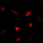
Cadherin‑12 in BG01V Human Embryonic Stem Cells.
Cadherin-12 was detected in immersion fixed BG01V human embryonic stem cells differentiated into neurons using Rat Anti-Human Cadherin-12 Monoclonal Antibody (Catalog # MAB2240) at 10 µg/mL for 3 hours at room temperature. Cells were stained using the NorthernLights™ 557-conjugated Anti-Rat IgG Secondary Antibody (red; Catalog # NL013) and counterstained with DAPI (blue). Specific staining was localized to cell membranes. View our protocol for Fluorescent ICC Staining of Stem Cells on Coverslips.Applications for Human Cadherin-12 Antibody (343621)
CyTOF-ready
Flow Cytometry
Sample:
Human neural progenitor cells differentiated by growth factor withdrawal
Note: Since classic Cadherins can be protected from trypsin treatment in the presence of Ca2+, cells in monolayer cultures are harvested with 0.01% Trypsin in the presence of 1-5 mM CaCl2 at 37° C. Flow cytometry can be performed according to the standard procedures, except that all the cell staining and washing steps are performed in the presence of Ca2+ and Mg2+ (e.g. using FACS buffer: PBS containing 1 mM CaCl2, 1 mM MgCl2, 2% FBS and 0.02% sodium azide).
Immunocytochemistry
Sample: Immersion fixed human neural progenitor cells differentiated by growth factor withdrawal and BG01V human embryonic stem cells differentiated into neurons
Western Blot
Sample: Recombinant Human Pro Cadherin‑12 Fc Chimera (Catalog # 2240-CA)
Reviewed Applications
Read 1 review rated 5 using MAB2240 in the following applications:
Flow Cytometry Panel Builder
Bio-Techne Knows Flow Cytometry
Save time and reduce costly mistakes by quickly finding compatible reagents using the Panel Builder Tool.
Advanced Features
- Spectra Viewer - Custom analysis of spectra from multiple fluorochromes
- Spillover Popups - Visualize the spectra of individual fluorochromes
- Antigen Density Selector - Match fluorochrome brightness with antigen density

Formulation, Preparation, and Storage
Purification
Reconstitution
Formulation
Shipping
Stability & Storage
- 12 months from date of receipt, -20 to -70 °C as supplied.
- 1 month, 2 to 8 °C under sterile conditions after reconstitution.
- 6 months, -20 to -70 °C under sterile conditions after reconstitution.
Calculators
Background: Cadherin-12
References
- Koch, A.W. et al. (2004) Cell. Mol. Life Sci. 61:1884.
- Angst, B.D. et al. (2001) J. Cell Sci. 114:629.
- Gessner, R. and R. Tauber (2000) Ann. N.Y. Acad. Sci. 915:136.
- Selig, S. et al. (1997) Proc. Natl. Acad. Sci. USA 94:2398.
- Tanihara, H. et al. (1994) Cell Adhes. Commun. 2:15.
- Shimoyama, Y. et al. (2000) Biochem. J. 349:159.
Alternate Names
Entrez Gene IDs
Gene Symbol
UniProt
Additional Cadherin-12 Products
Product Documents for Human Cadherin-12 Antibody (343621)
Certificate of Analysis
To download a Certificate of Analysis, please enter a lot or batch number in the search box below.
Note: Certificate of Analysis not available for kit components.
Product Specific Notices for Human Cadherin-12 Antibody (343621)
For research use only
Related Research Areas
Citations for Human Cadherin-12 Antibody (343621)
 Powered by Bioz
Customer Reviews for Human Cadherin-12 Antibody (343621) (1)
Have you used Human Cadherin-12 Antibody (343621)?
Submit a review and receive an Amazon gift card!
$25/€18/£15/$25CAN/¥2500 Yen for a review with an image
$10/€7/£6/$10CAN/¥1110 Yen for a review without an image
Submit a review
Customer Images
-
Application: Immunocytochemistry/ImmunofluorescenceSample Tested: Glioma cellsSpecies: HumanVerified Customer | Posted 10/07/2022

There are no reviews that match your criteria.
Protocols
Find general support by application which include: protocols, troubleshooting, illustrated assays, videos and webinars.
- 7-Amino Actinomycin D (7-AAD) Cell Viability Flow Cytometry Protocol
- Appropriate Fixation of IHC/ICC Samples
- Cellular Response to Hypoxia Protocols
- ClariTSA™ Fluorophore Kits
- Detection & Visualization of Antibody Binding
- Extracellular Membrane Flow Cytometry Protocol
- Flow Cytometry Protocol for Cell Surface Markers
- Flow Cytometry Protocol for Staining Membrane Associated Proteins
- Flow Cytometry Staining Protocols
- Flow Cytometry Troubleshooting Guide
- ICC Cell Smear Protocol for Suspension Cells
- ICC Immunocytochemistry Protocol Videos
- ICC for Adherent Cells
- Immunocytochemistry (ICC) Protocol
- Immunocytochemistry Troubleshooting
- Immunofluorescence of Organoids Embedded in Cultrex Basement Membrane Extract
- Immunohistochemistry (IHC) and Immunocytochemistry (ICC) Protocols
- Intracellular Flow Cytometry Protocol Using Alcohol (Methanol)
- Intracellular Flow Cytometry Protocol Using Detergents
- Intracellular Nuclear Staining Flow Cytometry Protocol Using Detergents
- Intracellular Staining Flow Cytometry Protocol Using Alcohol Permeabilization
- Intracellular Staining Flow Cytometry Protocol Using Detergents to Permeabilize Cells
- Preparing Samples for IHC/ICC Experiments
- Preventing Non-Specific Staining (Non-Specific Binding)
- Primary Antibody Selection & Optimization
- Propidium Iodide Cell Viability Flow Cytometry Protocol
- Protocol for Liperfluo
- Protocol for VisUCyte™ HRP Polymer Detection Reagent
- Protocol for the Characterization of Human Th22 Cells
- Protocol for the Characterization of Human Th9 Cells
- Protocol for the Fluorescent ICC Staining of Cell Smears - Graphic
- Protocol for the Fluorescent ICC Staining of Cultured Cells on Coverslips - Graphic
- Protocol for the Preparation and Fluorescent ICC Staining of Cells on Coverslips
- Protocol for the Preparation and Fluorescent ICC Staining of Non-adherent Cells
- Protocol for the Preparation and Fluorescent ICC Staining of Stem Cells on Coverslips
- Protocol for the Preparation of a Cell Smear for Non-adherent Cell ICC - Graphic
- Protocol: Annexin V and PI Staining by Flow Cytometry
- Protocol: Annexin V and PI Staining for Apoptosis by Flow Cytometry
- R&D Systems Quality Control Western Blot Protocol
- TUNEL and Active Caspase-3 Detection by IHC/ICC Protocol
- The Importance of IHC/ICC Controls
- Troubleshooting Guide: Fluorokine Flow Cytometry Kits
- Troubleshooting Guide: Western Blot Figures
- Western Blot Conditions
- Western Blot Protocol
- Western Blot Protocol for Cell Lysates
- Western Blot Troubleshooting
- Western Blot Troubleshooting Guide
- View all Protocols, Troubleshooting, Illustrated assays and Webinars
FAQs for Human Cadherin-12 Antibody (343621)
-
Q: Why does the staining protocol with this Cadherin antibody use buffers containing Ca2+ and Mg2+?
A: The staining protocol with this and other Cadherin antibodies uses buffer containing Ca2+ and Mg2+ because Cadherin function is Calcium-dependent.