
Human High-mobility group box 1 protein (HMGB1), previously known as HMG-1 or amphoterin, is a member of the high mobility group box family of non-histone chromosomal proteins (1‑3). Human HMGB1 is expressed as a 30 kDa, 215 amino acid (aa) single chain polypeptide containing three domains: two N-terminal globular, 70 aa positively charged DNA-binding domains (HMG boxes A and B), and a negatively charged 30 aa C-terminal region that contains only Asp and Glu (4, 5). Residues 27‑43 and 178‑184 contain a NLS. Posttranslational modifications of the molecule have been reported, with acetylation occurring on as many as 17 lysine residues (6). HMGB1 is expressed at high levels in almost all cells (2, 4). It was originally discovered as a nuclear protein that could bend DNA. Such bending stabilizes nucleosome formation and regulates the expression of select genes upon recruitment by DNA binding proteins (1, 7, 8). It is now known that HMGB1 can also act extracellularly, both as an inflammatory mediator that promotes monocyte migration and cytokine secretion, and as a mediator of T cell-dendritic cell interaction (1, 4, 7, 9, 10). The cytokine activity of HBMG1 is restricted to the HMG B box, (3) while the A box is associated with the helix-loop-helix domain of transcription factors (11). HMBG1 is released in response to cell death and as a secretion product. Although HMBG-1 does not possess a classic signal sequence, it appears to be secreted as an acetylated form via secretory endolysosome exocytosis (6, 12). Once secreted, HMGB1 transduces cellular signals through its high affinity receptor, RAGE and, possibly, TLR2 and TLR4 (1, 3, 4). Human HMGB1 is 100% aa identical to canine HMGB1 and 99% aa identical to mouse, rat, bovine and porcine HMGB1, respectively.
Human HMGB1/HMG-1 Antibody (115603) [PE]
R&D Systems | Catalog # IC1690P

Scientific images may be resized, cropped, or adjusted for brightness or contrast for presentation purposes. These updates are limited to presentation and do not affect the underlying scientific interpretation of the data. Where available, additional source data may be provided upon request.
Key Product Details
Species Reactivity
Validated:
Cited:
Applications
Validated:
Cited:
Label
Antibody Source
Product Specifications
Immunogen
Gly2-Glu215 (predicted)
Accession # P09429
Specificity
Clonality
Host
Isotype
Scientific Data Images for Human HMGB1/HMG-1 Antibody (115603) [PE]
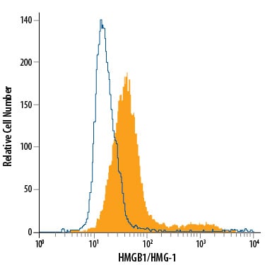
Detection of HMGB1/HMG-1 in HCT-116 Human Cell Line by Flow Cytometry.
HCT-116 human colorectal carcinoma cell line was stained with Mouse Anti-Human HMGB1/HMG-1 PE-conjugated Monoclonal Antibody (Catalog # IC1690P, filled histogram) or isotype control antibody (Catalog # IC0041P, open histogram). To facilitate intracellular staining, cells were fixed with Flow Cytometry Fixation Buffer (Catalog # FC004) and permeabilized with Flow Cytometry Permeabilization/Wash Buffer I (Catalog # FC005). View our protocol for Staining Intracellular Molecules.Applications for Human HMGB1/HMG-1 Antibody (115603) [PE]
Intracellular Staining by Flow Cytometry
Sample: HCT-116 human colorectal carcinoma cell line fixed with Flow Cytometry Fixation Buffer (Catalog # FC004) and permeabilized with Flow Cytometry Permeabilization/Wash Buffer I (Catalog # FC005).
Spectra Viewer
Plan Your Experiments
Use our spectra viewer to interactively plan your experiments, assessing multiplexing options. View the excitation and emission spectra for our fluorescent dye range and other commonly used dyes.
Spectra Viewer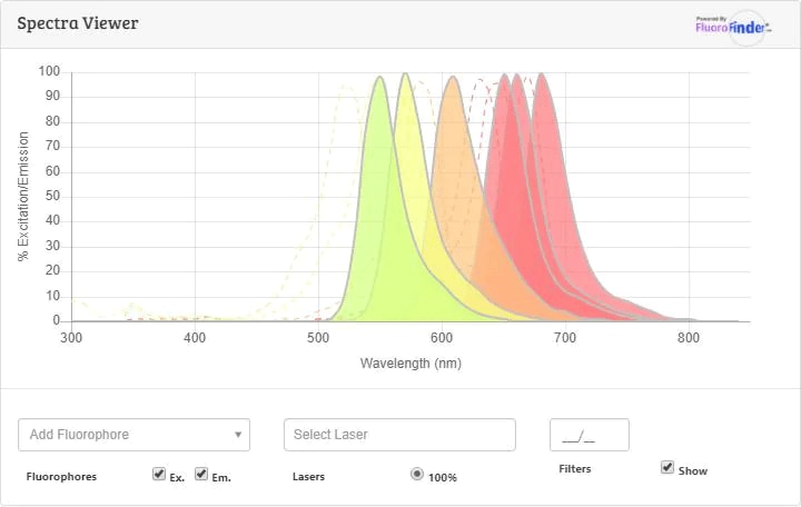
Flow Cytometry Panel Builder
Bio-Techne Knows Flow Cytometry
Save time and reduce costly mistakes by quickly finding compatible reagents using the Panel Builder Tool.
Advanced Features
- Spectra Viewer - Custom analysis of spectra from multiple fluorochromes
- Spillover Popups - Visualize the spectra of individual fluorochromes
- Antigen Density Selector - Match fluorochrome brightness with antigen density

Formulation, Preparation, and Storage
Purification
Formulation
Shipping
Stability & Storage
- 12 months from date of receipt, 2 to 8 °C as supplied.
Background: HMGB1/HMG-1
References
- Lotze, M.T. and K.J. Tracey (2005) Nat. Rev. Immunol. 5:331.
- Yang, H. et al. (2005) J. Leukoc. Biol. 78:1.
- Dumitriu, I.E. et al. (2005) Trends Immunol. 26:381.
- Degryse, B. and M. de Virgilio (2003) FEBS Lett. 553:11.
- Wen, L. et al. (1989) Nucleic Acids Res. 17:1197.
- Bonaldi, T. et al. (2003) EMBO J. 22:5551.
- Muller, S. et al. (2001) EMBO J. 20:4337.
- Bustin, M. (1999) Mol. Cell. Biol. 19:5237.
- Wang, H. et al. (1999) Science. 285:248.
- Dimitriu, I.E. et al. (2005) J. Immunol. 174:7506.
- Najima, Y. et al. (2005) J. Biol. Chem. 280:27523.
- Gardella, S. et al. (2002) EMBO Rep. 3:995.
Long Name
Alternate Names
Entrez Gene IDs
Gene Symbol
UniProt
Additional HMGB1/HMG-1 Products
Product Documents for Human HMGB1/HMG-1 Antibody (115603) [PE]
Certificate of Analysis
To download a Certificate of Analysis, please enter a lot or batch number in the search box below.
Note: Certificate of Analysis not available for kit components.
Product Specific Notices for Human HMGB1/HMG-1 Antibody (115603) [PE]
For research use only
Related Research Areas
Citations for Human HMGB1/HMG-1 Antibody (115603) [PE]
 Powered by Bioz
Customer Reviews for Human HMGB1/HMG-1 Antibody (115603) [PE]
There are currently no reviews for this product. Be the first to review Human HMGB1/HMG-1 Antibody (115603) [PE] and earn rewards!
Have you used Human HMGB1/HMG-1 Antibody (115603) [PE]?
Submit a review and receive an Amazon gift card!
$25/€18/£15/$25CAN/¥2500 for a review with an image
$10/€7/£6/$10CAN/¥1110 for a review without an image
Submit a review
Protocols
Find general support by application which include: protocols, troubleshooting, illustrated assays, videos and webinars.
- 7-Amino Actinomycin D (7-AAD) Cell Viability Flow Cytometry Protocol
- Extracellular Membrane Flow Cytometry Protocol
- Flow Cytometry Protocol for Cell Surface Markers
- Flow Cytometry Protocol for Staining Membrane Associated Proteins
- Flow Cytometry Staining Protocols
- Flow Cytometry Troubleshooting Guide
- Intracellular Flow Cytometry Protocol Using Alcohol (Methanol)
- Intracellular Flow Cytometry Protocol Using Detergents
- Intracellular Nuclear Staining Flow Cytometry Protocol Using Detergents
- Intracellular Staining Flow Cytometry Protocol Using Alcohol Permeabilization
- Intracellular Staining Flow Cytometry Protocol Using Detergents to Permeabilize Cells
- Propidium Iodide Cell Viability Flow Cytometry Protocol
- Protocol for Liperfluo
- Protocol for the Characterization of Human Th22 Cells
- Protocol for the Characterization of Human Th9 Cells
- Protocol: Annexin V and PI Staining by Flow Cytometry
- Protocol: Annexin V and PI Staining for Apoptosis by Flow Cytometry
- Troubleshooting Guide: Fluorokine Flow Cytometry Kits
- View all Protocols, Troubleshooting, Illustrated assays and Webinars