Human High-mobility group box 1 protein (HMGB1), previously known as HMG-1 or amphoterin, is a member of the high mobility group box family of non-histone chromosomal proteins (1‑3). Human HMGB1 is expressed as a 30 kDa, 215 amino acid (aa) single chain polypeptide containing three domains: two N-terminal globular, 70 aa positively charged DNA-binding domains (HMG boxes A and B), and a negatively charged 30 aa C-terminal region that contains only Asp and Glu (4, 5). Residues 27‑43 and 178‑184 contain a NLS. Posttranslational modifications of the molecule have been reported, with acetylation occurring on as many as 17 lysine residues (6). HMGB1 is expressed at high levels in almost all cells (2, 4). It was originally discovered as a nuclear protein that could bend DNA. Such bending stabilizes nucleosome formation and regulates the expression of select genes upon recruitment by DNA binding proteins (1, 7, 8). It is now known that HMGB1 can also act extracellularly, both as an inflammatory mediator that promotes monocyte migration and cytokine secretion, and as a mediator of T cell-dendritic cell interaction (1, 4, 7, 9, 10). The cytokine activity of HBMG1 is restricted to the HMG B box, (3) while the A box is associated with the helix-loop-helix domain of transcription factors (11). HMBG1 is released in response to cell death and as a secretion product. Although HMBG-1 does not possess a classic signal sequence, it appears to be secreted as an acetylated form via secretory endolysosome exocytosis (6, 12). Once secreted, HMGB1 transduces cellular signals through its high affinity receptor, RAGE and, possibly, TLR2 and TLR4 (1, 3, 4). Human HMGB1 is 100% aa identical to canine HMGB1 and 99% aa identical to mouse, rat, bovine and porcine HMGB1, respectively.
Human HMGB1/HMG-1 Antibody (115603)
R&D Systems | Catalog # MAB1690

Key Product Details
Validated by
Biological Validation
Species Reactivity
Validated:
Human
Cited:
Human, Mouse, Rat
Applications
Validated:
Immunohistochemistry, Western Blot, Intracellular Staining by Flow Cytometry, Immunocytochemistry, Chromatin Immunoprecipitation (ChIP), CyTOF-ready
Cited:
Immunohistochemistry, Immunohistochemistry-Paraffin, Western Blot, ELISA, Neutralization, Immunocytochemistry, Immunoprecipitation, ELISA Development, ELISA Standard, In vivo assay, Immunodepletion
Label
Unconjugated
Antibody Source
Monoclonal Mouse IgG2B Clone # 115603
Loading...
Product Specifications
Immunogen
E. coli-derived recombinant human HMGB1/HMG-1
Gly2-Glu215 (predicted)
Accession # P09429
Gly2-Glu215 (predicted)
Accession # P09429
Specificity
Detects human HMGB1/HMG-1 in direct ELISAs and Western blots.
Clonality
Monoclonal
Host
Mouse
Isotype
IgG2B
Scientific Data Images for Human HMGB1/HMG-1 Antibody (115603)
Detection of Human HMGB1/HMG‑1 by Western Blot.
Western blot shows lysates of HepG2 human hepatocellular carcinoma cell line, Jurkat human acute T cell leukemia cell line, and HEK293 human embryonic kidney cell line. PVDF membrane was probed with 0.05 µg/mL of Mouse Anti-Human HMGB1/HMG-1 Monoclonal Antibody (Catalog # MAB1690) followed by HRP-conjugated Anti-Mouse IgG Secondary Antibody (Catalog # HAF018). A specific band was detected for HMGB1/HMG-1 at approximately 26 kDa (as indicated). This experiment was conducted under reducing conditions and using Immunoblot Buffer Group 1.
Detection of HMGB1/HMG‑1-regulated Genes by Chromatin Immunoprecipitation.
HepG2 human hepatocellular carcinoma cell line was fixed using formaldehyde, resuspended in lysis buffer, and sonicated to shear chromatin. HMGB1/HMG-1/DNA complexes were immunoprecipitated using 5 µg Mouse Anti-Human HMGB1/HMG-1 PE-conjugated Monoclonal Antibody (Catalog # MAB1690) or control antibody (Catalog # MAB004) for 15 minutes in an ultrasonic bath, followed by Biotinylated Anti-Mouse IgG Secondary Antibody (Catalog # BAF007). Immunocomplexes were captured using 50 µL of MagCellect Streptavidin Ferrofluid (Catalog # MAG999) and DNA was purified using chelating resin solution. TheAFPpromoter was detected by standard PCR.
Detection of HMGB1 in HCT‑116 Human Cell Line by Flow Cytometry.
HCT-116 human colorectal carcinoma cell line was stained with Mouse Anti-Human HMGB1/HMG-1 Monoclonal Antibody (Catalog # MAB1690, filled histogram) or isotype control antibody (Catalog # MAB0041, open histogram), followed by Phycoerythrin-conjugated Anti-Mouse IgG F(ab')2Secondary Antibody (Catalog # F0102B). To facilitate intracellular staining, cells were fixed with Flow Cytometry Fixation Buffer (Catalog # FC004) and permeabilized with Flow Cytometry Permeabilization/Wash Buffer I (Catalog # FC005). View our protocol for Staining Intracellular Molecules.
HMGB1/HMG‑1 in CCD‑18Co Human Cell Line.
HMGB1/HMG-1 was detected in immersion fixed CCD-18Co human colonic fibroblast cell line using Mouse Anti-Human HMGB1/HMG-1 Monoclonal Antibody (Catalog # MAB1690) at 10 µg/mL for 3 hours at room temperature. Cells were stained using the NorthernLights™ 557-conjugated Anti-Mouse IgG Secondary Antibody (red, upper panel; Catalog # NL007) and counterstained with DAPI (blue, lower panel). Specific staining was localized to nuclei. View our protocol for Fluorescent ICC Staining of Cells on Coverslips.
HMGB1/HMG-1 in Human Breast Cancer Tissue.
HMGB1/HMG-1 was detected in immersion fixed paraffin-embedded sections of human breast cancer tissue using Mouse Anti-Human HMGB1/HMG-1 Monoclonal Antibody (Catalog # MAB1690) at 5 µg/mL overnight at 4 °C. Before incubation with the primary antibody, tissue was subjected to heat-induced epitope retrieval using Antigen Retrieval Reagent-Basic (Catalog # CTS013). Tissue was stained using the Anti-Mouse IgG VisUCyte™ HRP Polymer Antibody (brown; Catalog # VC001) and counterstained with hematoxylin (blue). Specific staining was localized to cell nuclei. View our protocol for IHC Staining with VisUCyte HRP Polymer Detection Reagents.
Detection of Rat HMGB1/HMG-1 by Western Blot
The effect of corticosterone on high-mobility group box-1 (HMGB1) release in primary cultured cortical astrocytes. (A) Increasing concentrations of corticosterone induced HMGB1 release from astrocytes. Cells were treated with the indicated concentrations of corticosterone for 24 h, and the amount of HMGB1 in the media was analyzed by Western blotting. Representative blots are shown (HMGB1: 30 kDa). The data are expressed as the mean ± SEM. * p < 0.05, ** p < 0.01 vs. basal (concentration “0”) (Dunnett’s test; n = 7–15). (B) Astrocytes were treated with 1 μM corticosterone for the indicated periods of time and the amount of HMGB1 in the media was analyzed by Western blotting. Representative blots are shown (HMGB1: 30 kDa). The data are expressed as the mean ± SEM. * p < 0.05, ** p < 0.01 vs. basal (time “0”) (Dunnett’s test; n = 3–15). (C) Astrocytes were treated with 1 μM corticosterone for 24 h, and media concentrations of HMGB1 were analyzed by ELISA. The data are expressed as the mean ± SEM. * p < 0.05 vs. vehicle (t-test; n = 5). Image collected and cropped by CiteAb from the following publication (https://pubmed.ncbi.nlm.nih.gov/32344830), licensed under a CC-BY license. Not internally tested by R&D Systems.
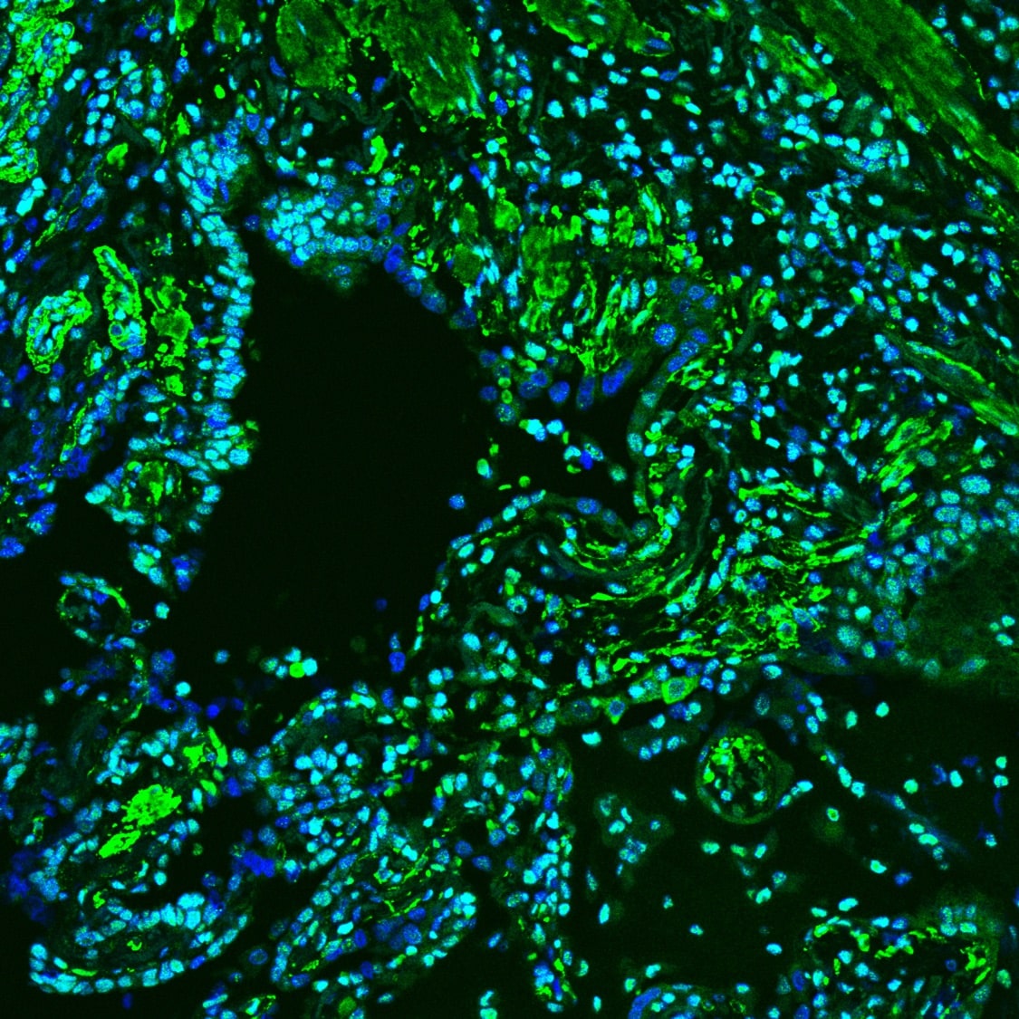
Detection of Human HMGB1/HMG-1 by Immunohistochemistry
RAGE and HMGB1 are expressed in myofibroblasts of endarterectomised chronic thromboembolic tissues of CTEPH patients.One representative patient is shown (12 out of 15 patients (80%) displayed analogous staining patterns. Photograph showing the macroscopic aspect of a representative PEA specimen (A). Scale bar: 6 cm. Immunohistochemical expression of RAGE (B, scale bar: 20 µm), HMGB1 (C), vimentin (D), alpha-smooth muscle actin (E) and CD34 (F) on adjacent tissue sections of the PEA specimen shown in (A). Scale bar: 40 µm. RAGE receptor for advanced glycation endproducts, HMGB1 high mobility group box 1, CTEPH chronic thromboembolic pulmonary hypertension, PEA pulmonary endarterectomy. Image collected and cropped by CiteAb from the following publication (https://dx.plos.org/10.1371/journal.pone.0106440), licensed under a CC-BY license. Not internally tested by R&D Systems.Applications for Human HMGB1/HMG-1 Antibody (115603)
Application
Recommended Usage
Chromatin Immunoprecipitation (ChIP)
5 µg/5 x 106 cells
Sample: HepG2 human hepatocellular carcinoma cell line chromatin, AFP promoter detected by standard PCR
Sample: HepG2 human hepatocellular carcinoma cell line chromatin, AFP promoter detected by standard PCR
CyTOF-ready
Ready to be labeled using established conjugation methods. No BSA or other carrier proteins that could interfere with conjugation.
Immunocytochemistry
8-25 µg/mL
Sample: Immersion-fixed CCD-18Co human colonic fibroblast cell line and MCF-7 human breast cancer cell line
Sample: Immersion-fixed CCD-18Co human colonic fibroblast cell line and MCF-7 human breast cancer cell line
Immunohistochemistry
5-25 µg/mL
Sample: Immersion fixed paraffin-embedded sections of human breast cancer tissue
Sample: Immersion fixed paraffin-embedded sections of human breast cancer tissue
Intracellular Staining by Flow Cytometry
2.5 µg/106 cells
Sample: HCT‑116 human colorectal carcinoma cell line fixed with Flow Cytometry Fixation Buffer (Catalog # FC004) and permeabilized with Flow Cytometry Permeabilization/Wash Buffer I (Catalog # FC005)
Sample: HCT‑116 human colorectal carcinoma cell line fixed with Flow Cytometry Fixation Buffer (Catalog # FC004) and permeabilized with Flow Cytometry Permeabilization/Wash Buffer I (Catalog # FC005)
Western Blot
0.05 µg/mL
Sample: HepG2 human hepatocellular carcinoma cell line, Jurkat human acute T cell leukemia cell line, and HEK293 human embryonic kidney cell line
Sample: HepG2 human hepatocellular carcinoma cell line, Jurkat human acute T cell leukemia cell line, and HEK293 human embryonic kidney cell line
Reviewed Applications
Read 3 reviews rated 4.3 using MAB1690 in the following applications:
Flow Cytometry Panel Builder
Bio-Techne Knows Flow Cytometry
Save time and reduce costly mistakes by quickly finding compatible reagents using the Panel Builder Tool.
Advanced Features
- Spectra Viewer - Custom analysis of spectra from multiple fluorochromes
- Spillover Popups - Visualize the spectra of individual fluorochromes
- Antigen Density Selector - Match fluorochrome brightness with antigen density

Formulation, Preparation, and Storage
Purification
Protein A or G purified from hybridoma culture supernatant
Reconstitution
Reconstitute at 0.5 mg/mL in sterile PBS. For liquid material, refer to CoA for concentration.
Loading...
Formulation
Lyophilized from a 0.2 μm filtered solution in PBS with Trehalose. *Small pack size (SP) is supplied either lyophilized or as a 0.2 µm filtered solution in PBS.
Shipping
Lyophilized product is shipped at ambient temperature. Liquid small pack size (-SP) is shipped with polar packs. Upon receipt, store immediately at the temperature recommended below.
Stability & Storage
Use a manual defrost freezer and avoid repeated freeze-thaw cycles.
- 12 months from date of receipt, -20 to -70 °C as supplied.
- 1 month, 2 to 8 °C under sterile conditions after reconstitution.
- 6 months, -20 to -70 °C under sterile conditions after reconstitution.
Calculators
Background: HMGB1/HMG-1
References
- Lotze, M.T. and K.J. Tracey (2005) Nat. Rev. Immunol. 5:331.
- Yang, H. et al. (2005) J. Leukoc. Biol. 78:1.
- Dumitriu, I.E. et al. (2005) Trends Immunol. 26:381.
- Degryse, B. and M. de Virgilio (2003) FEBS Lett. 553:11.
- Wen, L. et al. (1989) Nucleic Acids Res. 17:1197.
- Bonaldi, T. et al. (2003) EMBO J. 22:5551.
- Muller, S. et al. (2001) EMBO J. 20:4337.
- Bustin, M. (1999) Mol. Cell. Biol. 19:5237.
- Wang, H. et al. (1999) Science. 285:248.
- Dimitriu, I.E. et al. (2005) J. Immunol. 174:7506.
- Najima, Y. et al. (2005) J. Biol. Chem. 280:27523.
- Gardella, S. et al. (2002) EMBO Rep. 3:995.
Long Name
High Mobility Group Protein 1
Alternate Names
HMG-1, HMG1
Entrez Gene IDs
3146 (Human)
Gene Symbol
HMGB1
UniProt
Additional HMGB1/HMG-1 Products
Product Documents for Human HMGB1/HMG-1 Antibody (115603)
Certificate of Analysis
To download a Certificate of Analysis, please enter a lot or batch number in the search box below.
Note: Certificate of Analysis not available for kit components.
Product Specific Notices for Human HMGB1/HMG-1 Antibody (115603)
For research use only
Related Research Areas
Citations for Human HMGB1/HMG-1 Antibody (115603)
 Powered by Bioz
Customer Reviews for Human HMGB1/HMG-1 Antibody (115603) (3)
4.3 out of 5
3 Customer Ratings
Have you used Human HMGB1/HMG-1 Antibody (115603)?
Submit a review and receive an Amazon gift card!
$25/€18/£15/$25CAN/¥2500 Yen for a review with an image
$10/€7/£6/$10CAN/¥1110 Yen for a review without an image
Submit a review
Customer Images

Showing
1
-
3 of
3 reviews
Showing All
Filter By:
-
Application: Western BlotSample Tested: Cancer cell lysatesSpecies: HumanVerified Customer | Posted 10/17/2018

-
Application: Immunocytochemistry/ImmunofluorescenceSample Tested: fibrotic lungSpecies: MouseVerified Customer | Posted 10/13/2016
-
Application: Western BlotSample Tested: Cell lysateSpecies: HumanVerified Customer | Posted 01/23/2016I should say this is a good product. I purchased the small size, detected HMGB1 expression after TNF-a challenged. It is helpful.
There are no reviews that match your criteria.
Protocols
Find general support by application which include: protocols, troubleshooting, illustrated assays, videos and webinars.
- 7-Amino Actinomycin D (7-AAD) Cell Viability Flow Cytometry Protocol
- Antigen Retrieval Protocol (PIER)
- Antigen Retrieval for Frozen Sections Protocol
- Appropriate Fixation of IHC/ICC Samples
- Cellular Response to Hypoxia Protocols
- ChIP Protocol Video
- Chromatin Immunoprecipitation (ChIP) Protocol
- Chromatin Immunoprecipitation Protocol
- Chromogenic IHC Staining of Formalin-Fixed Paraffin-Embedded (FFPE) Tissue Protocol
- Chromogenic Immunohistochemistry Staining of Frozen Tissue
- ClariTSA™ Fluorophore Kits
- Detection & Visualization of Antibody Binding
- Extracellular Membrane Flow Cytometry Protocol
- Flow Cytometry Protocol for Cell Surface Markers
- Flow Cytometry Protocol for Staining Membrane Associated Proteins
- Flow Cytometry Staining Protocols
- Flow Cytometry Troubleshooting Guide
- Fluorescent IHC Staining of Frozen Tissue Protocol
- Graphic Protocol for Heat-induced Epitope Retrieval
- Graphic Protocol for the Preparation and Fluorescent IHC Staining of Frozen Tissue Sections
- Graphic Protocol for the Preparation and Fluorescent IHC Staining of Paraffin-embedded Tissue Sections
- Graphic Protocol for the Preparation of Gelatin-coated Slides for Histological Tissue Sections
- ICC Cell Smear Protocol for Suspension Cells
- ICC Immunocytochemistry Protocol Videos
- ICC for Adherent Cells
- IHC Sample Preparation (Frozen sections vs Paraffin)
- Immunocytochemistry (ICC) Protocol
- Immunocytochemistry Troubleshooting
- Immunofluorescence of Organoids Embedded in Cultrex Basement Membrane Extract
- Immunofluorescent IHC Staining of Formalin-Fixed Paraffin-Embedded (FFPE) Tissue Protocol
- Immunohistochemistry (IHC) and Immunocytochemistry (ICC) Protocols
- Immunohistochemistry Frozen Troubleshooting
- Immunohistochemistry Paraffin Troubleshooting
- Intracellular Flow Cytometry Protocol Using Alcohol (Methanol)
- Intracellular Flow Cytometry Protocol Using Detergents
- Intracellular Nuclear Staining Flow Cytometry Protocol Using Detergents
- Intracellular Staining Flow Cytometry Protocol Using Alcohol Permeabilization
- Intracellular Staining Flow Cytometry Protocol Using Detergents to Permeabilize Cells
- Preparing Samples for IHC/ICC Experiments
- Preventing Non-Specific Staining (Non-Specific Binding)
- Primary Antibody Selection & Optimization
- Propidium Iodide Cell Viability Flow Cytometry Protocol
- Protocol for Heat-Induced Epitope Retrieval (HIER)
- Protocol for Liperfluo
- Protocol for Making a 4% Formaldehyde Solution in PBS
- Protocol for VisUCyte™ HRP Polymer Detection Reagent
- Protocol for the Characterization of Human Th22 Cells
- Protocol for the Characterization of Human Th9 Cells
- Protocol for the Fluorescent ICC Staining of Cell Smears - Graphic
- Protocol for the Fluorescent ICC Staining of Cultured Cells on Coverslips - Graphic
- Protocol for the Preparation & Fixation of Cells on Coverslips
- Protocol for the Preparation and Chromogenic IHC Staining of Frozen Tissue Sections
- Protocol for the Preparation and Chromogenic IHC Staining of Frozen Tissue Sections - Graphic
- Protocol for the Preparation and Chromogenic IHC Staining of Paraffin-embedded Tissue Sections
- Protocol for the Preparation and Chromogenic IHC Staining of Paraffin-embedded Tissue Sections - Graphic
- Protocol for the Preparation and Fluorescent ICC Staining of Cells on Coverslips
- Protocol for the Preparation and Fluorescent ICC Staining of Non-adherent Cells
- Protocol for the Preparation and Fluorescent ICC Staining of Stem Cells on Coverslips
- Protocol for the Preparation and Fluorescent IHC Staining of Frozen Tissue Sections
- Protocol for the Preparation and Fluorescent IHC Staining of Paraffin-embedded Tissue Sections
- Protocol for the Preparation of Gelatin-coated Slides for Histological Tissue Sections
- Protocol for the Preparation of a Cell Smear for Non-adherent Cell ICC - Graphic
- Protocol: Annexin V and PI Staining by Flow Cytometry
- Protocol: Annexin V and PI Staining for Apoptosis by Flow Cytometry
- R&D Systems Quality Control Western Blot Protocol
- TUNEL and Active Caspase-3 Detection by IHC/ICC Protocol
- The Importance of IHC/ICC Controls
- Troubleshooting Guide: Fluorokine Flow Cytometry Kits
- Troubleshooting Guide: Immunohistochemistry
- Troubleshooting Guide: Western Blot Figures
- Western Blot Conditions
- Western Blot Protocol
- Western Blot Protocol for Cell Lysates
- Western Blot Troubleshooting
- Western Blot Troubleshooting Guide
- View all Protocols, Troubleshooting, Illustrated assays and Webinars
Loading...