Laminins are heterotrimeric, noncollagenous glycoproteins composed of alpha, beta and gamma chains. Through interactions with Integrins, Dystroglycan and other receptors, Laminins contribute to cell differentiation, cell shape and migration, and maintenance of tissue phenotypes and survival (1). Laminin-5, also known as Epiligrin, consists of an alpha 3, beta 3, and gamma 2 chain. It is abundant in transitional epithelium, stratified squamous epithelia, lung mucosa and other epithelial glands and plays an important role in the initiation and maintenance of epithelial cell anchorage to the underlying connective tissue (2, 3).
Human Laminin alpha 3/Laminin-5 Antibody (P3H9-2)
R&D Systems | Catalog # MAB2144

Discontinued Product
MAB2144 has been discontinued.
View all Laminin alpha 3/Laminin-5 products.
Key Product Details
Species Reactivity
Validated:
Human
Cited:
Human, Mouse
Applications
Validated:
Immunohistochemistry, Western Blot, Adhesion Blockade, Immunocytochemistry, Immunoprecipitation
Cited:
Immunohistochemistry, Neutralization, Immunocytochemistry/ Immunofluorescence
Label
Unconjugated
Antibody Source
Monoclonal Mouse IgG1 Clone # P3H9-2
Loading...
Product Specifications
Immunogen
Human keratinocytes
Specificity
Detects human Laminin alpha 3/Laminin‑5 in Western blots.
Clonality
Monoclonal
Host
Mouse
Isotype
IgG1
Scientific Data Images for Human Laminin alpha 3/Laminin-5 Antibody (P3H9-2)
Laminin alpha 3/Laminin-5 in MCF 10A Human Cell Line.
Laminin a3/Laminin-5 was detected in immersion fixed MCF 10A human breast epithelial cell line using 10 µg/mL Mouse Anti-Human Laminin a3/Laminin-5 Monoclonal Antibody (Catalog # MAB2144) for 3 hours at room temperature. Cells were stained with the NorthernLights™ 557-conjugated Anti-Mouse IgG Secondary Antibody (red; Catalog # NL007) and counterstained with DAPI (blue). View our protocol for Fluorescent ICC Staining of Cells on Coverslips.
Laminin alpha 3/Laminin‑5 in Human Skin.
Laminin a3/Laminin-5 was detected in immersion fixed paraffin-embedded sections of human skin using Mouse Anti-Human Laminin a3/Laminin-5 Monoclonal Antibody (Catalog # MAB2144) at 25 µg/mL overnight at 4 °C. Tissue was stained using the Anti-Mouse HRP-DAB Cell & Tissue Staining Kit (brown; Catalog # CTS002) and counterstained with hematoxylin (blue). Specific labeling was localized to cell in the epiderimis. View our protocol for Chromogenic IHC Staining of Paraffin-embedded Tissue Sections.Applications for Human Laminin alpha 3/Laminin-5 Antibody (P3H9-2)
Application
Recommended Usage
Adhesion Blockade
Gonzales, M. et al. (1999) Mol. Biol. Cell 10:259.
Immunocytochemistry
8-25 µg/mL
Sample: Immersion fixed MCF 10A human breast epithelial cell line
Sample: Immersion fixed MCF 10A human breast epithelial cell line
Immunohistochemistry
8-25 µg/mL
Sample: Immersion fixed paraffin-embedded sections of human skin
Sample: Immersion fixed paraffin-embedded sections of human skin
Immunoprecipitation
Gallatin, W.M. et al. (1989) Proc. Natl. Acad. Sci. 86:4654.
Western Blot
Wayner, E.A. (1993) J. Cell Biol. 121(5):1141; Gallatin, W.M. et al. (1989) Proc. Natl. Acad. Sci. USA 86(12):4654. This application was not tested by R&D Systems.
Reviewed Applications
Read 3 reviews rated 4.7 using MAB2144 in the following applications:
Formulation, Preparation, and Storage
Purification
Protein A or G purified from hybridoma culture supernatant
Reconstitution
Reconstitute at 0.5 mg/mL in sterile PBS. For liquid material, refer to CoA for concentration.
Formulation
Lyophilized from a 0.2 μm filtered solution in PBS with Trehalose. *Small pack size (SP) is supplied either lyophilized or as a 0.2 µm filtered solution in PBS.
Shipping
Lyophilized product is shipped at ambient temperature. Liquid small pack size (-SP) is shipped with polar packs. Upon receipt, store immediately at the temperature recommended below.
Stability & Storage
Use a manual defrost freezer and avoid repeated freeze-thaw cycles.
- 12 months from date of receipt, -20 to -70 °C as supplied.
- 1 month, 2 to 8 °C under sterile conditions after reconstitution.
- 6 months, -20 to -70 °C under sterile conditions after reconstitution.
Calculators
Background: Laminin alpha 3/Laminin-5
References
- Colognato, H. et al. (2000) Dev. Dyn. 218:213.
- Stahl, S. et al. (1997) J. Cell Sci. 110:55.
- Gonzales, M. et al. (1999) Mol. Biol. Cell 10:259.
Alternate Names
Epiligrin, LAMA3, Laminin-5, Laminin5
Entrez Gene IDs
3909 (Human)
Gene Symbol
LAMA3
Additional Laminin alpha 3/Laminin-5 Products
Product Documents for Human Laminin alpha 3/Laminin-5 Antibody (P3H9-2)
Certificate of Analysis
To download a Certificate of Analysis, please enter a lot or batch number in the search box below.
Note: Certificate of Analysis not available for kit components.
Product Specific Notices for Human Laminin alpha 3/Laminin-5 Antibody (P3H9-2)
For research use only
Related Research Areas
Citations for Human Laminin alpha 3/Laminin-5 Antibody (P3H9-2)
 Powered by Bioz
Customer Reviews for Human Laminin alpha 3/Laminin-5 Antibody (P3H9-2) (3)
4.7 out of 5
3 Customer Ratings
Have you used Human Laminin alpha 3/Laminin-5 Antibody (P3H9-2)?
Submit a review and receive an Amazon gift card!
$25/€18/£15/$25CAN/¥2500 Yen for a review with an image
$10/€7/£6/$10CAN/¥1110 Yen for a review without an image
Submit a review
Customer Images
Showing
1
-
3 of
3 reviews
Showing All
Filter By:
-
Application: Western BlotSample Tested: Colonic cancer cellsSpecies: HumanVerified Customer | Posted 07/22/2021

-
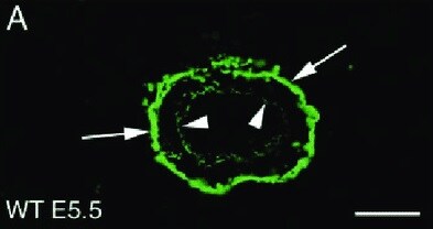
Application: ImmunohistochemistrySample Tested: Embryonic basement membraneSpecies: HumanVerified Customer | Posted 07/08/2021
-
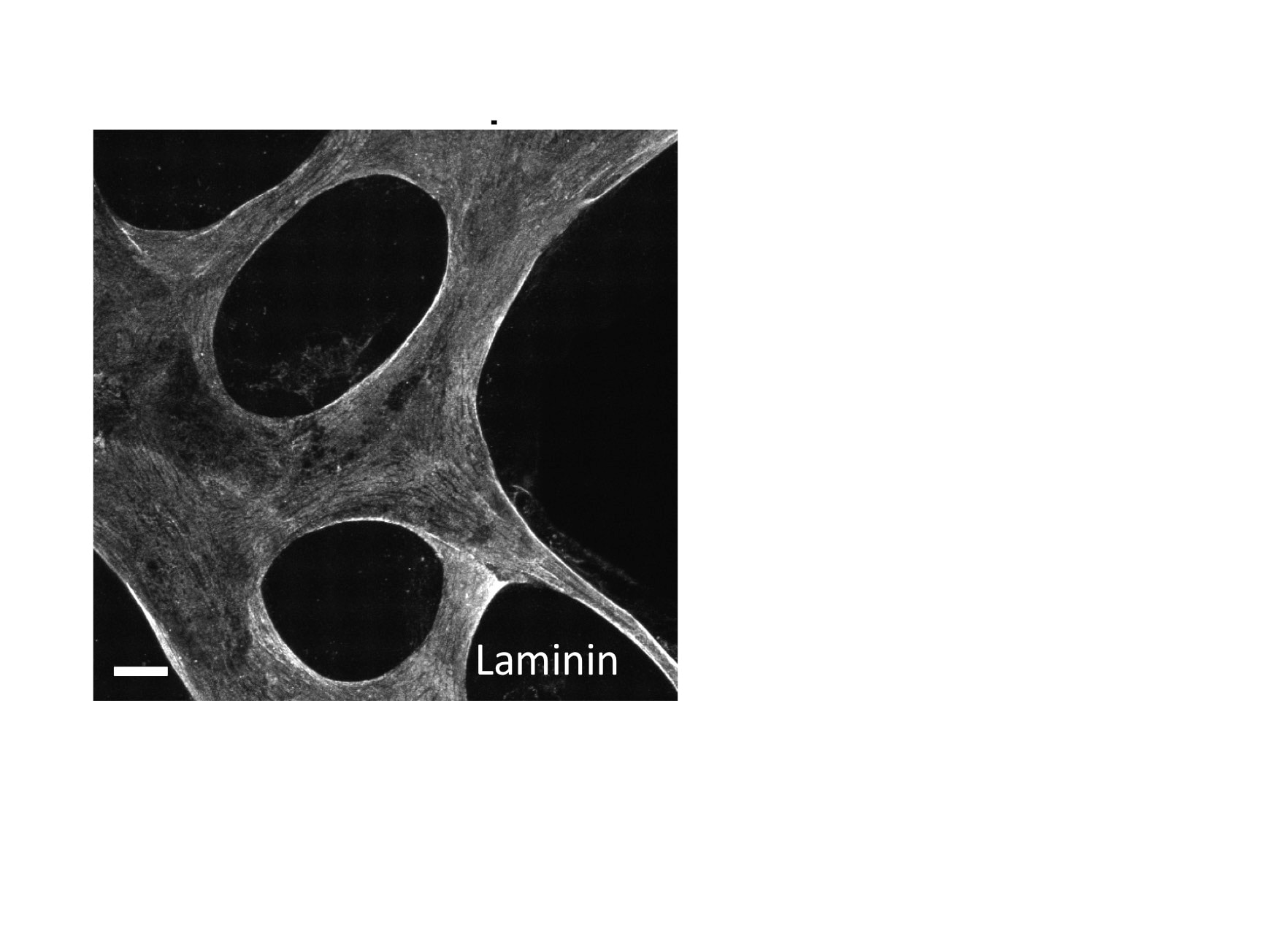
Application: Immunocytochemistry/ImmunofluorescenceSample Tested: HUVEC human umbilical vein endothelial cellsSpecies: HumanVerified Customer | Posted 04/26/2017
There are no reviews that match your criteria.
Protocols
Find general support by application which include: protocols, troubleshooting, illustrated assays, videos and webinars.
- Antigen Retrieval Protocol (PIER)
- Antigen Retrieval for Frozen Sections Protocol
- Appropriate Fixation of IHC/ICC Samples
- Cellular Response to Hypoxia Protocols
- Chromogenic IHC Staining of Formalin-Fixed Paraffin-Embedded (FFPE) Tissue Protocol
- Chromogenic Immunohistochemistry Staining of Frozen Tissue
- ClariTSA™ Fluorophore Kits
- Detection & Visualization of Antibody Binding
- Fluorescent IHC Staining of Frozen Tissue Protocol
- Graphic Protocol for Heat-induced Epitope Retrieval
- Graphic Protocol for the Preparation and Fluorescent IHC Staining of Frozen Tissue Sections
- Graphic Protocol for the Preparation and Fluorescent IHC Staining of Paraffin-embedded Tissue Sections
- Graphic Protocol for the Preparation of Gelatin-coated Slides for Histological Tissue Sections
- ICC Cell Smear Protocol for Suspension Cells
- ICC Immunocytochemistry Protocol Videos
- ICC for Adherent Cells
- IHC Sample Preparation (Frozen sections vs Paraffin)
- Immunocytochemistry (ICC) Protocol
- Immunocytochemistry Troubleshooting
- Immunofluorescence of Organoids Embedded in Cultrex Basement Membrane Extract
- Immunofluorescent IHC Staining of Formalin-Fixed Paraffin-Embedded (FFPE) Tissue Protocol
- Immunohistochemistry (IHC) and Immunocytochemistry (ICC) Protocols
- Immunohistochemistry Frozen Troubleshooting
- Immunohistochemistry Paraffin Troubleshooting
- Immunoprecipitation Protocol
- Preparing Samples for IHC/ICC Experiments
- Preventing Non-Specific Staining (Non-Specific Binding)
- Primary Antibody Selection & Optimization
- Protocol for Heat-Induced Epitope Retrieval (HIER)
- Protocol for Making a 4% Formaldehyde Solution in PBS
- Protocol for VisUCyte™ HRP Polymer Detection Reagent
- Protocol for the Fluorescent ICC Staining of Cell Smears - Graphic
- Protocol for the Fluorescent ICC Staining of Cultured Cells on Coverslips - Graphic
- Protocol for the Preparation & Fixation of Cells on Coverslips
- Protocol for the Preparation and Chromogenic IHC Staining of Frozen Tissue Sections
- Protocol for the Preparation and Chromogenic IHC Staining of Frozen Tissue Sections - Graphic
- Protocol for the Preparation and Chromogenic IHC Staining of Paraffin-embedded Tissue Sections
- Protocol for the Preparation and Chromogenic IHC Staining of Paraffin-embedded Tissue Sections - Graphic
- Protocol for the Preparation and Fluorescent ICC Staining of Cells on Coverslips
- Protocol for the Preparation and Fluorescent ICC Staining of Non-adherent Cells
- Protocol for the Preparation and Fluorescent ICC Staining of Stem Cells on Coverslips
- Protocol for the Preparation and Fluorescent IHC Staining of Frozen Tissue Sections
- Protocol for the Preparation and Fluorescent IHC Staining of Paraffin-embedded Tissue Sections
- Protocol for the Preparation of Gelatin-coated Slides for Histological Tissue Sections
- Protocol for the Preparation of a Cell Smear for Non-adherent Cell ICC - Graphic
- R&D Systems Quality Control Western Blot Protocol
- TUNEL and Active Caspase-3 Detection by IHC/ICC Protocol
- The Importance of IHC/ICC Controls
- Troubleshooting Guide: Immunohistochemistry
- Troubleshooting Guide: Western Blot Figures
- Western Blot Conditions
- Western Blot Protocol
- Western Blot Protocol for Cell Lysates
- Western Blot Troubleshooting
- Western Blot Troubleshooting Guide
- View all Protocols, Troubleshooting, Illustrated assays and Webinars
Loading...
Associated Pathways

