The immunoglobulin-like transcript (ILT) family of activating and inhibitory type immunoreceptors are expressed on many leukocyte subsets and function in the regulation of immune responses (1‑3). This family was also named leukocyte Ig-like receptors (LIR) and monocyte/macrophage Ig-like receptors (MIR). ILTs share significant homology with killer cell Ig-like receptors (KIR). The ILT genes are located on human chromosome 19q13.4 in the leukocyte receptor complex, which also include the genes encoding KIRs (4). With the exception of ILT-6, which is a soluble molecule, all ILT family members are type I transmembrane proteins having two or four extracellular Ig-like domains (2, 3). One subset of the ILT receptors (referred to as subfamily B of the LIRs) has long cytoplasmic tails containing immunoreceptor tyrosine-based inhibitory motifs (ITIMs) that inhibit signaling events by recruiting SH2-containing protein tyrosine phosphatase-1. Another subset of the ILT receptors (referred to as subfamily A of the LIRs) contains activating receptors with short cytoplasmic regions that lack signal transduction motifs. These receptors contain a basic arginine residue within their transmembrane domains, which allows association with Fc R gamma, an immunoreceptor tyrosine-based activation motif (ITAM)-bearing signal adapter protein (1‑3). ILT2, also known as LIR1, MIR7, and CD85j, is expressed on most monocytes, dendritic cells, and mature B cells (1‑3). It is also expressed on small percentages of T cells and NK cells. ILT2 has four extracellular Ig-like domains and three cytoplasmic ITIMs. It functions as an inhibitory receptor that prevents cellular activation. ILT2 has been shown to bind classical (HLA-A and -B) and nonclassical (HLA-G1, -E and -F) MHC class I molecules (MHCI) (1‑3). ILT2 also binds with high affinity to an MHC class I homologue from human cytomegalovirus (3). Ligation of ILT2 by MHC class I may function to poise cellular activation thresholds and inhibit various leukocyte effector mechanisms that are regulated by MHC class I molecules on target cells.
Human LILRB1/CD85j/ILT2 APC‑conjugated Antibody
R&D Systems | Catalog # FAB20171A

Key Product Details
Species Reactivity
Validated:
Cited:
Applications
Validated:
Cited:
Label
Antibody Source
Product Specifications
Immunogen
Gly24-His458
Accession # Q8NHL6
Specificity
Clonality
Host
Isotype
Scientific Data Images for Human LILRB1/CD85j/ILT2 APC‑conjugated Antibody
Detection of LILRB1/CD85j/ILT2 in Human Blood Monocytes by Flow Cytometry.
Human peripheral blood monocytes were stained with Mouse Anti-Human LILRB1/CD85j/ILT2 APC-conjugated Monoclonal Antibody (Catalog # FAB20171A, filled histogram) or isotype control antibody (Catalog # IC002A, open histogram). View our protocol for Staining Membrane-associated Proteins.Applications for Human LILRB1/CD85j/ILT2 APC‑conjugated Antibody
Flow Cytometry
Sample: Human peripheral blood monocytes
Reviewed Applications
Read 2 reviews rated 5 using FAB20171A in the following applications:
Spectra Viewer
Plan Your Experiments
Use our spectra viewer to interactively plan your experiments, assessing multiplexing options. View the excitation and emission spectra for our fluorescent dye range and other commonly used dyes.
Spectra Viewer
Flow Cytometry Panel Builder
Bio-Techne Knows Flow Cytometry
Save time and reduce costly mistakes by quickly finding compatible reagents using the Panel Builder Tool.
Advanced Features
- Spectra Viewer - Custom analysis of spectra from multiple fluorochromes
- Spillover Popups - Visualize the spectra of individual fluorochromes
- Antigen Density Selector - Match fluorochrome brightness with antigen density

Formulation, Preparation, and Storage
Purification
Formulation
Shipping
Stability & Storage
- 12 months from date of receipt, 2 to 8 °C as supplied.
Background: LILRB1/CD85j/ILT2
References
- Allen, D. et al. (2000) Immunobiol. 202:34.
- Colonna, M. et al. (1999) J. Leukocyte Biol. 66:375.
- Borges, L. and D. Cosman (2000) Cytokine Growth Factor Rev. 11:209.
- Young, N. et al. (2001) Immunogenetics 53:270.
Long Name
Alternate Names
Entrez Gene IDs
Gene Symbol
UniProt
Additional LILRB1/CD85j/ILT2 Products
Product Documents for Human LILRB1/CD85j/ILT2 APC‑conjugated Antibody
Certificate of Analysis
To download a Certificate of Analysis, please enter a lot or batch number in the search box below.
Note: Certificate of Analysis not available for kit components.
Product Specific Notices for Human LILRB1/CD85j/ILT2 APC‑conjugated Antibody
For research use only
Citations for Human LILRB1/CD85j/ILT2 APC‑conjugated Antibody
 Powered by Bioz
Customer Reviews for Human LILRB1/CD85j/ILT2 APC‑conjugated Antibody (2)
Have you used Human LILRB1/CD85j/ILT2 APC‑conjugated Antibody?
Submit a review and receive an Amazon gift card!
$25/€18/£15/$25CAN/¥2500 Yen for a review with an image
$10/€7/£6/$10CAN/¥1110 Yen for a review without an image
Submit a review
Customer Images
-
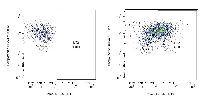
Application: Flow CytometrySample Tested: Dendritic cellsSpecies: HumanVerified Customer | Posted 07/05/2018

-
Application: Flow CytometrySample Tested: Human peripheral CD11c+ dendritic cells and Dendritic cellsSpecies: HumanVerified Customer | Posted 06/24/2018
There are no reviews that match your criteria.
Protocols
Find general support by application which include: protocols, troubleshooting, illustrated assays, videos and webinars.
- 7-Amino Actinomycin D (7-AAD) Cell Viability Flow Cytometry Protocol
- Extracellular Membrane Flow Cytometry Protocol
- Flow Cytometry Protocol for Cell Surface Markers
- Flow Cytometry Protocol for Staining Membrane Associated Proteins
- Flow Cytometry Staining Protocols
- Flow Cytometry Troubleshooting Guide
- Intracellular Flow Cytometry Protocol Using Alcohol (Methanol)
- Intracellular Flow Cytometry Protocol Using Detergents
- Intracellular Nuclear Staining Flow Cytometry Protocol Using Detergents
- Intracellular Staining Flow Cytometry Protocol Using Alcohol Permeabilization
- Intracellular Staining Flow Cytometry Protocol Using Detergents to Permeabilize Cells
- Propidium Iodide Cell Viability Flow Cytometry Protocol
- Protocol for Liperfluo
- Protocol for the Characterization of Human Th22 Cells
- Protocol for the Characterization of Human Th9 Cells
- Protocol: Annexin V and PI Staining by Flow Cytometry
- Protocol: Annexin V and PI Staining for Apoptosis by Flow Cytometry
- Troubleshooting Guide: Fluorokine Flow Cytometry Kits
- View all Protocols, Troubleshooting, Illustrated assays and Webinars