Sheep anti-Mouse IgG (H+L) Secondary Antibody [DyLight 800]
Novus Biologicals | Catalog # NBP1-72928
Key Product Details
Species Reactivity
Mouse
Applications
Western Blot, Fluorophore-linked immunosorbent assay, Immunocytochemistry/ Immunofluorescence
Label
DyLight 800 (Excitation = 777 nm, Emission = 794 nm)
Antibody Source
Polyclonal Sheep IgG
Loading...
Product Specifications
Immunogen
Mouse IgG whole molecule
Specificity
This antibody will react with heavy chains of Mouse IgG and with light chains of most Mouse immunoglobulins.
Clonality
Polyclonal
Host
Sheep
Isotype
IgG
Description
Store vial at 4C prior to restoration. For extended storage aliquot contents and freeze at -20C or below. Avoid cycles of freezing and thawing. Centrifuge product if not completely clear after standing at room temperature. This product is stable for several weeks at 4C as an undiluted liquid. Dilute only prior to immediate use.
This product was prepared from monospecific antiserum by immunoaffinity chromatography using Mouse IgG coupled to agarose beads followed by solid phase adsorption(s) to remove any unwanted reactivities. Assay by immunoelectrophoresis resulted in a single precipitin arc against anti-Sheep Serum, Mouse IgG and Mouse Serum
This product was prepared from monospecific antiserum by immunoaffinity chromatography using Mouse IgG coupled to agarose beads followed by solid phase adsorption(s) to remove any unwanted reactivities. Assay by immunoelectrophoresis resulted in a single precipitin arc against anti-Sheep Serum, Mouse IgG and Mouse Serum
Scientific Data Images for Sheep anti-Mouse IgG (H+L) Secondary Antibody [DyLight 800]
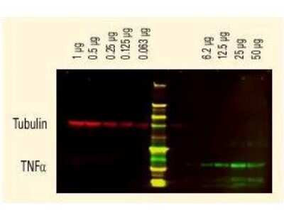
Western Blot: Sheep anti-Mouse IgG (H+L) Secondary Antibody [DyLight 800] [NBP1-72928] - DyLight (TM) dyes can be used for two-color Western Blot detection with low background and high signal. Anti-tubulin was detected using a DyLight (TM) 680 conjugate. Anti-TNFa was detected using a DyLight (TM) 800 conjugate. The image was captured using the Odyssey(R) Infrared Imaging System developed by LI-COR.
Applications for Sheep anti-Mouse IgG (H+L) Secondary Antibody [DyLight 800]
Application
Recommended Usage
Fluorophore-linked immunosorbent assay
1:20000
Immunocytochemistry/ Immunofluorescence
1:5000
Western Blot
1:10000
Application Notes
This product has been tested by dot blot and is designed for immunofluorescence microscopy, fluorescence based plate assays (FLISA) and fluorescent western blotting. This product is also suitable for multiplex analysis, including multicolor imaging, utilizing various commercial platforms. The emission spectra for this DyLight(TM) conjugate match the principle output wavelengths of most common fluorescence instrumentation.
Formulation, Preparation, and Storage
Purification
Multi-step
Reconstitution
Reconstitute with 100 ul deionized water (or equivalent).
Formulation
Lyophilized from 0.02 M Potassium Phosphate, 0.15 M Sodium Chloride, pH 7.2, 10 mg/mL Bovine Serum Albumin (BSA) - Immunoglobulin and Protease free
Preservative
0.01% Sodium Azide
Concentration
LYOPH mg/ml
Shipping
The product is shipped with polar packs. Upon receipt, store it immediately at the temperature recommended below.
Stability & Storage
Store lyophilized antibody at 4C in the dark. Aliquot reconstituted liquid and store at -20C. Avoid freeze-thaw cycles.
Calculators
Background: IgG (H+L)
The 4 IgG subclasses, sharing 95% amino acid identity, include IgG1, IgG2, IgG3, and IgG4 for humans and IgG1, IgG2a, IgG2b, and IgG3 for mice. The relative abundance of each human subclass is 60% for IgG1, 32% for IgG2, 4% for IgG3, and 4% for IgG4. In an IgG deficiency, there may be a shortage of one or more subclasses (4).
References
1. Painter RH. (1998) Encyclopedia of Immunology (Second Edition). Elsevier. 1208-1211
2. Chapter 9 - Antibodies. (2012) Immunology for Pharmacy. Mosby 70-78
3. Schroeder H, Cavacini, L. (2010) Structure and Function of Immunoglobulins. J Allergy Clin Immunol. 125(2 0 2): S41-S52. PMID: 20176268
4. Vidarsson G, Dekkers G, Rispens T. (2014) IgG subclasses and allotypes: from structure to effector functions. Front Immunol. 5:520. PMID: 25368619
Additional IgG (H+L) Products
Product Documents for Sheep anti-Mouse IgG (H+L) Secondary Antibody [DyLight 800]
Certificate of Analysis
To download a Certificate of Analysis, please enter a lot or batch number in the search box below.
Product Specific Notices for Sheep anti-Mouse IgG (H+L) Secondary Antibody [DyLight 800]
This product is for research use only and is not approved for use in humans or in clinical diagnosis. Secondary Antibodies are guaranteed for 1 year from date of receipt.
Citations for Sheep anti-Mouse IgG (H+L) Secondary Antibody [DyLight 800]
 Powered by Bioz
Customer Reviews for Sheep anti-Mouse IgG (H+L) Secondary Antibody [DyLight 800]
There are currently no reviews for this product. Be the first to review Sheep anti-Mouse IgG (H+L) Secondary Antibody [DyLight 800] and earn rewards!
Have you used Sheep anti-Mouse IgG (H+L) Secondary Antibody [DyLight 800]?
Submit a review and receive an Amazon gift card!
$25/€18/£15/$25CAN/¥2500 Yen for a review with an image
$10/€7/£6/$10CAN/¥1110 Yen for a review without an image
Submit a review
Protocols
Find general support by application which include: protocols, troubleshooting, illustrated assays, videos and webinars.
- Appropriate Fixation of IHC/ICC Samples
- Cellular Response to Hypoxia Protocols
- ClariTSA™ Fluorophore Kits
- Detection & Visualization of Antibody Binding
- ELISA Sample Preparation & Collection Guide
- ELISA Troubleshooting Guide
- How to Run an R&D Systems DuoSet ELISA
- How to Run an R&D Systems Quantikine ELISA
- How to Run an R&D Systems Quantikine™ QuicKit™ ELISA
- ICC Cell Smear Protocol for Suspension Cells
- ICC Immunocytochemistry Protocol Videos
- ICC for Adherent Cells
- Immunocytochemistry (ICC) Protocol
- Immunocytochemistry Troubleshooting
- Immunofluorescence of Organoids Embedded in Cultrex Basement Membrane Extract
- Immunohistochemistry (IHC) and Immunocytochemistry (ICC) Protocols
- Preparing Samples for IHC/ICC Experiments
- Preventing Non-Specific Staining (Non-Specific Binding)
- Primary Antibody Selection & Optimization
- Protocol for VisUCyte™ HRP Polymer Detection Reagent
- Protocol for the Fluorescent ICC Staining of Cell Smears - Graphic
- Protocol for the Fluorescent ICC Staining of Cultured Cells on Coverslips - Graphic
- Protocol for the Preparation and Fluorescent ICC Staining of Cells on Coverslips
- Protocol for the Preparation and Fluorescent ICC Staining of Non-adherent Cells
- Protocol for the Preparation and Fluorescent ICC Staining of Stem Cells on Coverslips
- Protocol for the Preparation of a Cell Smear for Non-adherent Cell ICC - Graphic
- Quantikine HS ELISA Kit Assay Principle, Alkaline Phosphatase
- Quantikine HS ELISA Kit Principle, Streptavidin-HRP Polymer
- R&D Systems Quality Control Western Blot Protocol
- Sandwich ELISA (Colorimetric) – Biotin/Streptavidin Detection Protocol
- Sandwich ELISA (Colorimetric) – Direct Detection Protocol
- TUNEL and Active Caspase-3 Detection by IHC/ICC Protocol
- The Importance of IHC/ICC Controls
- Troubleshooting Guide: ELISA
- Troubleshooting Guide: Western Blot Figures
- Western Blot Conditions
- Western Blot Protocol
- Western Blot Protocol for Cell Lysates
- Western Blot Troubleshooting
- Western Blot Troubleshooting Guide
- View all Protocols, Troubleshooting, Illustrated assays and Webinars
Loading...