Integrin beta 2/CD18 Antibody (YFC118.3) - BSA Free
Novus Biologicals | Catalog # NB200-610
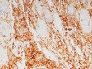
![Immunocytochemistry/ Immunofluorescence: Integrin beta 2/CD18 Antibody (YFC118.3) - BSA Free [NB200-610]](https://resources.rndsystems.com/images/products/Integrin-beta-2-CD18-Antibody-YFC118-3-Immunocytochemistry-Immunofluorescence-NB200-610-img0011.jpg "Immunocytochemistry/ Immunofluorescence: Integrin beta 2/CD18 Antibody (YFC118.3) - BSA Free [NB200-610]")
Key Product Details
Species Reactivity
Applications
Label
Antibody Source
Format
Product Specifications
Immunogen
Reactivity Notes
Specificity
Clonality
Host
Isotype
Scientific Data Images for Integrin beta 2/CD18 Antibody (YFC118.3) - BSA Free
Immunocytochemistry/ Immunofluorescence: Integrin beta 2/CD18 Antibody (YFC118.3) - BSA Free [NB200-610]
Immunocytochemistry/Immunofluorescence: Integrin beta 2/CD18 Antibody (YFC118.3) [NB200-610] - A-D Confocal microscopy of IBA-1 (green staining) immunohistochemistry of RPE flatmounts (RPE autofluorescence visible as orange due to its autofluorescence in the red and green channel) from a healthy donor (A), a geographic atrophy lesion (B), and large drusen (C and D). (A, B, D): orthogonal Z-stack projection; (C): oblique Z-stack projection and dissecting microscope appearance of postmortem large drusen after removal of the overlaying retina (inset). E-G Double-labeling on the subretinal side of the retina (to avoid masking by RPE autofluorescence) of IBA-1+ (E, green fluorescence) and CD18 (F, red fluorescence; G, merge). H. Orthogonal and lateral Z-stack of a subretinal IBA-1+ (green fluorescence) MPs adjacent to the RPE (orange autofluorescence) in the vicinity of a large drusen.![Immunohistochemistry-Frozen: Integrin beta 2/CD18 Antibody (YFC118.3) - BSA Free [NB200-610]](https://resources.rndsystems.com/images/products/Integrin-beta-2-CD18-Antibody-YFC118-3-Immunohistochemistry-Frozen-NB200-610-img0018.jpg "Immunohistochemistry-Frozen: Integrin beta 2/CD18 Antibody (YFC118.3) - BSA Free [NB200-610]")
Immunohistochemistry-Frozen: Integrin beta 2/CD18 Antibody (YFC118.3) - BSA Free [NB200-610]
Immunohistochemistry-Frozen: Integrin beta 2/CD18 Antibody (YFC118.3) [NB200-610] - Canine histiocytic sarcoma stained with Integrin beta 2/CD18 Antibody (YFC118.3). IHC-Fr image submitted by a verified customer review.![Flow Cytometry: Integrin beta 2/CD18 Antibody (YFC118.3) - BSA Free [NB200-610]](https://resources.rndsystems.com/images/products/Integrin-beta-2-CD18-Antibody-YFC118-3-Flow-Cytometry-NB200-610-img0017.jpg "Flow Cytometry: Integrin beta 2/CD18 Antibody (YFC118.3) - BSA Free [NB200-610]")
Flow Cytometry: Integrin beta 2/CD18 Antibody (YFC118.3) - BSA Free [NB200-610]
Flow Cytometry: Integrin beta 2/CD18 Antibody (YFC118.3) [NB200-610] - Staining of human peripheral blood granulocytes with RAT ANTI HUMAN CD18: FITC.![Immunocytochemistry/ Immunofluorescence: Integrin beta 2/CD18 Antibody (YFC118.3) - BSA Free [NB200-610]](https://resources.rndsystems.com/images/products/Integrin-beta-2-CD18-Antibody-YFC118-3-Immunocytochemistry-Immunofluorescence-NB200-610-img0007.jpg "Immunocytochemistry/ Immunofluorescence: Integrin beta 2/CD18 Antibody (YFC118.3) - BSA Free [NB200-610]")
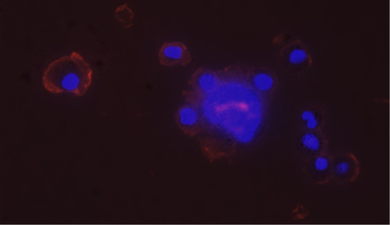
Immunocytochemistry/ Immunofluorescence: Integrin beta 2/CD18 Antibody (YFC118.3) - BSA Free [NB200-610]
Immunocytochemistry/Immunofluorescence: Integrin beta 2/CD18 Antibody (YFC118.3) [NB200-610] - Analysis of canine thoracic fluid samples using anti-Integrin beta 2 antibody. The antibody showing positive cell surface staining. ICC/IF image submitted by a verified customer review.![Flow Cytometry: Integrin beta 2/CD18 Antibody (YFC118.3) - BSA Free [NB200-610]](https://resources.rndsystems.com/images/products/Integrin-beta-2-CD18-Antibody-YFC118-3-Flow-Cytometry-NB200-610-img0001.jpg "Flow Cytometry: Integrin beta 2/CD18 Antibody (YFC118.3) - BSA Free [NB200-610]")
Flow Cytometry: Integrin beta 2/CD18 Antibody (YFC118.3) - BSA Free [NB200-610]
Flow Cytometry: Integrin beta 2/CD18 Antibody (YFC118.3) [NB200-610] - Staining of human peripheral blood lymphocytes with Rat anti Human CD18.![Flow Cytometry: Integrin beta 2/CD18 Antibody (YFC118.3) - BSA Free [NB200-610]](https://resources.rndsystems.com/images/products/Integrin-beta-2-CD18-Antibody-YFC118-3-Flow-Cytometry-NB200-610-img0008.jpg "Flow Cytometry: Integrin beta 2/CD18 Antibody (YFC118.3) - BSA Free [NB200-610]")
Flow Cytometry: Integrin beta 2/CD18 Antibody (YFC118.3) - BSA Free [NB200-610]
Flow Cytometry: Integrin beta 2/CD18 Antibody (YFC118.3) [NB200-610] - A) Isotype control with macrophages showing delineation of the M1 marker based on the negative population. B and C represent expression of CD18 (B) or CD11b (C) before incubation with Leishmania. D and E represent expression of CD18 (D) or CD11b (E) after association with Leishmania in a SD assay. In all cases the percent positive cells within the M1 marker are identified. This Figure is a representative diagram of 4 experiments with similar findings.![Flow Cytometry: Integrin beta 2/CD18 Antibody (YFC118.3) - BSA Free [NB200-610]](https://resources.rndsystems.com/images/products/Integrin-beta-2-CD18-Antibody-YFC118-3-Flow-Cytometry-NB200-610-img0015.jpg "Flow Cytometry: Integrin beta 2/CD18 Antibody (YFC118.3) - BSA Free [NB200-610]")
Flow Cytometry: Integrin beta 2/CD18 Antibody (YFC118.3) - BSA Free [NB200-610]
Flow Cytometry: Integrin beta 2/CD18 Antibody (YFC118.3) [NB200-610] - LFA-1 activation analyses by Flow Cytometry. (A) Profiles of CD11a, CD18 activation epitope (CD18act, representing high affinity conformation) and CD18 (CD18tot) expression by Teff and Treg#1 cells pre-incubated or not with indicated antibodies. Anti-CD28 and anti-CTLA-4 were used at 10 ug/ml. (B) Histograms of Mean Fluorescent Intensity (MFI) of CD11a, CD18act and CD18tot expressed on Teff and Treg#1 cells. Ratio CD18act MFI: CD18tot was established to analyze LFA-1 high affinity conformation in indicated conditions. Data are representative of more than three different experiments. Filled gray, negative control; Red line, Teff and Treg cells alone; Green line, cells with APC; Blue line, cells with APC and anti-CD28; and Black line: cells with APC, anti-CD28 and anti-CTLA-4.Applications for Integrin beta 2/CD18 Antibody (YFC118.3) - BSA Free
Flow Cytometry
Immunohistochemistry
Immunohistochemistry-Frozen
Immunoprecipitation
Reviewed Applications
Read 2 reviews rated 4.5 using NB200-610 in the following applications:
Flow Cytometry Panel Builder
Bio-Techne Knows Flow Cytometry
Save time and reduce costly mistakes by quickly finding compatible reagents using the Panel Builder Tool.
Advanced Features
- Spectra Viewer - Custom analysis of spectra from multiple fluorochromes
- Spillover Popups - Visualize the spectra of individual fluorochromes
- Antigen Density Selector - Match fluorochrome brightness with antigen density

Formulation, Preparation, and Storage
Purification
Formulation
Format
Preservative
Concentration
Shipping
Stability & Storage
Background: Integrin beta 2/CD18
Alternate Names
Gene Symbol
UniProt
Additional Integrin beta 2/CD18 Products
Product Documents for Integrin beta 2/CD18 Antibody (YFC118.3) - BSA Free
Certificate of Analysis
To download a Certificate of Analysis, please enter a lot or batch number in the search box below.
Product Specific Notices for Integrin beta 2/CD18 Antibody (YFC118.3) - BSA Free
This product is for research use only and is not approved for use in humans or in clinical diagnosis. Primary Antibodies are guaranteed for 1 year from date of receipt.
Customer Reviews for Integrin beta 2/CD18 Antibody (YFC118.3) - BSA Free (2)
Have you used Integrin beta 2/CD18 Antibody (YFC118.3) - BSA Free?
Submit a review and receive an Amazon gift card!
$25/€18/£15/$25CAN/¥2500 Yen for a review with an image
$10/€7/£6/$10CAN/¥1110 Yen for a review without an image
Submit a review
Customer Images
-
Application: Immunohistochemistry-FrozenSample Tested: histiocytic sarcoma cell line and histiocytic sarcomaSpecies: CanineVerified Customer | Posted 09/04/2021Integrin beta 2/CD18 Antibody in canine histiocytic sarcoma

-
Application: ImmunocytochemistrySample Tested:Species: OtherVerified Customer | Posted 01/23/2015
There are no reviews that match your criteria.
Protocols
Find general support by application which include: protocols, troubleshooting, illustrated assays, videos and webinars.
- 7-Amino Actinomycin D (7-AAD) Cell Viability Flow Cytometry Protocol
- Antigen Retrieval Protocol (PIER)
- Antigen Retrieval for Frozen Sections Protocol
- Appropriate Fixation of IHC/ICC Samples
- Cellular Response to Hypoxia Protocols
- Chromogenic IHC Staining of Formalin-Fixed Paraffin-Embedded (FFPE) Tissue Protocol
- Chromogenic Immunohistochemistry Staining of Frozen Tissue
- ClariTSA™ Fluorophore Kits
- Detection & Visualization of Antibody Binding
- Extracellular Membrane Flow Cytometry Protocol
- Flow Cytometry Protocol for Cell Surface Markers
- Flow Cytometry Protocol for Staining Membrane Associated Proteins
- Flow Cytometry Staining Protocols
- Flow Cytometry Troubleshooting Guide
- Fluorescent IHC Staining of Frozen Tissue Protocol
- Graphic Protocol for Heat-induced Epitope Retrieval
- Graphic Protocol for the Preparation and Fluorescent IHC Staining of Frozen Tissue Sections
- Graphic Protocol for the Preparation and Fluorescent IHC Staining of Paraffin-embedded Tissue Sections
- Graphic Protocol for the Preparation of Gelatin-coated Slides for Histological Tissue Sections
- IHC Sample Preparation (Frozen sections vs Paraffin)
- Immunofluorescent IHC Staining of Formalin-Fixed Paraffin-Embedded (FFPE) Tissue Protocol
- Immunohistochemistry (IHC) and Immunocytochemistry (ICC) Protocols
- Immunohistochemistry Frozen Troubleshooting
- Immunohistochemistry Paraffin Troubleshooting
- Immunoprecipitation Protocol
- Intracellular Flow Cytometry Protocol Using Alcohol (Methanol)
- Intracellular Flow Cytometry Protocol Using Detergents
- Intracellular Nuclear Staining Flow Cytometry Protocol Using Detergents
- Intracellular Staining Flow Cytometry Protocol Using Alcohol Permeabilization
- Intracellular Staining Flow Cytometry Protocol Using Detergents to Permeabilize Cells
- Preparing Samples for IHC/ICC Experiments
- Preventing Non-Specific Staining (Non-Specific Binding)
- Primary Antibody Selection & Optimization
- Propidium Iodide Cell Viability Flow Cytometry Protocol
- Protocol for Heat-Induced Epitope Retrieval (HIER)
- Protocol for Liperfluo
- Protocol for Making a 4% Formaldehyde Solution in PBS
- Protocol for VisUCyte™ HRP Polymer Detection Reagent
- Protocol for the Characterization of Human Th22 Cells
- Protocol for the Characterization of Human Th9 Cells
- Protocol for the Preparation & Fixation of Cells on Coverslips
- Protocol for the Preparation and Chromogenic IHC Staining of Frozen Tissue Sections
- Protocol for the Preparation and Chromogenic IHC Staining of Frozen Tissue Sections - Graphic
- Protocol for the Preparation and Chromogenic IHC Staining of Paraffin-embedded Tissue Sections
- Protocol for the Preparation and Chromogenic IHC Staining of Paraffin-embedded Tissue Sections - Graphic
- Protocol for the Preparation and Fluorescent IHC Staining of Frozen Tissue Sections
- Protocol for the Preparation and Fluorescent IHC Staining of Paraffin-embedded Tissue Sections
- Protocol for the Preparation of Gelatin-coated Slides for Histological Tissue Sections
- Protocol: Annexin V and PI Staining by Flow Cytometry
- Protocol: Annexin V and PI Staining for Apoptosis by Flow Cytometry
- TUNEL and Active Caspase-3 Detection by IHC/ICC Protocol
- The Importance of IHC/ICC Controls
- Troubleshooting Guide: Fluorokine Flow Cytometry Kits
- Troubleshooting Guide: Immunohistochemistry
- View all Protocols, Troubleshooting, Illustrated assays and Webinars
FAQs for Integrin beta 2/CD18 Antibody (YFC118.3) - BSA Free
-
Q: Can you please provide me the immunogen sequence?
A: NB200-610 is a licensed clone and all of our available information comes from the clone inventor. Unfortunately, there is no additional information on the immunogen available.
Associated Pathways


