LPCAT2 Antibody - BSA Free
Novus Biologicals | Catalog # NBP1-88921
![Western Blot: LPCAT2 Antibody [NBP1-88921]](https://resources.rndsystems.com/images/products/LPCAT2-Antibody-Western-Blot-NBP1-88921-img0010.jpg "Western Blot: LPCAT2 Antibody [NBP1-88921]")
Key Product Details
Species Reactivity
Validated:
Cited:
Applications
Validated:
Cited:
Label
Antibody Source
Format
Product Specifications
Immunogen
Reactivity Notes
Clonality
Host
Isotype
Scientific Data Images for LPCAT2 Antibody - BSA Free
Western Blot: LPCAT2 Antibody [NBP1-88921]
Western Blot: LPCAT2 Antibody [NBP1-88921] - Analysis in mouse cell line NIH-3T3 and rat cell line NBT-II.![Immunohistochemistry-Paraffin: LPCAT2 Antibody [NBP1-88921]](https://resources.rndsystems.com/images/products/LPCAT2-Antibody-Immunohistochemistry-Paraffin-NBP1-88921-img0022.jpg "Immunohistochemistry-Paraffin: LPCAT2 Antibody [NBP1-88921]")
Immunohistochemistry-Paraffin: LPCAT2 Antibody [NBP1-88921]
Immunohistochemistry-Paraffin: LPCAT2 Antibody [NBP1-88921] - Analysis in human thyroid gland and skeletal muscle tissues using NBP1-88921 antibody. Corresponding LPCAT2 RNA-seq data are presented for the same tissues.![Western Blot: LPCAT2 Antibody [NBP1-88921]](https://resources.rndsystems.com/images/products/LPCAT2-Antibody-Western-Blot-NBP1-88921-img0009.jpg "Western Blot: LPCAT2 Antibody [NBP1-88921]")
Western Blot: LPCAT2 Antibody [NBP1-88921]
Western Blot: LPCAT2 Antibody [NBP1-88921] - Analysis in human thyroid gland tissue.![Immunohistochemistry-Frozen: LPCAT2 Antibody [NBP1-88921]](https://resources.rndsystems.com/images/products/LPCAT2-Antibody-Immunohistochemistry-Frozen-NBP1-88921-img0007.jpg "Immunohistochemistry-Frozen: LPCAT2 Antibody [NBP1-88921]")
Immunohistochemistry-Frozen: LPCAT2 Antibody [NBP1-88921]
Immunohistochemistry-Frozen: LPCAT2 Antibody [NBP1-88921] - Staining of LPCAT2 in rat neuronal tissue using anti-LPCAT2 antibody. Image from verified customer review.![Immunohistochemistry-Paraffin: LPCAT2 Antibody [NBP1-88921]](https://resources.rndsystems.com/images/products/LPCAT2-Antibody-Immunohistochemistry-Paraffin-NBP1-88921-img0018.jpg "Immunohistochemistry-Paraffin: LPCAT2 Antibody [NBP1-88921]")
Immunohistochemistry-Paraffin: LPCAT2 Antibody [NBP1-88921]
Immunohistochemistry-Paraffin: LPCAT2 Antibody [NBP1-88921] - Staining of human skeletal muscle shows very weak positivity in myocytes.![Immunohistochemistry-Paraffin: LPCAT2 Antibody [NBP1-88921]](https://resources.rndsystems.com/images/products/LPCAT2-Antibody-Immunohistochemistry-Paraffin-NBP1-88921-img0019.jpg "Immunohistochemistry-Paraffin: LPCAT2 Antibody [NBP1-88921]")
Immunohistochemistry-Paraffin: LPCAT2 Antibody [NBP1-88921]
Immunohistochemistry-Paraffin: LPCAT2 Antibody [NBP1-88921] - Staining of human small intestine shows weak membranous positivity in glandular cells.![Immunohistochemistry-Paraffin: LPCAT2 Antibody [NBP1-88921]](https://resources.rndsystems.com/images/products/LPCAT2-Antibody-Immunohistochemistry-Paraffin-NBP1-88921-img0020.jpg "Immunohistochemistry-Paraffin: LPCAT2 Antibody [NBP1-88921]")
Immunohistochemistry-Paraffin: LPCAT2 Antibody [NBP1-88921]
Immunohistochemistry-Paraffin: LPCAT2 Antibody [NBP1-88921] - Staining of human testis shows weak granular cytoplasmic positivity in Leydig cells.![Immunohistochemistry-Paraffin: LPCAT2 Antibody [NBP1-88921]](https://resources.rndsystems.com/images/products/LPCAT2-Antibody-Immunohistochemistry-Paraffin-NBP1-88921-img0021.jpg "Immunohistochemistry-Paraffin: LPCAT2 Antibody [NBP1-88921]")
Immunohistochemistry-Paraffin: LPCAT2 Antibody [NBP1-88921]
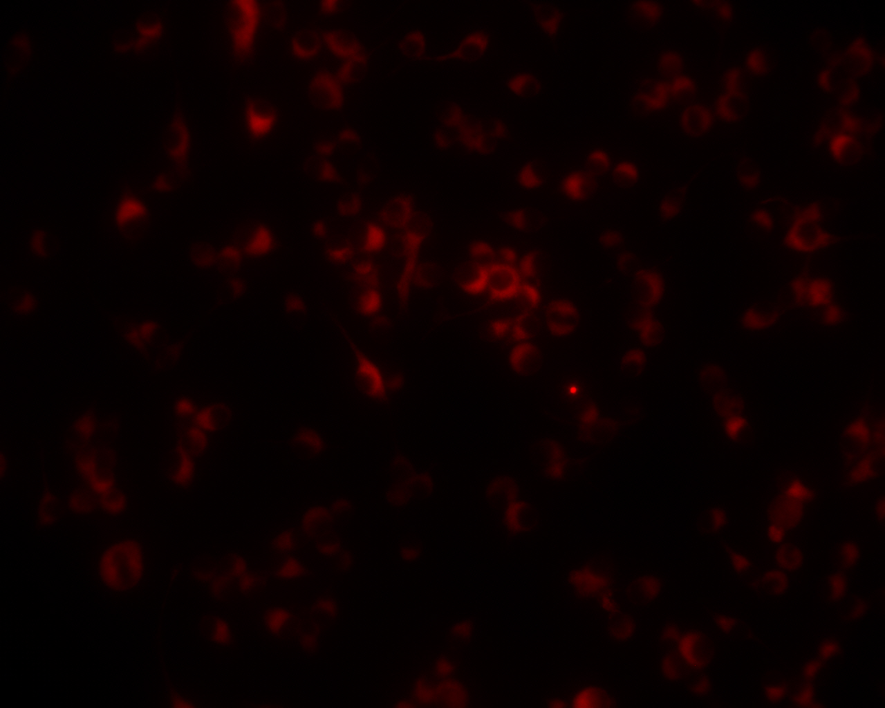
Immunohistochemistry-Paraffin: LPCAT2 Antibody [NBP1-88921] - Staining of human thyroid gland shows strong granular cytoplasmic positivity in glandular cells.![LPCAT2 Antibody - BSA Free Immunocytochemistry/ Immunofluorescence: LPCAT2 Antibody [NBP1-88921]](https://resources.rndsystems.com/images/products/nbp1-88921_-immunocytochemistry-immunofluorescence-639174076736043673.jpg "Immunocytochemistry/ Immunofluorescence: LPCAT2 Antibody [NBP1-88921]")
Immunocytochemistry/ Immunofluorescence: LPCAT2 Antibody [NBP1-88921]
Staining of human cell line A-431 shows localization to endoplasmic reticulum & lipid droplets.Applications for LPCAT2 Antibody - BSA Free
Immunocytochemistry/ Immunofluorescence
Immunohistochemistry
Immunohistochemistry-Frozen
Immunohistochemistry-Paraffin
Western Blot
Reviewed Applications
Read 2 reviews rated 4.5 using NBP1-88921 in the following applications:
Formulation, Preparation, and Storage
Purification
Formulation
Format
Preservative
Concentration
Shipping
Stability & Storage
Background: LPCAT2
Long Name
Alternate Names
Gene Symbol
Additional LPCAT2 Products
Product Documents for LPCAT2 Antibody - BSA Free
Certificate of Analysis
To download a Certificate of Analysis, please enter a lot or batch number in the search box below.
Product Specific Notices for LPCAT2 Antibody - BSA Free
This product is for research use only and is not approved for use in humans or in clinical diagnosis. Primary Antibodies are guaranteed for 1 year from date of receipt.
Citations for LPCAT2 Antibody - BSA Free
 Powered by Bioz
Customer Reviews for LPCAT2 Antibody - BSA Free (2)
Have you used LPCAT2 Antibody - BSA Free?
Submit a review and receive an Amazon gift card!
$25/€18/£15/$25CAN/¥2500 Yen for a review with an image
$10/€7/£6/$10CAN/¥1110 Yen for a review without an image
Submit a review
Customer Images

-
Application: Immunohistochemistry-FrozenSample Tested: Rat neuronal tissueSpecies: RatVerified Customer | Posted 02/20/2015LPCAT2 staining in rat tissue

-
Application: ImmunocytochemistrySample Tested: RAW264.7 cellsSpecies: MouseVerified Customer | Posted 02/12/2013RAW264.7 cells on mouse tissue
There are no reviews that match your criteria.
Protocols
Find general support by application which include: protocols, troubleshooting, illustrated assays, videos and webinars.
- Antigen Retrieval Protocol (PIER)
- Antigen Retrieval for Frozen Sections Protocol
- Appropriate Fixation of IHC/ICC Samples
- Cellular Response to Hypoxia Protocols
- Chromogenic IHC Staining of Formalin-Fixed Paraffin-Embedded (FFPE) Tissue Protocol
- Chromogenic Immunohistochemistry Staining of Frozen Tissue
- ClariTSA™ Fluorophore Kits
- Detection & Visualization of Antibody Binding
- Fluorescent IHC Staining of Frozen Tissue Protocol
- Graphic Protocol for Heat-induced Epitope Retrieval
- Graphic Protocol for the Preparation and Fluorescent IHC Staining of Frozen Tissue Sections
- Graphic Protocol for the Preparation and Fluorescent IHC Staining of Paraffin-embedded Tissue Sections
- Graphic Protocol for the Preparation of Gelatin-coated Slides for Histological Tissue Sections
- ICC Cell Smear Protocol for Suspension Cells
- ICC Immunocytochemistry Protocol Videos
- ICC for Adherent Cells
- IHC Sample Preparation (Frozen sections vs Paraffin)
- Immunocytochemistry (ICC) Protocol
- Immunocytochemistry Troubleshooting
- Immunofluorescence of Organoids Embedded in Cultrex Basement Membrane Extract
- Immunofluorescent IHC Staining of Formalin-Fixed Paraffin-Embedded (FFPE) Tissue Protocol
- Immunohistochemistry (IHC) and Immunocytochemistry (ICC) Protocols
- Immunohistochemistry Frozen Troubleshooting
- Immunohistochemistry Paraffin Troubleshooting
- Preparing Samples for IHC/ICC Experiments
- Preventing Non-Specific Staining (Non-Specific Binding)
- Primary Antibody Selection & Optimization
- Protocol for Heat-Induced Epitope Retrieval (HIER)
- Protocol for Making a 4% Formaldehyde Solution in PBS
- Protocol for VisUCyte™ HRP Polymer Detection Reagent
- Protocol for the Fluorescent ICC Staining of Cell Smears - Graphic
- Protocol for the Fluorescent ICC Staining of Cultured Cells on Coverslips - Graphic
- Protocol for the Preparation & Fixation of Cells on Coverslips
- Protocol for the Preparation and Chromogenic IHC Staining of Frozen Tissue Sections
- Protocol for the Preparation and Chromogenic IHC Staining of Frozen Tissue Sections - Graphic
- Protocol for the Preparation and Chromogenic IHC Staining of Paraffin-embedded Tissue Sections
- Protocol for the Preparation and Chromogenic IHC Staining of Paraffin-embedded Tissue Sections - Graphic
- Protocol for the Preparation and Fluorescent ICC Staining of Cells on Coverslips
- Protocol for the Preparation and Fluorescent ICC Staining of Non-adherent Cells
- Protocol for the Preparation and Fluorescent ICC Staining of Stem Cells on Coverslips
- Protocol for the Preparation and Fluorescent IHC Staining of Frozen Tissue Sections
- Protocol for the Preparation and Fluorescent IHC Staining of Paraffin-embedded Tissue Sections
- Protocol for the Preparation of Gelatin-coated Slides for Histological Tissue Sections
- Protocol for the Preparation of a Cell Smear for Non-adherent Cell ICC - Graphic
- R&D Systems Quality Control Western Blot Protocol
- TUNEL and Active Caspase-3 Detection by IHC/ICC Protocol
- The Importance of IHC/ICC Controls
- Troubleshooting Guide: Immunohistochemistry
- Troubleshooting Guide: Western Blot Figures
- Western Blot Conditions
- Western Blot Protocol
- Western Blot Protocol for Cell Lysates
- Western Blot Troubleshooting
- Western Blot Troubleshooting Guide
- View all Protocols, Troubleshooting, Illustrated assays and Webinars
FAQs for LPCAT2 Antibody - BSA Free
-
Q: Do you have an estimated band size for the LPCAT2 antibody (NBP1-88921) for Western blotting?
A: According to Uniprot, human LPCAT2 has a predicted molecular weight of approximately 60kDa. Please see UniProt entry Q7L5N7.