Best Seller
Luciferase Antibody - BSA Free
Novus Biologicals | Catalog # NB100-1677

Key Product Details
Species Reactivity
Validated:
Firefly
Cited:
Firefly
Applications
Validated:
Western Blot, ELISA
Cited:
Immunohistochemistry, Immunohistochemistry-Paraffin, Immunohistochemistry-Frozen, Western Blot, Immunocytochemistry/ Immunofluorescence, IF/IHC, Microarray
Label
Unconjugated
Antibody Source
Polyclonal Goat IgG
Format
BSA Free
Loading...
Product Specifications
Immunogen
Luciferase [Photinus pyralis (Firefly)]
Reactivity Notes
No reactivity is observed against Sea pansy (Renilla reniformis) luciferase.
Specificity
No reactivity is observed against Sea pansy (Renilla reniformis) luciferase.
Clonality
Polyclonal
Host
Goat
Isotype
IgG
Description
This product is an IgG fraction antibody purified from monospecific antiserum by a multi-step process which includes delipidation, salt fractionation and ion exchange chromatography followed by extensive dialysis against the buffer stated above. Assay by immunoelectrophoresis resulted in a single precipitin arc against anti-Goat Serum as well as purified and partially purified Luciferase [Photinus pyralis (Firefly)]
Store vial at -20C prior to opening. Aliquot contents and freeze at -20C or below for extended storage. Avoid cycles of freezing and thawing. Centrifuge product if not completely clear after standing at room temperature. This product is stable for several weeks at 4C as an undiluted liquid. Dilute only prior to immediate use.
Store vial at -20C prior to opening. Aliquot contents and freeze at -20C or below for extended storage. Avoid cycles of freezing and thawing. Centrifuge product if not completely clear after standing at room temperature. This product is stable for several weeks at 4C as an undiluted liquid. Dilute only prior to immediate use.
Scientific Data Images for Luciferase Antibody - BSA Free
Immunohistochemical Analysis of Luciferase in Human Tumor Xenograft
Human tumor xenograft expressing firefly luciferase intracranially injected into a mouse brain. Image provided via product review by verified customer.
Use of Luciferase Antibody in Western Blot
Lane 1: Luciferase, 50ng.Luciferase antibody at 1:1000 overnight at 4C. Secondary antibody: Peroxidase goat secondary antibody at 1:40,000 for 30 min at RT. Block: incubated with blocking buffer for 30 min at RT. Predicted/Observed size: 60 kDa for Luciferase. Other band(s): None.
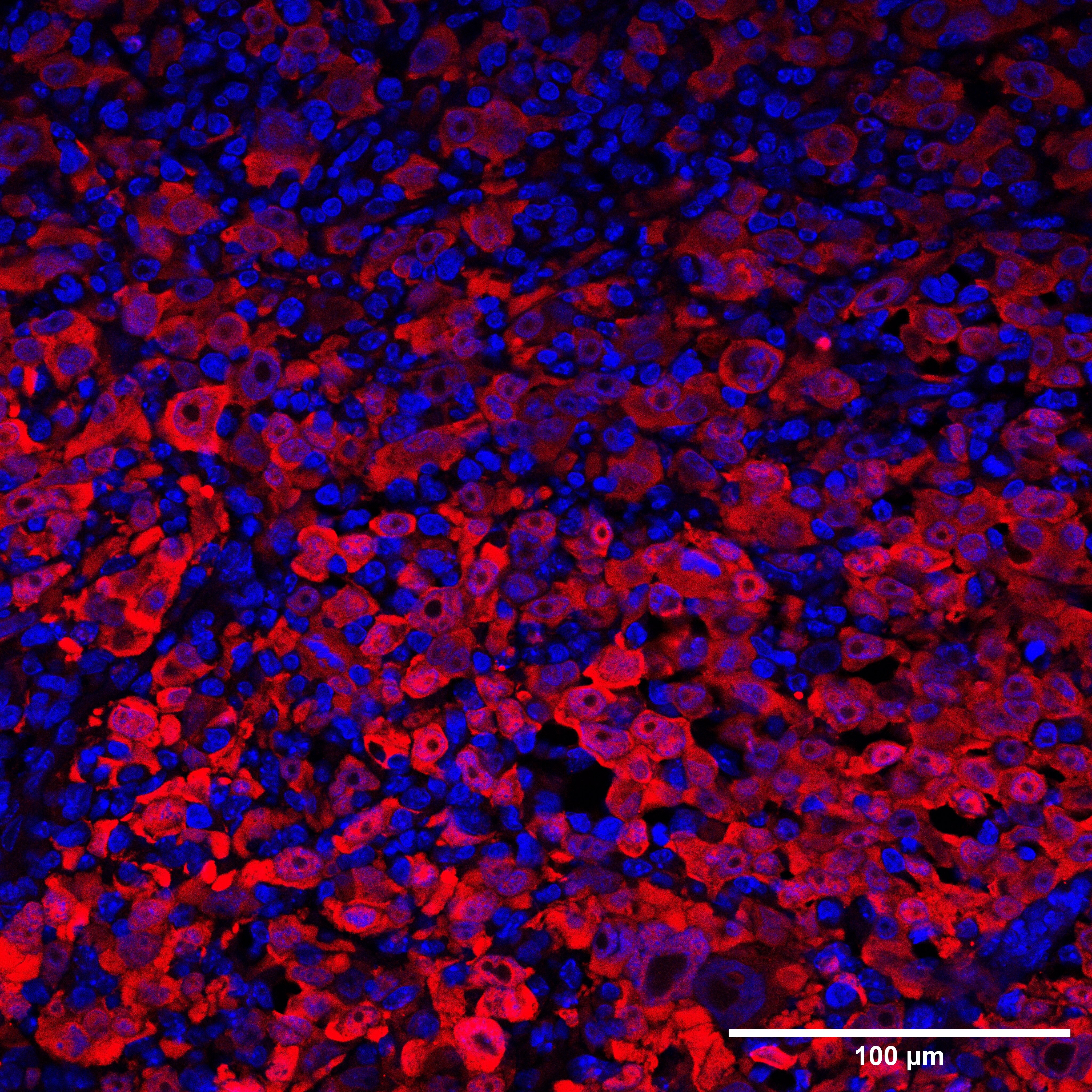
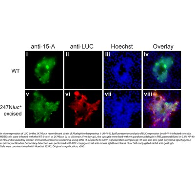
Immunocytochemistry/Immunofluorescence Staining of Luciferase in AIHV-1 Infected MDBK Cells
In vitro expression of Luciferase by 247Nluc+ recombinant strain of Alcelaphine herpesvirus 1 (AIHV-1). Epifluoresence expression of Luciferase by AIHV-1 infected syncytia. MDBK cells were infected with the WT (i to iv) or 247Nluc+ (v to viii) strain. Five days p.i. the the syncytia were fixed with 4% paraformaldehyde in PBS, permeabilized in 0.1% NP-40 in PBS and revealed by indirect IF co-staining. See image for additional details. Image provided via product review by verified customer.
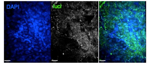
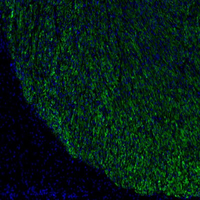
Immunohistochemical Staining of Luciferase in Paraffin Embedded Human Breast Tumor Xenograft
Analysis of human breast tumor xenograft expressing firefly luciferase subcutaneously injected into a mouse using Luciferase antibody. Primary antibody dilution 1:200. Image from verified customer review.
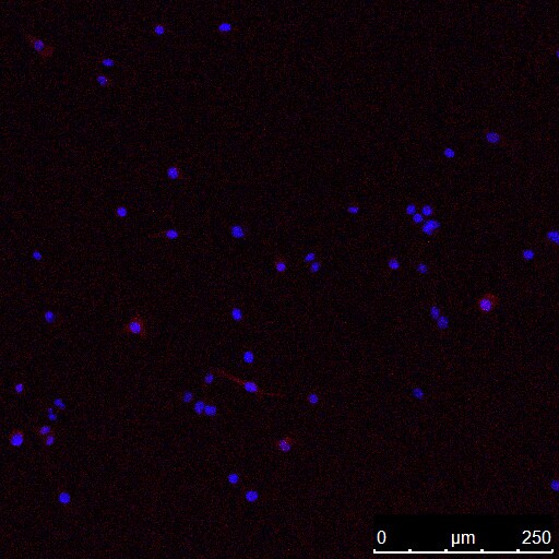
Staining of Luciferase in pLuc Transfected NIH3T3 Cells
NIH3T3 cells. Cells (25,000/well) transfected with pLuc plasmid were stained with 20ug/ml NB100-1677 and 1:200 dilution of donkey anti-goat IgG-FITC (green). Cells were mounted with DAPI (blue) and visualized at 200X magnification.
Immunohistochemical Staining of Luciferase in Paraffin Embedded Mouse Tibias
Luciferase immunostaining was analyzed in tibias from ovx ERE-luciferase mice taken 24 h after a 17-E2 injection. Positive luciferase staining was identified in hypertrophic chondrocytes (hc) (A), lining cells (lc) (B), osteoblasts (ob) (C), a subpopulation (10%) of osteocytes (ot) (upper arrow points at a positively stained osteocyte, whereas the lower depicts a negative osteocyte) (D), and megakaryocytes (E). Faint staining was found on the periosteal surface (p) (F). No background staining was seen when omitting the primary antibody (data not shown). The bar in the lower left corner represents 25 um. pc, Proliferative chondrocyte.
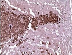
Immunohistochemical Detection of Luciferase in Paraffin Embedded Mouse Brain Tissue
Luciferase expression in mouse brain tissue bearing human glioma cells expressing fire fly luciferase. Image from verified customer review.
Use of Anti-Luciferase Antibody in Western Blot
Western Blot of Goat anti-Luciferase antibody. Lane 1 Marker: Opal Pre-stained ladderApplications for Luciferase Antibody - BSA Free
Application
Recommended Usage
ELISA
1:200-1:1000
Western Blot
1:1000-1:5000
Application Notes
This antibody has been tested in Western Blot. Expect a band ~60kDa in appropriate cell lysates. Although not tested, this antibody would be useful in immunofluorescence, immunoprecipitation, immunocytochemistry, and most immunological methods requiring high titers and specificity.
Reviewed Applications
Read 7 reviews rated 4.9 using NB100-1677 in the following applications:
Formulation, Preparation, and Storage
Purification
Multi-step
Formulation
0.02 M Potassium Phosphate, 0.15 M Sodium Chloride, pH 7.2
Format
BSA Free
Preservative
0.01% Sodium Azide
Concentration
Please see the vial label for concentration. If unlisted please contact technical services.
Shipping
The product is shipped with polar packs. Upon receipt, store it immediately at the temperature recommended below.
Stability & Storage
Store at 4C short term. Store at -20C long term. Avoid freeze-thaw cycles.
Background: Luciferase
The luciferase assay is fast and sensitive, differentiating itself from the CAT (chloramphenicol acetyltransferase) assay because it does not require a radioactive substrate.
References
1. Eun, H. (1996). Marker/Reporter enzymes. Enzymology Primer for Recombinant DNA Technology, 567-645. doi:10.1016/b978-012243740-3/50011-9
2. McNabb, D. S., Reed, R., & Marciniak, R. A. (2005). Dual luciferase assay system for rapid assessment of gene expression in Saccharomyces cerevisiae. Eukaryotic Cell, 4(9), 1539-1549. doi:10.1128/ec.4.9.1539-1549.2005
3. Fraga, H. (2008). Firefly luminescence: A historical perspective and recent developments. Photochemical & Photobiological Sciences, 7(2), 146-158. doi:10.1039/b719181b
4. Younes, A., Lukyanenko, Y. O., Lyashkov, A. E., Lakatta, E. G., & Sollott, S. J. (2011). A bioluminescence method for direct measurement of phosphodiesterase activity. Analytical Biochemistry, 417(1), 36-40. doi:10.1016/j.ab.2011.05.036
Alternate Names
LuC, luciferin 4 monooxygenase, Luciferin 4-monooxygenase
UniProt
Additional Luciferase Products
Product Documents for Luciferase Antibody - BSA Free
Certificate of Analysis
To download a Certificate of Analysis, please enter a lot or batch number in the search box below.
Product Specific Notices for Luciferase Antibody - BSA Free
This product is for research use only and is not approved for use in humans or in clinical diagnosis. Primary Antibodies are guaranteed for 1 year from date of receipt.
Citations for Luciferase Antibody - BSA Free
 Powered by Bioz
Customer Reviews for Luciferase Antibody - BSA Free (7)
4.9 out of 5
7 Customer Ratings
Have you used Luciferase Antibody - BSA Free?
Submit a review and receive an Amazon gift card!
$25/€18/£15/$25CAN/¥2500 Yen for a review with an image
$10/€7/£6/$10CAN/¥1110 Yen for a review without an image
Submit a review
Customer Images
Showing
1
-
5 of
7 reviews
Showing All
Filter By:
-
Application: ImmunocytochemistrySample Tested: E0771Species: MouseVerified Customer | Posted 12/30/2022E0771 was transducted with Luciferase-contained lentivirus. ICC was performed to validate the transduction rate. Anti-luciferase antibody was applied at a concentration of 2.5 ug/ml. Alexa 649 conjugated anti-goat IgG was used.
 Bio-Techne ResponseThis review was submitted through the legacy Novus Innovators Program, reflecting a new species or application tested on a primary antibody.
 -
Application: Immunohistochemistry-ParaffinSample Tested: Breast cancer cellsSpecies: HumanVerified Customer | Posted 11/25/2022Immunocytochemistry/Immunofluorescence: Luciferase Antibody [NB100-1677] - Human breast tumor xenograft expressing firefly luciferase subcutaneously injected into a mouse. Antibody diluted at 1:200. FFPE tumor.
-
Application: Western BlotSample Tested: Mouse Bone marrow derived macrophage and osteoclastSpecies: MouseVerified Customer | Posted 04/05/2018
-
Application: Immunocytochemistry/ImmunofluorescenceSample Tested: mouse prostateSpecies: MouseVerified Customer | Posted 10/10/2016
-
Application: Immunohistochemistry-ParaffinSample Tested: Mouse brain tissue bearing human glioma cells expressing fire fly luciferaseSpecies: OtherVerified Customer | Posted 06/18/2016Luciferase expression IHC in human glioma xenograft in mouse brain
-
Application: Immunohistochemistry-ParaffinSample Tested: Human tumor xenograft expressing firefly luciferase intracranially injected into a mouse brainSpecies: OtherVerified Customer | Posted 06/21/2013
-
Application: ImmunocytochemistrySample Tested: virus expressing luciferase transgeneSpecies: OtherVerified Customer | Posted 10/04/2011
There are no reviews that match your criteria.
Protocols
Find general support by application which include: protocols, troubleshooting, illustrated assays, videos and webinars.
- Cellular Response to Hypoxia Protocols
- ELISA Sample Preparation & Collection Guide
- ELISA Troubleshooting Guide
- How to Run an R&D Systems DuoSet ELISA
- How to Run an R&D Systems Quantikine ELISA
- How to Run an R&D Systems Quantikine™ QuicKit™ ELISA
- Quantikine HS ELISA Kit Assay Principle, Alkaline Phosphatase
- Quantikine HS ELISA Kit Principle, Streptavidin-HRP Polymer
- R&D Systems Quality Control Western Blot Protocol
- Sandwich ELISA (Colorimetric) – Biotin/Streptavidin Detection Protocol
- Sandwich ELISA (Colorimetric) – Direct Detection Protocol
- Troubleshooting Guide: ELISA
- Troubleshooting Guide: Western Blot Figures
- Western Blot Conditions
- Western Blot Protocol
- Western Blot Protocol for Cell Lysates
- Western Blot Troubleshooting
- Western Blot Troubleshooting Guide
- View all Protocols, Troubleshooting, Illustrated assays and Webinars
FAQs for Luciferase Antibody - BSA Free
Showing
1
-
2 of
2 FAQs
Showing All
-
Q: Hello, can I use this antibody (NB100-1677) for immunohistochemistry on rat frozen brain sections?
A: This antibody will work in IHC on frozen and paraffin embedded tissues, provided that they express Firefly Luciferase from Photinus pyralis.
-
Q: One of our customers would like additional information about your goat anti-luciferase (catalog #NB100-1677). They want to use this antibody for immunohistochemistry of paraffin-embedded sections, and would like to know if you recommend an antigen retrieval step? If so, which one is best?
A: For IHC-P with NB100-1677, we would recommend 10 mm citrate buffer (pH 6.0) based heat induced antigen retrieval at 95-99 C.
-
Q: Hello, can I use this antibody (NB100-1677) for immunohistochemistry on rat frozen brain sections?
A: This antibody will work in IHC on frozen and paraffin embedded tissues, provided that they express Firefly Luciferase from Photinus pyralis.
-
Q: One of our customers would like additional information about your goat anti-luciferase (catalog #NB100-1677). They want to use this antibody for immunohistochemistry of paraffin-embedded sections, and would like to know if you recommend an antigen retrieval step? If so, which one is best?
A: For IHC-P with NB100-1677, we would recommend 10 mm citrate buffer (pH 6.0) based heat induced antigen retrieval at 95-99 C.
Loading...