mCherry Antibody (1C51) - BSA Free
Novus Biologicals | Catalog # NBP1-96752
![Immunohistochemistry: mCherry Antibody (1C51) [NBP1-96752]](https://resources.rndsystems.com/images/products/mCherry-Antibody-1C51-Immunohistochemistry-NBP1-96752-img0006.jpg "Immunohistochemistry: mCherry Antibody (1C51) [NBP1-96752]")
Key Product Details
Species Reactivity
Validated:
Non-species specific
Cited:
Human, Mouse, Rat, Fish - Danio rerio (Zebrafish), Insect, Insect - Drosophila, Rabbit
Applications
Validated:
Knockout Validated, Immunohistochemistry, Immunohistochemistry-Paraffin, Immunohistochemistry-Frozen, Western Blot, Flow Cytometry, Immunocytochemistry/ Immunofluorescence, Immunoprecipitation, Single Cell Western
Cited:
Knockout Validated, Immunohistochemistry-Paraffin, Immunohistochemistry-Frozen, Western Blot, Flow Cytometry, Immunocytochemistry, Immunocytochemistry/ Immunofluorescence, Immunoprecipitation, Chromatin Immunoprecipitation, IF/IHC
Label
Unconjugated
Antibody Source
Monoclonal Mouse IgG2A Clone # 1C51
Format
BSA Free
Loading...
Product Specifications
Immunogen
This mCherry Antibody (1C51) was developed against recombinant full-length mCherry purified from E. coli.
Specificity
This mCherry Antibody (1C51) does not cross react with GFP.
Clonality
Monoclonal
Host
Mouse
Isotype
IgG2A
Theoretical MW
27 kDa.
Disclaimer note: The observed molecular weight of the protein may vary from the listed predicted molecular weight due to post translational modifications, post translation cleavages, relative charges, and other experimental factors.
Disclaimer note: The observed molecular weight of the protein may vary from the listed predicted molecular weight due to post translational modifications, post translation cleavages, relative charges, and other experimental factors.
Scientific Data Images for mCherry Antibody (1C51) - BSA Free
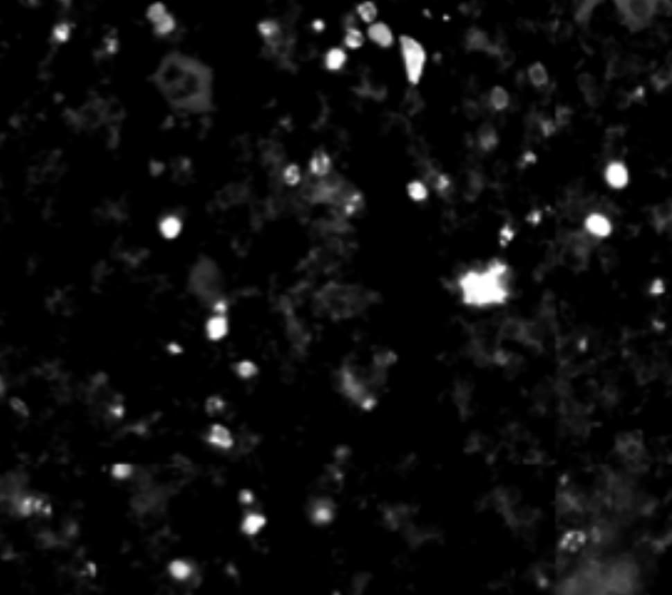
Immunohistochemistry: mCherry Antibody (1C51) [NBP1-96752]
mCherry-Antibody-1C51-Immunohistochemistry-NBP1-96752-img0006.jpg![Western Blot: mCherry Antibody (1C51) [NBP1-96752]](https://resources.rndsystems.com/images/products/mCherry-Antibody-1C51-Western-Blot-NBP1-96752-img0005.jpg "Western Blot: mCherry Antibody (1C51) [NBP1-96752]")
Western Blot: mCherry Antibody (1C51) [NBP1-96752]
Western Blot: mCherry Antibody (1C51) [NBP1-96752] - Analysis of HEK293 cell lysates and recombinant protein solutions using mCherry antibody, dilution 1:1,000 (Green). [1] protein standard, [2] HEK293, [3] HEK293 cells transfected with mCherry-HA construct, [4] mCherry recombinant protein, [5] GFP recombinant protein, and [6] HEK293 transfected with GFP construct. Major band at about 30 kDa corresponds to mCherry protein (predicted molecular weight: 27 kDa). mCherry antibody does not react with GFP protein. The same blot was simultaneously probed with chicken HSP60 pAb, dilution 1:5,000 in red which reveals band at 60 kDa seen only in cell lysates.![Western Blot: mCherry Antibody (1C51) [NBP1-96752]](https://resources.rndsystems.com/images/products/mCherry-Antibody-1C51-Western-Blot-NBP1-96752-img0008.jpg "Western Blot: mCherry Antibody (1C51) [NBP1-96752]")
Western Blot: mCherry Antibody (1C51) [NBP1-96752]
mCherry-Antibody-1C51-Western-Blot-NBP1-96752-img0008.jpg![Western Blot: mCherry Antibody (1C51) [NBP1-96752]](https://resources.rndsystems.com/images/products/mCherry-Antibody-1C51-Western-Blot-NBP1-96752-img0007.jpg "Western Blot: mCherry Antibody (1C51) [NBP1-96752]")
Western Blot: mCherry Antibody (1C51) [NBP1-96752]
mCherry-Antibody-1C51-Western-Blot-NBP1-96752-img0007.jpg![Immunocytochemistry/ Immunofluorescence: mCherry Antibody (1C51) [NBP1-96752]](https://resources.rndsystems.com/images/products/mCherry-Antibody-1C51-Immunocytochemistry-Immunofluorescence-NBP1-96752-img0003.jpg "Immunocytochemistry/ Immunofluorescence: mCherry Antibody (1C51) [NBP1-96752]")
Immunocytochemistry/ Immunofluorescence: mCherry Antibody (1C51) [NBP1-96752]
Immunocytochemistry/Immunofluorescence: mCherry Antibody (1C51) [NBP1-96752] - HEK293 cells transfected with mCherry and visualized in red. The cells were stained with NBP1-96752 in the green channel, and visualized using a confocal microscope. Transfected cells are yellow, showing overlap of the mCherry and NBP1-96752. Untransfected HEK293 cells do not express Cherry and do not stain with the antibody, but their nuclei can be visualized using a DNA stain (blue).![Western Blot: mCherry Antibody (1C51) [NBP1-96752]](https://resources.rndsystems.com/images/products/mCherry-Antibody-1C51-Western-Blot-NBP1-96752-img0002.jpg "Western Blot: mCherry Antibody (1C51) [NBP1-96752]")
Western Blot: mCherry Antibody (1C51) [NBP1-96752]
Western Blot: mCherry Antibody (1C51) [NBP1-96752] - WB assay of the crude extract of HEK293 cells transfected with pFin-EF1-mCherry vector (lane +) and an equal amount of protein extract from untransfected HEK293 cells (lane -). NBP1-96752 binds a major band running at about 28 kDa (observed molecular weight) corresponding to intact full-length mCherry. The two other bands are clearly processed forms of mCherry as they are not present in non-transfected HEK293 cells.![Immunohistochemistry-Frozen: mCherry Antibody (1C51) [NBP1-96752]](https://resources.rndsystems.com/images/products/mCherry-Antibody-1C51-Immunohistochemistry-Frozen-NBP1-96752-img0004.jpg "Immunohistochemistry-Frozen: mCherry Antibody (1C51) [NBP1-96752]")
Immunohistochemistry-Frozen: mCherry Antibody (1C51) [NBP1-96752]
Immunohistochemistry-Frozen: mCherry Antibody (1C51) [NBP1-96752] - Mouse Bone Marrow Sections (Femur). Fixed-frozen and decalcified. tdTomato reporter transgenic mice. tdTomato in hematopoietic cells were detected by anti-mCherry antibody. Antibody is cross-reactive and works well for fixed-frozen bone marrow. Background is low. IHC-Fr image submitted by a verified customer review. [NBP1-96752] -")
Western Blot: mCherry Antibody (1C51) [NBP1-96752] -
Western Blot: mCherry Antibody (1C51) [NBP1-96752] - Mis18 alpha :Mis18 beta -hexamer mediates dimerization of M18BP1.(A) Analytical SEC results of M18BP11–140-MBP (cyan), M18BP11–228-MBP (red), Mis18 alpha :Mis18 beta :M18BP11–140-MBP (purple), Mis18 alpha :Mis18 beta :M18BP11–228-MBP (green). The elution volumes of thyroglobulin (670 kD), ferritin (440 kD), catalase (240 kD) & ovalbumin (44 kD) are shown as standards. Red lines indicate fractions collected for Tricine–SDS-PAGE analyses. Gels were stained with CBB. (B) Sedimentation velocity AUC results of the same samples used in the analytical SEC experiments (panel A). The best-fit size distributions are shown with the colors indicated in panel A. Data profiles used for curve-fitting analyses are shown in Figure 7—figure supplement 1. (C) Summary table of the results obtained from the AUC experiments of panel B. Sed. coef., sedimentation coefficient; MWobs., observed molecular weight; MWtheo., theoretical molecular weight. (D) Western blot results of co-immunoprecipitation experiments using GFP-Trap_A beads. HeLa CENP-A-SNAP + EGFP-M18BP11–140-P2A-T2A-mCherry-M18BP11–140, EGFP-M18BP11–140/T40D/S110E-P2A-T2A-mCherry-M18BP11–140/T40D/S110E, GST-EGFP-M18BP11–140-P2A-T2A-GST-mCherry-M18BP11–140, or GST-EGFP-M18BP11–140/T40D/S110E-P2A-T2A-GST-mCherry-M18BP11–140/T40D/S110E were analyzed.DOI:http://dx.doi.org/10.7554/eLife.23352.014Data profiles for AUC experiments.Best-fitting results of the sedimentation velocity AUC data of M18BP11–140-MBP, M18BP11–228-MBP, Mis18 alpha :Mis18 beta :M18BP11–140-MBP, and Mis18 alpha :Mis18 beta :M18BP11–228-MBP. Residuals represent the deviation of the continuous c(s) distribution model from the observed signals. The values of RMSD for data fitting are shown.DOI:http://dx.doi.org/10.7554/eLife.23352.015 Image collected & cropped by CiteAb from the following publication (https://pubmed.ncbi.nlm.nih.gov/28059702), licensed under a CC-BY license. Not internally tested by Novus Biologicals. [NBP1-96752] -")
Western Blot: mCherry Antibody (1C51) [NBP1-96752] -
Western Blot: mCherry Antibody (1C51) [NBP1-96752] - The oligomerization state of overexpressed Cav1 varies as a function of its tag. COS-7 cells expressing the indicated constructs were lysed in digitonin & subjected to BN-PAGE followed by western blotting for Cav1 (red) & either GFP, mCherry or myc (green). A) Cells were either left untransfected (‘control’) or transfected with EGFP, Cav1-GFP or P132L-GFP. B) As in (A) except cells were transfected with the indicated mCherry constructs. C) As in (A) except cells were transfected with Cav1-myc or P132L-myc. Figures are representative of two independent experiments. Red arrows indicate the high molecular weight band positive for both tag antibodies & Cav1 antibodies (h1-97 or 2297). Black arrows indicate the high molecular weight band only positive for Cav1 antibodies (h1-97 or 2297). Green arrows indicate the low molecular weight bands only positive for FP tag antibodies. Image collected & cropped by CiteAb from the following publication (https://pubmed.ncbi.nlm.nih.gov/25639341), licensed under a CC-BY license. Not internally tested by Novus Biologicals.Applications for mCherry Antibody (1C51) - BSA Free
Application
Recommended Usage
Immunocytochemistry/ Immunofluorescence
1:500
Immunohistochemistry
1:500
Single Cell Western
100 ug/mL
Western Blot
1:1000 - 1:2000
Application Notes
Use in Flow reported in scientific literature (PMID:33335127). Use in IHC and IHC-P reported in scientific literature (PMID: 27396338 and 27716840 respectively).
mCherry antibody validated for IHC-Frozen from a verified customer review.
Use in Immunoprecipitation reported in scientific literature (PMID: 33008892).
Use in Knockout Validation was reported in scientific literature (PMID: 32547960).
mCherry antibody validated for IHC-Frozen from a verified customer review.
Use in Immunoprecipitation reported in scientific literature (PMID: 33008892).
Use in Knockout Validation was reported in scientific literature (PMID: 32547960).
Reviewed Applications
Read 3 reviews rated 4.3 using NBP1-96752 in the following applications:
Flow Cytometry Panel Builder
Bio-Techne Knows Flow Cytometry
Save time and reduce costly mistakes by quickly finding compatible reagents using the Panel Builder Tool.
Advanced Features
- Spectra Viewer - Custom analysis of spectra from multiple fluorochromes
- Spillover Popups - Visualize the spectra of individual fluorochromes
- Antigen Density Selector - Match fluorochrome brightness with antigen density

Formulation, Preparation, and Storage
Purification
Protein G purified
Formulation
50% PBS, 50% glycerol
Format
BSA Free
Preservative
0.035% Sodium Azide
Concentration
1 mg/ml
Shipping
The product is shipped with polar packs. Upon receipt, store it immediately at the temperature recommended below.
Stability & Storage
Store at 4C short term. Aliquot and store at -20C long term. Avoid freeze-thaw cycles.
Background: mCherry
mCherry can be used as a long-wavelength hetero-FRET (fluorescence resonance energy transfer) acceptor and probe for homoFRET experiments given its high peak molar absorptivity, folding efficiency, and superior spectral properties (4). Additionally, because mCherry does not interfere with other plasmids or alter the growth of Legionella species during intracellular growth, it can be used for constitutive gene expression in a variety of gram-negative bacterial species (5). For example, a plasmid developed to constitutively express mCherry under the Ptac promoter has been used in several Legionella species including L. pneumophila, the causative agent of Legionnaires' disease (5).
References
1. Shaner, N. C., Steinbach, P. A., & Tsien, R. Y. (2005). A guide to choosing fluorescent proteins. Nature Methods, 2(12), 905-909. doi:10.1038/nmeth819
2. Bevis, B. J., & Glick, B. S. (2002). Rapidly maturing variants of the Discosoma red fluorescent protein (DsRed). Nature Biotechnology, 20(1), 83-87. https://doi.org/10.1038/nbt0102-83
3. Wall, M. A., Socolich, M., & Ranganathan, R. (2000). The structural basis for red fluorescence in the tetrameric GFP homolog DsRed. Nature Structural Biology, 7(12), 1133-1138. https://doi.org/10.1038/81992
4. Akrap, N., Seidel, T., & Barisas, B. G. (2010). Forster distances for fluorescence resonant energy transfer between mCherry and other visible fluorescent proteins. Analytical Biochemistry, 402(1), 105-106. https://doi.org/10.1016/j.ab.2010.03.026
5. Gebhardt, M. J., Jacobson, R. K., & Shuman, H. A. (2017). Seeing red; the development of pON.mCherry, a broad-host range constitutive expression plasmid for Gram-negative bacteria. Plos One, 12(3), e0173116. https://doi.org/10.1371/journal.pone.0173116
Long Name
mCherry
Alternate Names
DSRED, red fluorescent protein mCherry, Red Fluoroscent Protein
Additional mCherry Products
Product Documents for mCherry Antibody (1C51) - BSA Free
Certificate of Analysis
To download a Certificate of Analysis, please enter a lot or batch number in the search box below.
Product Specific Notices for mCherry Antibody (1C51) - BSA Free
This product is for research use only and is not approved for use in humans or in clinical diagnosis. Primary Antibodies are guaranteed for 1 year from date of receipt.
Citations for mCherry Antibody (1C51) - BSA Free
 Powered by Bioz
Customer Reviews for mCherry Antibody (1C51) - BSA Free (3)
4.3 out of 5
3 Customer Ratings
Have you used mCherry Antibody (1C51) - BSA Free?
Submit a review and receive an Amazon gift card!
$25/€18/£15/$25CAN/¥2500 Yen for a review with an image
$10/€7/£6/$10CAN/¥1110 Yen for a review without an image
Submit a review
Customer Images

Showing
1
-
3 of
3 reviews
Showing All
Filter By:
-
Application: Western BlotSample Tested: 293T lysateSpecies: HumanVerified Customer | Posted 04/19/2019Samples reflect varying amounts of mCherry expression in lysates in lanes 2-9. Lanes 1 and 10 contain a standard ladder (250-150-100-75-50-37 etc). Image was obtained via Licor imagingmCherry under a CMV promoter was tested by standard western blot at a 1:1000 dilution in 5% milk. Blot showed good bands with no non-specific bands. Note: This antibody does not detect TdTomato by Western Blot.

-
Application: Immunofluorescence - fixed-frozenSample Tested: bone marrowSpecies: MouseVerified Customer | Posted 10/21/2018tdTomato in hematopoietic cells was detected by anti-mCherry antibody. Antibody is cross-reactive and works well for fixed-frozen bone marrow. Background is low.Whole Mouse Bone Marrow Sections (Femur) - Fixed-frozen and decalcified. tdTomato reporter transgenic mice
-
Application: Western BlotSample Tested: Hela whole cell lysate, HeLa transfected with mCherry containing compound and HeLa transfected with mCherry-tagged compoundSpecies: HumanVerified Customer | Posted 03/17/2017
There are no reviews that match your criteria.
Protocols
Find general support by application which include: protocols, troubleshooting, illustrated assays, videos and webinars.
- 7-Amino Actinomycin D (7-AAD) Cell Viability Flow Cytometry Protocol
- Antigen Retrieval Protocol (PIER)
- Antigen Retrieval for Frozen Sections Protocol
- Appropriate Fixation of IHC/ICC Samples
- Cellular Response to Hypoxia Protocols
- Chromogenic IHC Staining of Formalin-Fixed Paraffin-Embedded (FFPE) Tissue Protocol
- Chromogenic Immunohistochemistry Staining of Frozen Tissue
- ClariTSA™ Fluorophore Kits
- Detection & Visualization of Antibody Binding
- Extracellular Membrane Flow Cytometry Protocol
- Flow Cytometry Protocol for Cell Surface Markers
- Flow Cytometry Protocol for Staining Membrane Associated Proteins
- Flow Cytometry Staining Protocols
- Flow Cytometry Troubleshooting Guide
- Fluorescent IHC Staining of Frozen Tissue Protocol
- Graphic Protocol for Heat-induced Epitope Retrieval
- Graphic Protocol for the Preparation and Fluorescent IHC Staining of Frozen Tissue Sections
- Graphic Protocol for the Preparation and Fluorescent IHC Staining of Paraffin-embedded Tissue Sections
- Graphic Protocol for the Preparation of Gelatin-coated Slides for Histological Tissue Sections
- ICC Cell Smear Protocol for Suspension Cells
- ICC Immunocytochemistry Protocol Videos
- ICC for Adherent Cells
- IHC Sample Preparation (Frozen sections vs Paraffin)
- Immunocytochemistry (ICC) Protocol
- Immunocytochemistry Troubleshooting
- Immunofluorescence of Organoids Embedded in Cultrex Basement Membrane Extract
- Immunofluorescent IHC Staining of Formalin-Fixed Paraffin-Embedded (FFPE) Tissue Protocol
- Immunohistochemistry (IHC) and Immunocytochemistry (ICC) Protocols
- Immunohistochemistry Frozen Troubleshooting
- Immunohistochemistry Paraffin Troubleshooting
- Immunoprecipitation Protocol
- Intracellular Flow Cytometry Protocol Using Alcohol (Methanol)
- Intracellular Flow Cytometry Protocol Using Detergents
- Intracellular Nuclear Staining Flow Cytometry Protocol Using Detergents
- Intracellular Staining Flow Cytometry Protocol Using Alcohol Permeabilization
- Intracellular Staining Flow Cytometry Protocol Using Detergents to Permeabilize Cells
- Preparing Samples for IHC/ICC Experiments
- Preventing Non-Specific Staining (Non-Specific Binding)
- Primary Antibody Selection & Optimization
- Propidium Iodide Cell Viability Flow Cytometry Protocol
- Protocol for Heat-Induced Epitope Retrieval (HIER)
- Protocol for Liperfluo
- Protocol for Making a 4% Formaldehyde Solution in PBS
- Protocol for VisUCyte™ HRP Polymer Detection Reagent
- Protocol for the Characterization of Human Th22 Cells
- Protocol for the Characterization of Human Th9 Cells
- Protocol for the Fluorescent ICC Staining of Cell Smears - Graphic
- Protocol for the Fluorescent ICC Staining of Cultured Cells on Coverslips - Graphic
- Protocol for the Preparation & Fixation of Cells on Coverslips
- Protocol for the Preparation and Chromogenic IHC Staining of Frozen Tissue Sections
- Protocol for the Preparation and Chromogenic IHC Staining of Frozen Tissue Sections - Graphic
- Protocol for the Preparation and Chromogenic IHC Staining of Paraffin-embedded Tissue Sections
- Protocol for the Preparation and Chromogenic IHC Staining of Paraffin-embedded Tissue Sections - Graphic
- Protocol for the Preparation and Fluorescent ICC Staining of Cells on Coverslips
- Protocol for the Preparation and Fluorescent ICC Staining of Non-adherent Cells
- Protocol for the Preparation and Fluorescent ICC Staining of Stem Cells on Coverslips
- Protocol for the Preparation and Fluorescent IHC Staining of Frozen Tissue Sections
- Protocol for the Preparation and Fluorescent IHC Staining of Paraffin-embedded Tissue Sections
- Protocol for the Preparation of Gelatin-coated Slides for Histological Tissue Sections
- Protocol for the Preparation of a Cell Smear for Non-adherent Cell ICC - Graphic
- Protocol: Annexin V and PI Staining by Flow Cytometry
- Protocol: Annexin V and PI Staining for Apoptosis by Flow Cytometry
- R&D Systems Quality Control Western Blot Protocol
- TUNEL and Active Caspase-3 Detection by IHC/ICC Protocol
- The Importance of IHC/ICC Controls
- Troubleshooting Guide: Fluorokine Flow Cytometry Kits
- Troubleshooting Guide: Immunohistochemistry
- Troubleshooting Guide: Western Blot Figures
- Western Blot Conditions
- Western Blot Protocol
- Western Blot Protocol for Cell Lysates
- Western Blot Troubleshooting
- Western Blot Troubleshooting Guide
- View all Protocols, Troubleshooting, Illustrated assays and Webinars
FAQs for mCherry Antibody (1C51) - BSA Free
Showing
1
-
5 of
6 FAQs
Showing All
-
Q: Do you have any data on the use of NBP1-96752 for immunohistochemistry?
A:
At this time we do not have any date on the use of NBP1-96752 in Immunohistochemistry. If you would be interested in testing this antibody, I would invite you to take a look at our Innovator's Reward Program.
-
Q: Does this antibody cross-react with GFP epitopes? As I would like to use both GFP and mCherry antibodies during histochemistry I would not like them to cross-react.
A: mCherry and GFP share just 29% sequence similarity, so this antibody is not predicted to cross-react to GFP and has never shown any ability to detect GFP in testing.
-
Q: I'm looking for an mCherry antibody to use for WB and got a paper that references your NBP1-96752 and was thinking to buy it. But then I saw your WB picture (image 6) in your webpage, and I don't really get why the antibody does not see only one band (about 28kDA) for the mCherry. What are these "different processed forms" supposed to be?
A:
mCherry differs from other GFP-derived proteins by maturing extremely rapidly, and is highly photostable and resists photobleaching. (http://www.ncbi.nlm.nih.gov/pmc/articles/PMC2910338/).
In short this protein does not naturally occur in the higher mammals, and this is relevant to answer your concern. Since not present in the higher mammals, the only way for our lab to easily assess whether NBP1-96752 was specific to mCharry protein was using transiently transfection of its gene (in pFin-EF1-mCherry vector) into the host of interests, in this case HEK293 cells. Therefore, the crude extract of HEk293 cells transfected with the gene (lane + on the posted WB image) could reveal the mCherry species by the antibody, but an equal amount of protein extract from untransfected HEK293 cells (lane -) could not.
In these experiments, it was not the endogenous protein was detected. The transiently expressed protein may be associated with many detection artifacts, namely, degradation easier, pre-maturation termination of transcription/translation, aggression of protein due to over expression of the proteins in situ, and many others. All of these might be contributing to the detection of the mCherry bands, as those see on the image.
Since this antibody have been cited in many publications (9), 8 of which were using the antibody in WB, we are confident that the quality of NBP1-96752 is good. In fact we 100% guarantee it to produce the positive results in WB. Or we will refund or free replacement of any primary antibody with the similar price. -
Q: Regarding RFP Antibody (NBP1-97373). Does it recognize dTomato?
A: I am sorry, but product NBP1-97373 has been discontinued. However, I have now received confirmation from the lab that mCherry Antibody (1C51) NBP1-96752 does indeed bind to dTomato, and so we would recommend this antibody for you.
-
Q: Regarding the mCherry antibody (NBP1-96752) I would like to know if it also detects RFP
A: We have not tested our mCherry antibody with catalogue number NBP1-96752 against RFP, however since the two proteins share a high degree of sequence homology the antibody is likely to recognise RFP. As we have not performed this testing in house however, we cannot guarantee that NBP1-96752 will or will not cross-react with RFP.
-
Q: Would this antibody detect DsRed?
A: NBP1-45840, NBP1-97373 and NBP1-97371 will all recognize DsRed. I have no information about its cross-reactivity of DsRed with NBP1-96752.
-
Q: Do you have any data on the use of NBP1-96752 for immunohistochemistry?
A:
At this time we do not have any date on the use of NBP1-96752 in Immunohistochemistry. If you would be interested in testing this antibody, I would invite you to take a look at our Innovator's Reward Program.
-
Q: Does this antibody cross-react with GFP epitopes? As I would like to use both GFP and mCherry antibodies during histochemistry I would not like them to cross-react.
A: mCherry and GFP share just 29% sequence similarity, so this antibody is not predicted to cross-react to GFP and has never shown any ability to detect GFP in testing.
-
Q: I'm looking for an mCherry antibody to use for WB and got a paper that references your NBP1-96752 and was thinking to buy it. But then I saw your WB picture (image 6) in your webpage, and I don't really get why the antibody does not see only one band (about 28kDA) for the mCherry. What are these "different processed forms" supposed to be?
A:
mCherry differs from other GFP-derived proteins by maturing extremely rapidly, and is highly photostable and resists photobleaching. (http://www.ncbi.nlm.nih.gov/pmc/articles/PMC2910338/).
In short this protein does not naturally occur in the higher mammals, and this is relevant to answer your concern. Since not present in the higher mammals, the only way for our lab to easily assess whether NBP1-96752 was specific to mCharry protein was using transiently transfection of its gene (in pFin-EF1-mCherry vector) into the host of interests, in this case HEK293 cells. Therefore, the crude extract of HEk293 cells transfected with the gene (lane + on the posted WB image) could reveal the mCherry species by the antibody, but an equal amount of protein extract from untransfected HEK293 cells (lane -) could not.
In these experiments, it was not the endogenous protein was detected. The transiently expressed protein may be associated with many detection artifacts, namely, degradation easier, pre-maturation termination of transcription/translation, aggression of protein due to over expression of the proteins in situ, and many others. All of these might be contributing to the detection of the mCherry bands, as those see on the image.
Since this antibody have been cited in many publications (9), 8 of which were using the antibody in WB, we are confident that the quality of NBP1-96752 is good. In fact we 100% guarantee it to produce the positive results in WB. Or we will refund or free replacement of any primary antibody with the similar price. -
Q: Regarding RFP Antibody (NBP1-97373). Does it recognize dTomato?
A: I am sorry, but product NBP1-97373 has been discontinued. However, I have now received confirmation from the lab that mCherry Antibody (1C51) NBP1-96752 does indeed bind to dTomato, and so we would recommend this antibody for you.
-
Q: Regarding the mCherry antibody (NBP1-96752) I would like to know if it also detects RFP
A: We have not tested our mCherry antibody with catalogue number NBP1-96752 against RFP, however since the two proteins share a high degree of sequence homology the antibody is likely to recognise RFP. As we have not performed this testing in house however, we cannot guarantee that NBP1-96752 will or will not cross-react with RFP.
-
Q: Would this antibody detect DsRed?
A: NBP1-45840, NBP1-97373 and NBP1-97371 will all recognize DsRed. I have no information about its cross-reactivity of DsRed with NBP1-96752.
Loading...