Key Product Details
Species Reactivity
Validated:
Human
Cited:
Human
Applications
Immunohistochemistry, Immunohistochemistry-Paraffin, Western Blot
Label
Unconjugated
Antibody Source
Monoclonal Mouse IgG2a Kappa Clone # 2F6
Loading...
Product Specifications
Immunogen
Partial recombinant MEKK1 (aa1077-1176) (SKNSMTLDLNSSSKCDDSFGCSSNSSNAVIPSDETVFTP-VEEKCRLDVNTELNSSIEDLLEASMPSSDTTVTFKSEVAVLSPEKAENDDTYKDDVNHNQK) (Uniprot: Q13233)
Localization
Cytoplasmic
Clonality
Monoclonal
Host
Mouse
Isotype
IgG2a Kappa
Description
200ug/ml of antibody purified from Bioreactor Concentrate by Protein A or G. Prepared in 10 mM PBS with 0.05% BSA & 0.05% azide. Also available WITHOUT BSA & azide at 1.0 mg/ml. (NBP2-47810)
Antibody with azide - store at 2 to 8C. Antibody without azide - store at -20 to -80C.
Antibody with azide - store at 2 to 8C. Antibody without azide - store at -20 to -80C.
Scientific Data Images for MEKK1 Antibody (2F6)
![Immunohistochemistry-Paraffin: MEKK1 Antibody (2F6) [NBP2-44405]](https://resources.rndsystems.com/images/products/MEKK1-Antibody-2F6-Immunohistochemistry-Paraffin-NBP2-44405-img0003.jpg "Immunohistochemistry-Paraffin: MEKK1 Antibody (2F6) [NBP2-44405]")
Immunohistochemistry-Paraffin: MEKK1 Antibody (2F6) [NBP2-44405]
Immunohistochemistry-Paraffin: MEKK1 Antibody (2F6) [NBP2-44405] - Human Uterine Carcinoma stained with MAP3K1 Monoclonal Antibody (2F6).![Immunohistochemistry-Paraffin: MEKK1 Antibody (2F6) [NBP2-44405]](https://resources.rndsystems.com/images/products/MEKK1-Antibody-2F6-Immunohistochemistry-Paraffin-NBP2-44405-img0001.jpg "Immunohistochemistry-Paraffin: MEKK1 Antibody (2F6) [NBP2-44405]")
Immunohistochemistry-Paraffin: MEKK1 Antibody (2F6) [NBP2-44405]
Immunohistochemistry-Paraffin: MEKK1 Antibody (2F6) [NBP2-44405] - Human Cervical Carcinoma stained with MAP3K1 Monoclonal Antibody (2F6).![Immunohistochemistry-Paraffin: MEKK1 Antibody (2F6) [NBP2-44405]](https://resources.rndsystems.com/images/products/MEKK1-Antibody-2F6-Immunohistochemistry-Paraffin-NBP2-44405-img0002.jpg "Immunohistochemistry-Paraffin: MEKK1 Antibody (2F6) [NBP2-44405]")
Immunohistochemistry-Paraffin: MEKK1 Antibody (2F6) [NBP2-44405]
Immunohistochemistry-Paraffin: MEKK1 Antibody (2F6) [NBP2-44405] - Human Thyroid Carcinoma stained with MAP3K1 Monoclonal Antibody (2F6).Applications for MEKK1 Antibody (2F6)
Application
Recommended Usage
Immunohistochemistry-Paraffin
1-2 ug/ml
Western Blot
1-2 ug/ml
Application Notes
Immunohistochemistry (Formalin-fixed): 1-2ug/ml for 30 minutes at RT. Staining of formalin-fixed tissues requires heating tissue sections in 10mM Tris Buffer with 1mM EDTA, pH 9.0, for 45 min at 95C followed by cooling at RT for 20 minutes.
Optimal dilution for a specific application should be determined.
Optimal dilution for a specific application should be determined.
Reviewed Applications
Read 1 review rated 5 using NBP2-44405 in the following applications:
Formulation, Preparation, and Storage
Purification
Protein A or G purified
Formulation
10 mM PBS with 0.05% BSA
Preservative
0.05% Sodium Azide
Concentration
0.2 mg/ml
Shipping
The product is shipped with polar packs. Upon receipt, store it immediately at the temperature recommended below.
Stability & Storage
Store at 4C.
Background: MEKK1
Alternate Names
Map3k1, MAPK/ERK kinase kinase 1, MAPKKK1MAP/ERK kinase kinase 1, MEK kinase 1, MEKK1EC 2.7.11.25, MEKKMEKK 1, mitogen-activated protein kinase kinase kinase 1
Gene Symbol
MAP3K1
UniProt
Additional MEKK1 Products
Product Documents for MEKK1 Antibody (2F6)
Certificate of Analysis
To download a Certificate of Analysis, please enter a lot or batch number in the search box below.
Product Specific Notices for MEKK1 Antibody (2F6)
This product is for research use only and is not approved for use in humans or in clinical diagnosis. Primary Antibodies are guaranteed for 1 year from date of receipt.
Citations for MEKK1 Antibody (2F6)
 Powered by Bioz
Customer Reviews for MEKK1 Antibody (2F6) (1)
5 out of 5
1 Customer Rating
Have you used MEKK1 Antibody (2F6)?
Submit a review and receive an Amazon gift card!
$25/€18/£15/$25CAN/¥2500 Yen for a review with an image
$10/€7/£6/$10CAN/¥1110 Yen for a review without an image
Submit a review
Customer Images
Showing
1
-
1 of
1 review
Showing All
Filter By:
-
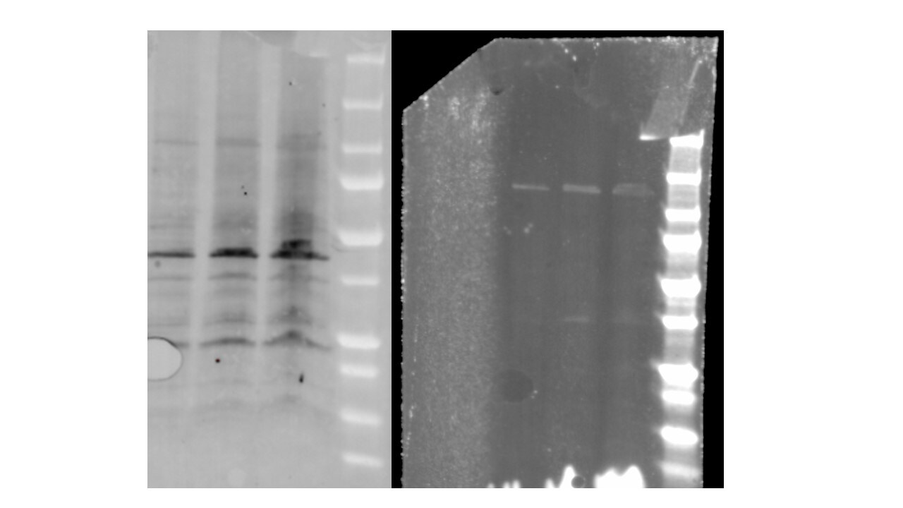
Application: Western BlotSample Tested: fibroblastsSpecies: HumanVerified Customer | Posted 12/04/2024Standard curve of MEKK1 antibody in valvular interstitial cells. The intensity of the band correlates with the amount of total protein of each band.The MEKK1 (NBP2-44405) antibody was used on valvular interstitial cells (fibroblasts) with 5, 10 and 15 µg of total proteins at a concentration of 1:1000.

There are no reviews that match your criteria.
Protocols
Find general support by application which include: protocols, troubleshooting, illustrated assays, videos and webinars.
- Antigen Retrieval Protocol (PIER)
- Antigen Retrieval for Frozen Sections Protocol
- Appropriate Fixation of IHC/ICC Samples
- Cellular Response to Hypoxia Protocols
- Chromogenic IHC Staining of Formalin-Fixed Paraffin-Embedded (FFPE) Tissue Protocol
- Chromogenic Immunohistochemistry Staining of Frozen Tissue
- ClariTSA™ Fluorophore Kits
- Detection & Visualization of Antibody Binding
- Fluorescent IHC Staining of Frozen Tissue Protocol
- Graphic Protocol for Heat-induced Epitope Retrieval
- Graphic Protocol for the Preparation and Fluorescent IHC Staining of Frozen Tissue Sections
- Graphic Protocol for the Preparation and Fluorescent IHC Staining of Paraffin-embedded Tissue Sections
- Graphic Protocol for the Preparation of Gelatin-coated Slides for Histological Tissue Sections
- IHC Sample Preparation (Frozen sections vs Paraffin)
- Immunofluorescent IHC Staining of Formalin-Fixed Paraffin-Embedded (FFPE) Tissue Protocol
- Immunohistochemistry (IHC) and Immunocytochemistry (ICC) Protocols
- Immunohistochemistry Frozen Troubleshooting
- Immunohistochemistry Paraffin Troubleshooting
- Preparing Samples for IHC/ICC Experiments
- Preventing Non-Specific Staining (Non-Specific Binding)
- Primary Antibody Selection & Optimization
- Protocol for Heat-Induced Epitope Retrieval (HIER)
- Protocol for Making a 4% Formaldehyde Solution in PBS
- Protocol for VisUCyte™ HRP Polymer Detection Reagent
- Protocol for the Preparation & Fixation of Cells on Coverslips
- Protocol for the Preparation and Chromogenic IHC Staining of Frozen Tissue Sections
- Protocol for the Preparation and Chromogenic IHC Staining of Frozen Tissue Sections - Graphic
- Protocol for the Preparation and Chromogenic IHC Staining of Paraffin-embedded Tissue Sections
- Protocol for the Preparation and Chromogenic IHC Staining of Paraffin-embedded Tissue Sections - Graphic
- Protocol for the Preparation and Fluorescent IHC Staining of Frozen Tissue Sections
- Protocol for the Preparation and Fluorescent IHC Staining of Paraffin-embedded Tissue Sections
- Protocol for the Preparation of Gelatin-coated Slides for Histological Tissue Sections
- R&D Systems Quality Control Western Blot Protocol
- TUNEL and Active Caspase-3 Detection by IHC/ICC Protocol
- The Importance of IHC/ICC Controls
- Troubleshooting Guide: Immunohistochemistry
- Troubleshooting Guide: Western Blot Figures
- Western Blot Conditions
- Western Blot Protocol
- Western Blot Protocol for Cell Lysates
- Western Blot Troubleshooting
- Western Blot Troubleshooting Guide
- View all Protocols, Troubleshooting, Illustrated assays and Webinars
Loading...