MUC2 Antibody (996/1) - BSA Free
Novus Biologicals | Catalog # NB120-11197
![Immunohistochemistry: MUC2 Antibody (996/1) - BSA Free [NB120-11197]](https://resources.rndsystems.com/images/products/MUC2-Antibody-996-1-Immunohistochemistry-NB120-11197-img0009.jpg "Immunohistochemistry: MUC2 Antibody (996/1) - BSA Free [NB120-11197]")
Key Product Details
Validated by
Biological Validation
Species Reactivity
Validated:
Human, Mouse
Cited:
Human, Mouse
Applications
Validated:
Immunohistochemistry, Immunohistochemistry-Paraffin, Immunohistochemistry-Frozen, Western Blot, Flow Cytometry, Flow (Intracellular), ICC/IF (Negative)
Cited:
Immunohistochemistry-Frozen, Western Blot, Flow Cytometry, IF/IHC
Label
Unconjugated
Antibody Source
Monoclonal Mouse IgG1 Clone # 996/1
Format
BSA Free
Loading...
Product Specifications
Immunogen
This MUC2 Antibody (996/1) was developed against MUC2 tandem repeat peptide
Reactivity Notes
Please note that this antibody is reactive to Mouse and derived from the same host, Mouse. Mouse-On-Mouse blocking reagent may be needed for IHC and ICC experiments to reduce high background signal. You can find these reagents under catalog numbers PK-2200-NB and MP-2400-NB. Please contact Technical Support if you have any questions. Mouse reactivity reported in scientific literature (PMID: 24045942).
Localization
Secreted
Specificity
MUC2 Antibody (996/1) recognizes the human MUC2 mucin, and shows no cross-reactivity with MUC1, MUC3 or MUC4 mucins. In tissue sections colon, liver and prostate stain strongly. It recognizes malignant colonic mucosa and normal mucosa.
Clonality
Monoclonal
Host
Mouse
Isotype
IgG1
Theoretical MW
540 kDa.
Disclaimer note: The observed molecular weight of the protein may vary from the listed predicted molecular weight due to post translational modifications, post translation cleavages, relative charges, and other experimental factors.
Disclaimer note: The observed molecular weight of the protein may vary from the listed predicted molecular weight due to post translational modifications, post translation cleavages, relative charges, and other experimental factors.
Scientific Data Images for MUC2 Antibody (996/1) - BSA Free
![Immunohistochemistry: MUC2 Antibody (996/1) - BSA Free [NB120-11197]](https://resources.rndsystems.com/images/products/MUC2-Antibody-996-1-Immunohistochemistry-NB120-11197-img0011.jpg "Immunohistochemistry: MUC2 Antibody (996/1) - BSA Free [NB120-11197]")
Immunohistochemistry: MUC2 Antibody (996/1) - BSA Free [NB120-11197]
MUC2-Antibody-996-1-Immunohistochemistry-NB120-11197-img0011.jpg![Flow Cytometry: MUC2 Antibody (996/1) - BSA Free [NB120-11197]](https://resources.rndsystems.com/images/products/MUC2-Antibody-996-1-Flow-Cytometry-NB120-11197-img0006.jpg "Flow Cytometry: MUC2 Antibody (996/1) - BSA Free [NB120-11197]")
Flow Cytometry: MUC2 Antibody (996/1) - BSA Free [NB120-11197]
Flow Cytometry: MUC2 Antibody (996/1) [NB120-11197] - An intracellular stain was performed on HeLa cells with MUC2 Antibody [996/1] NB120-11197AF647 (blue) and a matched isotype control (orange). Cells were fixed with 4% PFA and then permeabilized with 0.1% saponin. Cells were incubated in an antibody dilution of 2.5 ug/mL for 30 minutes at room temperature. Both antibodies were conjugated to Alexa Fluor 647.![Flow Cytometry: MUC2 Antibody (996/1) - BSA Free [NB120-11197]](https://resources.rndsystems.com/images/products/MUC2-Antibody-996-1-Flow-Cytometry-NB120-11197-img0003.jpg "Flow Cytometry: MUC2 Antibody (996/1) - BSA Free [NB120-11197]")
Flow Cytometry: MUC2 Antibody (996/1) - BSA Free [NB120-11197]
Flow Cytometry: MUC2 Antibody (996/1) [NB120-11197] - Analysis of Allophycocyanin conjugate of NB120-11197. An intracellular stain was performed on HeLa cells with MUC2 (996/1) antibody NB120-11197APC (blue) and a matched isotype control NBP2-27287APC (orange). Cells were fixed with 4% PFA and then permeabli![Flow Cytometry: MUC2 Antibody (996/1) - BSA Free [NB120-11197]](https://resources.rndsystems.com/images/products/MUC2-Antibody-996-1-Flow-Cytometry-NB120-11197-img0004.jpg "Flow Cytometry: MUC2 Antibody (996/1) - BSA Free [NB120-11197]")
Flow Cytometry: MUC2 Antibody (996/1) - BSA Free [NB120-11197]
Flow Cytometry: MUC2 Antibody (996/1) [NB120-11197] - Analysis of Allophycocyanin conjugate of NB120-11197. An intracellular stain was performed on NTERA-2 cells with MUC2 (996/1) antibody NB120-11197APC (blue) and a matched isotype control NBP2-27287APC (orange). Cells were fixed with 4% PFA and then permea![Flow (Intracellular): MUC2 Antibody (996/1) - BSA Free [NB120-11197]](https://resources.rndsystems.com/images/products/MUC2-Antibody-996-1-Flow-Intracellular-NB120-11197-img0005.jpg "Flow (Intracellular): MUC2 Antibody (996/1) - BSA Free [NB120-11197]")
Flow (Intracellular): MUC2 Antibody (996/1) - BSA Free [NB120-11197]
Flow (Intracellular): MUC2 Antibody (996/1) [NB120-11197] - An intracellular stain was performed on HeLa cells with MUC2 Antibody (996/1) NB120-11197AF488 (blue) and a matched isotype control (orange). Cells were fixed with 4% PFA and then permeabilized with 0.1% saponin. Cells were incubated in an antibody dilution of 10 ug/mL for 30 minutes at room temperature. Both antibodies were conjugated to Alexa Fluor 488. [NB120-11197] -")
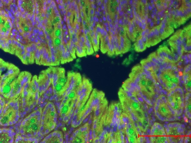
Immunohistochemistry-Paraffin: Mouse Monoclonal MUC2 Antibody (996/1) [NB120-11197] -
Immunohistochemistry-Paraffin: Mouse Monoclonal MUC2 Antibody (996/1) [NB120-11197] - Staining of mouse colon tissue using MUC2 Antibody. Colon, MUC2 -Green, DAPI-Blue. Antibody dilution - 1:100. Image from a verified customer review. - BSA Free [NB120-11197] -")
Western Blot: MUC2 Antibody (996/1) - BSA Free [NB120-11197] -
MDA5-mediated dsRNA sensing is required for intestinal Adar deficiency-induced bowel inflammation and organoid death.a Representative images of Adar (green) and J2 (red) staining in organoids isolated from the ileum of Villin-CreERT2 Adarfl/fl mice and treated with or without 4-OHT (200 nM) for 3 days.Scale bars: 100 μm. b Representative images of J2 staining (green) on ileal sections of Adarfl/fl and Adari delta gut mice (3 dpi). Scale bars: 100 μm. c Survival curves of Adarfl/fl, Adari delta gut, and Adari delta gutMda5−/−mice after tamoxifen induction (n = 5 for Adarfl/fl, n = 8 for Adari delta gut, n = 6 for Adari delta gutMda5−/−). d, hAdarfl/fl, Adari delta gut and Adari delta gutMda5−/− mice after tamoxifen induction at 3-dpi. d Small intestine and colon length (n = 5 for Adarfl/fl, n = 7 for Adari delta gut, n = 6 for Adari delta gutMda5−/−). e H&E staining of intestinal sections. Scale bars: 100 μm. Shown on the right are quantifications of histopathological scores, and lengths of ileal crypts and villi (n = 5). f Representative images of TUNEL staining (green) on the ileal sections. Scale bars: 100 μm. g Immunofluorescence of E-cadherin (red) staining. Scale bars: 100 μm. h Immunofluorescence of Lysozyme (green) and Muc2 (red) staining. Scale bars: 100 μm. i Western blot analysis of ileal samples (n = 3). j Representative images of the growth process of organoids isolated from the ileum of Adari delta gut and Adari delta gutMda5−/− mice treated with or without 4-OHT. Scale bars: 100 μm. Data are presented as the mean +/- SEM. The statistical significance was analyzed using Log-rank (Mantel-Cox) test (Fig. 5c). The remaining statistical differences were determined using one-way ANOVA with multiple comparisons. Source data are provided in the Source data file. Image collected and cropped by CiteAb from the following open publication (https://www.nature.com/articles/s41467-025-63554-4), licensed under a CC-BY license. Not internally tested by Novus Biologicals. - BSA Free [NB120-11197] -")
Immunocytochemistry/ Immunofluorescence: MUC2 Antibody (996/1) - BSA Free [NB120-11197] -
MS-induced intestinal epithelium injury was CRHR1 dependent.Photomicrographs of hematoxylin and eosin (H&E) stained (A–E) and immunofluorescence of Mucin 2 (Muc2; mucous-forming protein) (F–J) in proximal colon in all experimental groups. Histological scores (K) were highest in MS, demonstrated injury in MS compared to control. Treatment with Antalarmin and Astressin prevented this MS-induced colonic injury, but not by Astressin-2 beta. Crypt length in μm (L) (red lines in photomicrographs A–E) and the number of Muc2+ goblet cells per crypt (M) were reduced by MS compared to control, and restored to control levels following Antalarmin and Astressin treatment. Astressin-2 beta did not prevent these MS-induced effects. Myeloperoxidase (MPO; μmol/mg protein) expression was increased in MS group and was reduced to a level similar to control by treatment with Antalarmin but not by treatment with Astressin or Astressin-2 beta (N). Western blot analysis of NF-kappa B showed an increase in the phosphorylated expression of NF-kappa B in MS, which was prevented by Antalarmin administration, but not by Astressin or Astressin-2 beta (O,P). Trans-cellular flux of HRP (ng/ml.cm2.min; Q) measured by Ussing Chamber was increased in MS and MS + Astressin-2 beta groups, compared to control, but not in MS + Antalarmin and MS + Astressin groups (P). Results are means, +/-SD. p < 0.05 was considered significant. Image collected and cropped by CiteAb from the following open publication (https://pubmed.ncbi.nlm.nih.gov/28492284), licensed under a CC-BY license. Not internally tested by Novus Biologicals. - BSA Free [NB120-11197] -")
Immunocytochemistry/ Immunofluorescence: MUC2 Antibody (996/1) - BSA Free [NB120-11197] -
MS-induced intestinal epithelium injury was CRHR1 dependent.Photomicrographs of hematoxylin and eosin (H&E) stained (A–E) and immunofluorescence of Mucin 2 (Muc2; mucous-forming protein) (F–J) in proximal colon in all experimental groups. Histological scores (K) were highest in MS, demonstrated injury in MS compared to control. Treatment with Antalarmin and Astressin prevented this MS-induced colonic injury, but not by Astressin-2 beta. Crypt length in μm (L) (red lines in photomicrographs A–E) and the number of Muc2+ goblet cells per crypt (M) were reduced by MS compared to control, and restored to control levels following Antalarmin and Astressin treatment. Astressin-2 beta did not prevent these MS-induced effects. Myeloperoxidase (MPO; μmol/mg protein) expression was increased in MS group and was reduced to a level similar to control by treatment with Antalarmin but not by treatment with Astressin or Astressin-2 beta (N). Western blot analysis of NF-kappa B showed an increase in the phosphorylated expression of NF-kappa B in MS, which was prevented by Antalarmin administration, but not by Astressin or Astressin-2 beta (O,P). Trans-cellular flux of HRP (ng/ml.cm2.min; Q) measured by Ussing Chamber was increased in MS and MS + Astressin-2 beta groups, compared to control, but not in MS + Antalarmin and MS + Astressin groups (P). Results are means, +/-SD. p < 0.05 was considered significant. Image collected and cropped by CiteAb from the following open publication (https://pubmed.ncbi.nlm.nih.gov/28492284), licensed under a CC-BY license. Not internally tested by Novus Biologicals.Applications for MUC2 Antibody (996/1) - BSA Free
Application
Recommended Usage
Flow Cytometry
1:10-1:1000
Immunohistochemistry
1:10-1:500
Immunohistochemistry-Frozen
1:10-1:500
Immunohistochemistry-Paraffin
1:10-1:500
Western Blot
1:100-1:2000
Application Notes
Membrane permeabilization is required for Flow Cytometry.
Reviewed Applications
Read 2 reviews rated 4 using NB120-11197 in the following applications:
Flow Cytometry Panel Builder
Bio-Techne Knows Flow Cytometry
Save time and reduce costly mistakes by quickly finding compatible reagents using the Panel Builder Tool.
Advanced Features
- Spectra Viewer - Custom analysis of spectra from multiple fluorochromes
- Spillover Popups - Visualize the spectra of individual fluorochromes
- Antigen Density Selector - Match fluorochrome brightness with antigen density

Formulation, Preparation, and Storage
Purification
Protein A or G purified
Formulation
PBS
Format
BSA Free
Preservative
0.09% Sodium Azide
Concentration
1.0 mg/ml
Shipping
The product is shipped with polar packs. Upon receipt, store it immediately at the temperature recommended below.
Stability & Storage
Store at 4C short term. Aliquot and store at -20C long term. Avoid freeze-thaw cycles.
Background: MUC2
Changes or perturbations to MUC2 expression has been associated with a number of disease pathologies (1-4, 6). Specifically, altered MUC2 composition has been indicated in colorectal cancer, inflammatory bowel diseases (IBD) including ulcerative colitis and Chron's disease, and chronic obstructive pulmonary disease (COPD) (1-4, 6). In general, decreased or reduced MUC2 expression is associated with colorectal cancer disease progression and development of IBD (1-4, 6). Additionally, studies have found that upon intestinal infection due to parasites or bacteria MUC2 expression is increased, as is overall mucus secretion (3, 6).
References
1. Pothuraju, R., Krishn, S. R., Gautam, S. K., Pai, P., Ganguly, K., Chaudhary, S., Rachagani, S., Kaur, S., & Batra, S. K. (2020). Mechanistic and Functional Shades of Mucins and Associated Glycans in Colon Cancer. Cancers. https://doi.org/10.3390/cancers12030649
2. Ballester, B., Milara, J., & Cortijo, J. (2019). Mucins as a New Frontier in Pulmonary Fibrosis. Journal of Clinical Medicine. https://doi.org/10.3390/jcm8091447
3. Liu, Y., Yu, X., Zhao, J., Zhang, H., Zhai, Q., & Chen, W. (2020). The role of MUC2 mucin in intestinal homeostasis and the impact of dietary components on MUC2 expression. International Journal of Biological Macromolecules. https://doi.org/10.1016/j.ijbiomac.2020.07.191
4. Allen, A., Hutton, D. A., & Pearson, J. P. (1998). The MUC2 gene product: a human intestinal mucin. The International Journal of Biochemistry & Cell Biology. https://doi.org/10.1016/s1357-2725(98)00028-4
5. Uniprot (Q02817)
6. Kim, Y. S., & Ho, S. B. (2010). Intestinal goblet cells and mucins in health and disease: recent insights and progress. Current Gastroenterology Reports. https://doi.org/10.1007/s11894-010-0131-2
Alternate Names
Intestinal mucin-2, MLP, MUC2, MUC-2, mucin 2, intestinal/tracheal, mucin 2, oligomeric mucus/gel-forming, mucin-2, SMUC
Entrez Gene IDs
4583 (Human)
Gene Symbol
MUC2
UniProt
Additional MUC2 Products
Product Documents for MUC2 Antibody (996/1) - BSA Free
Certificate of Analysis
To download a Certificate of Analysis, please enter a lot or batch number in the search box below.
Product Specific Notices for MUC2 Antibody (996/1) - BSA Free
This product is for research use only and is not approved for use in humans or in clinical diagnosis. Primary Antibodies are guaranteed for 1 year from date of receipt.
Citations for MUC2 Antibody (996/1) - BSA Free
 Powered by Bioz
Customer Reviews for MUC2 Antibody (996/1) - BSA Free (2)
4 out of 5
2 Customer Ratings
Have you used MUC2 Antibody (996/1) - BSA Free?
Submit a review and receive an Amazon gift card!
$25/€18/£15/$25CAN/¥2500 Yen for a review with an image
$10/€7/£6/$10CAN/¥1110 Yen for a review without an image
Submit a review
Customer Images
Showing
1
-
2 of
2 reviews
Showing All
Filter By:
-
Application: Immunohistochemistry-ParaffinSample Tested: Colon tissueSpecies: MouseVerified Customer | Posted 05/10/2024Colon, MUC2 -Green, DAPI-Blue. Antibody dilution - 1:100

-
Application: Immunohistochemistry-ParaffinSample Tested: pig lungSpecies: OtherVerified Customer | Posted 03/11/2009
There are no reviews that match your criteria.
Protocols
Find general support by application which include: protocols, troubleshooting, illustrated assays, videos and webinars.
- 7-Amino Actinomycin D (7-AAD) Cell Viability Flow Cytometry Protocol
- Antigen Retrieval Protocol (PIER)
- Antigen Retrieval for Frozen Sections Protocol
- Appropriate Fixation of IHC/ICC Samples
- Cellular Response to Hypoxia Protocols
- Chromogenic IHC Staining of Formalin-Fixed Paraffin-Embedded (FFPE) Tissue Protocol
- Chromogenic Immunohistochemistry Staining of Frozen Tissue
- ClariTSA™ Fluorophore Kits
- Detection & Visualization of Antibody Binding
- Extracellular Membrane Flow Cytometry Protocol
- Flow Cytometry Protocol for Cell Surface Markers
- Flow Cytometry Protocol for Staining Membrane Associated Proteins
- Flow Cytometry Staining Protocols
- Flow Cytometry Troubleshooting Guide
- Fluorescent IHC Staining of Frozen Tissue Protocol
- Graphic Protocol for Heat-induced Epitope Retrieval
- Graphic Protocol for the Preparation and Fluorescent IHC Staining of Frozen Tissue Sections
- Graphic Protocol for the Preparation and Fluorescent IHC Staining of Paraffin-embedded Tissue Sections
- Graphic Protocol for the Preparation of Gelatin-coated Slides for Histological Tissue Sections
- IHC Sample Preparation (Frozen sections vs Paraffin)
- Immunofluorescent IHC Staining of Formalin-Fixed Paraffin-Embedded (FFPE) Tissue Protocol
- Immunohistochemistry (IHC) and Immunocytochemistry (ICC) Protocols
- Immunohistochemistry Frozen Troubleshooting
- Immunohistochemistry Paraffin Troubleshooting
- Intracellular Flow Cytometry Protocol Using Alcohol (Methanol)
- Intracellular Flow Cytometry Protocol Using Detergents
- Intracellular Nuclear Staining Flow Cytometry Protocol Using Detergents
- Intracellular Staining Flow Cytometry Protocol Using Alcohol Permeabilization
- Intracellular Staining Flow Cytometry Protocol Using Detergents to Permeabilize Cells
- Preparing Samples for IHC/ICC Experiments
- Preventing Non-Specific Staining (Non-Specific Binding)
- Primary Antibody Selection & Optimization
- Propidium Iodide Cell Viability Flow Cytometry Protocol
- Protocol for Heat-Induced Epitope Retrieval (HIER)
- Protocol for Liperfluo
- Protocol for Making a 4% Formaldehyde Solution in PBS
- Protocol for VisUCyte™ HRP Polymer Detection Reagent
- Protocol for the Characterization of Human Th22 Cells
- Protocol for the Characterization of Human Th9 Cells
- Protocol for the Preparation & Fixation of Cells on Coverslips
- Protocol for the Preparation and Chromogenic IHC Staining of Frozen Tissue Sections
- Protocol for the Preparation and Chromogenic IHC Staining of Frozen Tissue Sections - Graphic
- Protocol for the Preparation and Chromogenic IHC Staining of Paraffin-embedded Tissue Sections
- Protocol for the Preparation and Chromogenic IHC Staining of Paraffin-embedded Tissue Sections - Graphic
- Protocol for the Preparation and Fluorescent IHC Staining of Frozen Tissue Sections
- Protocol for the Preparation and Fluorescent IHC Staining of Paraffin-embedded Tissue Sections
- Protocol for the Preparation of Gelatin-coated Slides for Histological Tissue Sections
- Protocol: Annexin V and PI Staining by Flow Cytometry
- Protocol: Annexin V and PI Staining for Apoptosis by Flow Cytometry
- R&D Systems Quality Control Western Blot Protocol
- TUNEL and Active Caspase-3 Detection by IHC/ICC Protocol
- The Importance of IHC/ICC Controls
- Troubleshooting Guide: Fluorokine Flow Cytometry Kits
- Troubleshooting Guide: Immunohistochemistry
- Troubleshooting Guide: Western Blot Figures
- Western Blot Conditions
- Western Blot Protocol
- Western Blot Protocol for Cell Lysates
- Western Blot Troubleshooting
- Western Blot Troubleshooting Guide
- View all Protocols, Troubleshooting, Illustrated assays and Webinars
Loading...