![Immunohistochemistry-Paraffin: MUM1 Antibody (MUM1p) [NB200-356]](https://resources.rndsystems.com/images/products/MUM1-Antibody-MUM1p-Immunohistochemistry-Paraffin-NB200-356-img0002.jpg "Immunohistochemistry-Paraffin: MUM1 Antibody (MUM1p) [NB200-356]")
Loading...
Key Product Details
Species Reactivity
Human, Alpaca, Canine, Equine, Feline
Applications
Multiplex Immunofluorescence, Immunohistochemistry, Immunohistochemistry-Paraffin, Immunohistochemistry-Frozen
Label
Unconjugated
Antibody Source
Monoclonal Mouse IgG1 Clone # MUM1p
Loading...
Product Specifications
Immunogen
Recombinant GST-MUM1 protein.
Reactivity Notes
Alpaca, Feline, Canine, and Equine reactivity reported from a verified customer review. Cross-reacts with Human. Not yet tested in other species.
Localization
Cytoplasmic and Nuclear
Specificity
Multiple myeloma oncogene-1 (MUM1) is a 50kDa protein encoded by the MUM1 gene. This antibody stains MUM1 protein, which is expressed in a subset of B cells in the light zone of the germinal center, plasma cells, activated T cells and a wide spectrum of related hematolymphoid neoplasms. This antibody is useful in the sub classification of lymphoid malignancies
Clonality
Monoclonal
Host
Mouse
Isotype
IgG1
Scientific Data Images for MUM1 Antibody (MUM1p)
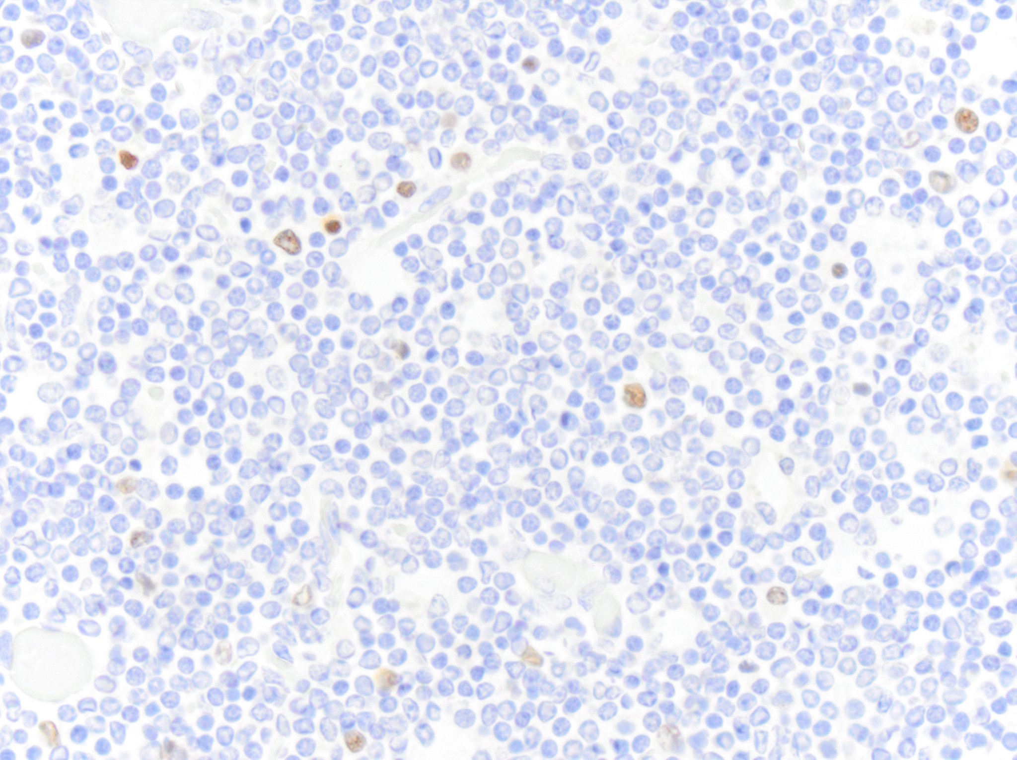
Immunohistochemistry-Paraffin: MUM1 Antibody (MUM1p) [NB200-356]
Immunohistochemistry-Paraffin: MUM1 Antibody (MUM1p) [NB200-356] - Formalin fixed paraffin embedded human tonsil stained with MUM1 protein antibody (NB200-356).![Immunohistochemistry-Paraffin: MUM1 Antibody (MUM1p) [NB200-356]](https://resources.rndsystems.com/images/products/MUM1-Antibody-MUM1p-Immunohistochemistry-Paraffin-NB200-356-img0001.jpg "Immunohistochemistry-Paraffin: MUM1 Antibody (MUM1p) [NB200-356]")
Immunohistochemistry-Paraffin: MUM1 Antibody (MUM1p) [NB200-356]
Immunohistochemistry-Paraffin: MUM1 Antibody (MUM1p) [NB200-356] - Formalin fixed paraffin embedded human tonsil stained with MUM1 protein antibody. [NB200-356]")
Immunohistochemistry-Paraffin: Mouse Monoclonal MUM1 Antibody (MUM1p) [NB200-356]
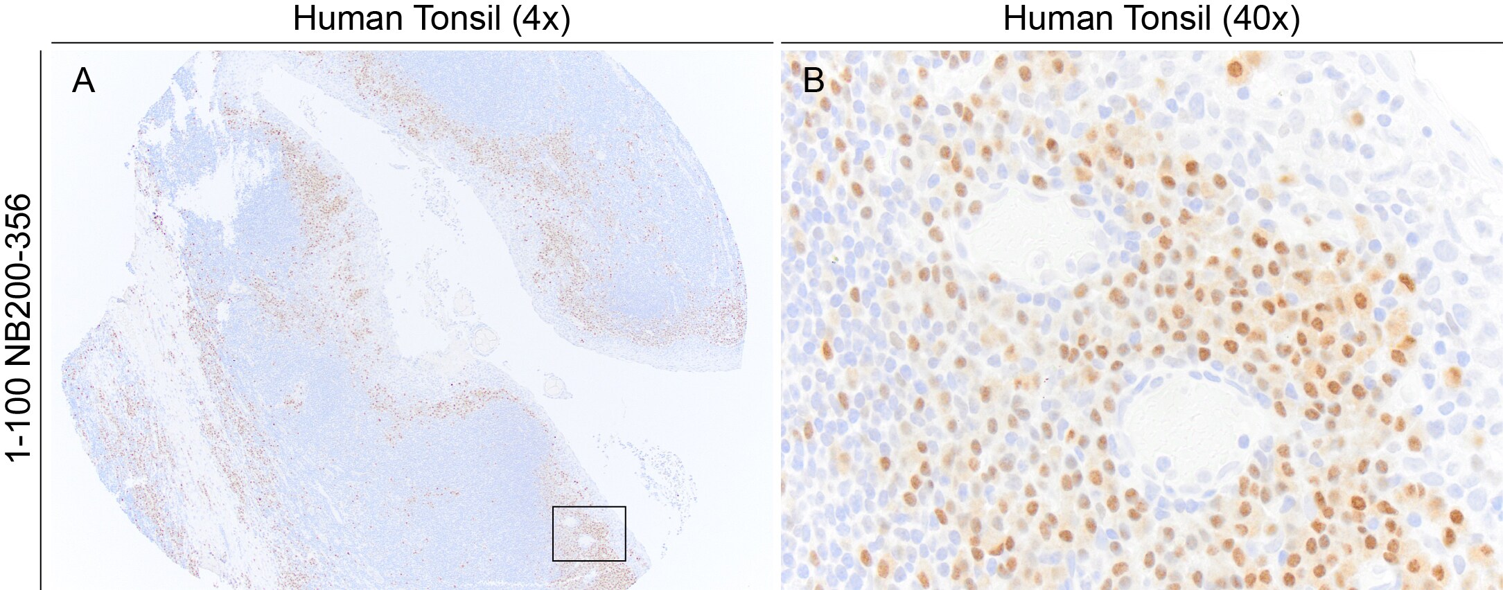
Image demonstrating MUM1 immunoreactivity in an FFPE section of human tonsil. NB200-356 was diluted 1:100 and was left on tissue sections for 30m at room temperature. Heat induced epitope retrieval with a citrate-based buffer was used. Image from a verified customer review. [NB200-356]")
Immunohistochemistry-Paraffin: Mouse Monoclonal MUM1 Antibody (MUM1p) [NB200-356]
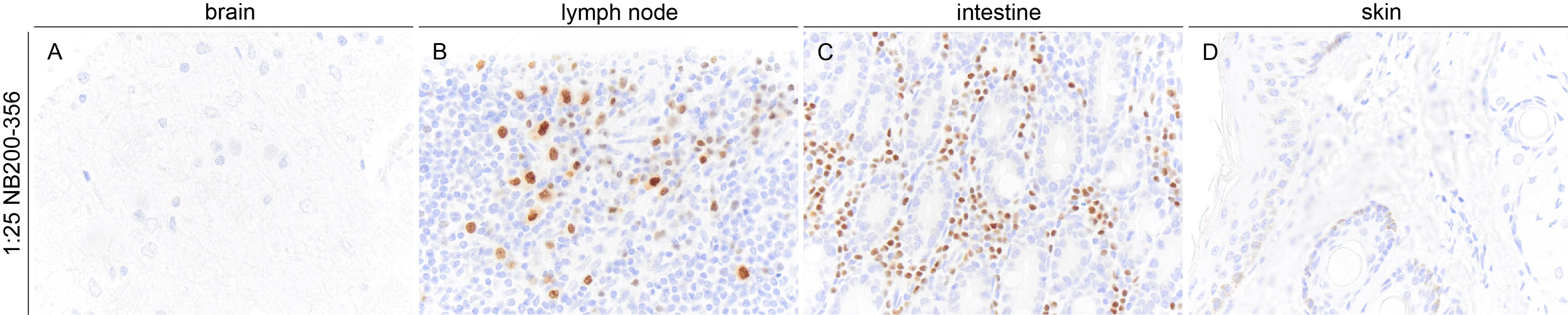
Images demonstrating MUM1 immunoreactivity in a variety of horse FFPE tissue sections. NB200-356 was diluted 1:25 and was left on tissue sections for 1hr at room temperature. Heat induced epitope retrieval with a high pH buffer was used. Image from a verified customer review. [NB200-356]")
Immunohistochemistry-Paraffin: Mouse Monoclonal MUM1 Antibody (MUM1p) [NB200-356]
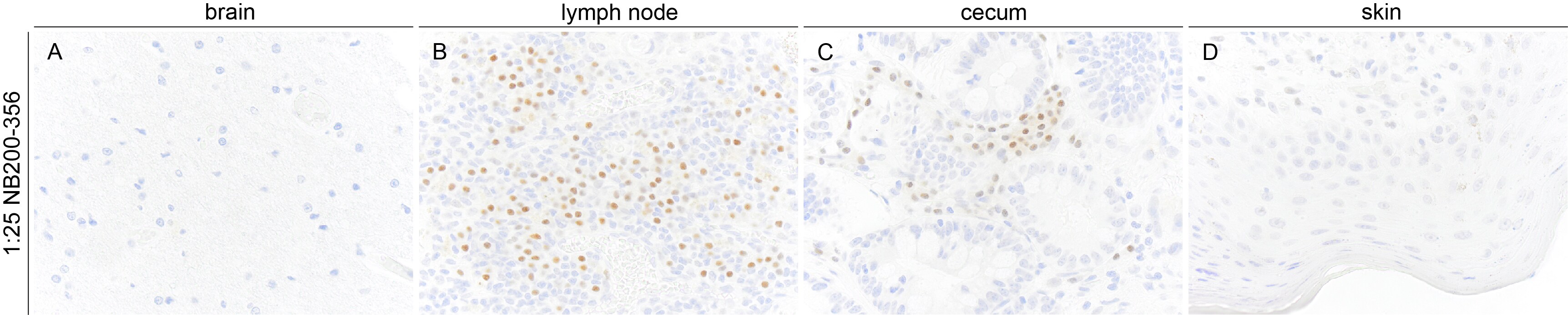
Images demonstrating MUM1 immunoreactivity in a variety of dog FFPE tissue sections. NB200-356 was diluted 1:25 and was left on tissue sections for 1hr at room temperature. Heat induced epitope retrieval with a high pH buffer was used. Image from a verified customer review. [NB200-356]")
Immunohistochemistry-Paraffin: Mouse Monoclonal MUM1 Antibody (MUM1p) [NB200-356]
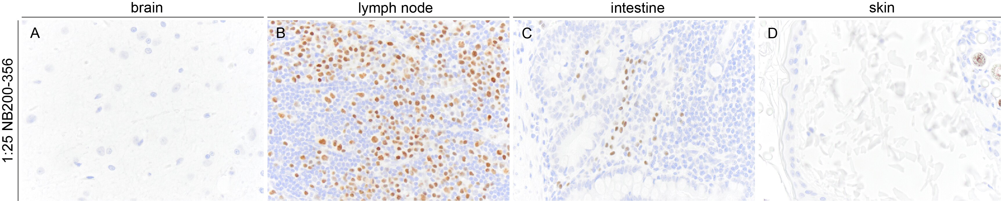
Images demonstrating MUM1 immunoreactivity in a variety of cat FFPE tissue sections. NB200-356 was diluted 1:25 and was left on tissue sections for 1hr at room temperature. Heat induced epitope retrieval with a high pH buffer was used. Image from a verified customer review. [NB200-356]")
Immunohistochemistry-Paraffin: Mouse Monoclonal MUM1 Antibody (MUM1p) [NB200-356]
Image showing MUM1 immunoreactivity in an FFPE section of alpaca lymph node. NB200-356 was diluted 1-25 and was left on tissue sections for 1hr at room temperature. Image from a verified customer review.Applications for MUM1 Antibody (MUM1p)
Application
Recommended Usage
Immunohistochemistry
1:10-1:25
Immunohistochemistry-Frozen
1:10-1:500
Immunohistochemistry-Paraffin
1:10-1:25
Application Notes
IHC-P: recommended pretreatment of EDTA buffer, pH 8.0. Recommended incubation time of 30 min at RT.
Reviewed Applications
Read 5 reviews rated 5 using NB200-356 in the following applications:
Formulation, Preparation, and Storage
Purification
Tissue culture supernatant
Formulation
Tissue culture supernatant
Preservative
0.05% Sodium Azide
Concentration
This product is unpurified. The exact concentration of antibody is not quantifiable.
Shipping
The product is shipped with polar packs. Upon receipt, store it immediately at the temperature recommended below.
Stability & Storage
Store at 4C.
Background: MUM1
Alternate Names
EXPAND1, FLJ14868, FLJ22283, melanoma associated antigen (mutated) 1, melanoma ubiquitous mutated protein, MGC131891, MGC163315, MUM-1HSPC211, Mutated melanoma-associated antigen 1, Protein expandere, PWWP domain-containing protein MUM1
Gene Symbol
PWWP3A
Additional MUM1 Products
Product Documents for MUM1 Antibody (MUM1p)
Certificate of Analysis
To download a Certificate of Analysis, please enter a lot or batch number in the search box below.
Product Specific Notices for MUM1 Antibody (MUM1p)
This product is for research use only and is not approved for use in humans or in clinical diagnosis. Primary Antibodies are guaranteed for 1 year from date of receipt.
Citations for MUM1 Antibody (MUM1p)
 Powered by Bioz
Customer Reviews for MUM1 Antibody (MUM1p) (5)
5 out of 5
5 Customer Ratings
Have you used MUM1 Antibody (MUM1p)?
Submit a review and receive an Amazon gift card!
$25/€18/£15/$25CAN/¥2500 Yen for a review with an image
$10/€7/£6/$10CAN/¥1110 Yen for a review without an image
Submit a review
Customer Images
Showing
1
-
5 of
5 reviews
Showing All
Filter By:
-
Application: Immunohistochemistry-ParaffinSample Tested: TonsilSpecies: HumanVerified Customer | Posted 06/23/2025Image demonstrating MUM1 immunoreactivity in an FFPE section of human tonsil. NB200-356 was diluted 1:100 and was left on tissue sections for 30m at room temperature. Heat induced epitope retrieval with a citrate-based buffer was used.

-
Application: Immunohistochemistry-ParaffinSample Tested: Multiple TissuesSpecies: HorseVerified Customer | Posted 06/23/2025Images demonstrating MUM1 immunoreactivity in a variety of horse FFPE tissue sections. NB200-356 was diluted 1:25 and was left on tissue sections for 1hr at room temperature. Heat induced epitope retrieval with a high pH buffer was used.
-
Application: Immunohistochemistry-ParaffinSample Tested: Multiple TissuesSpecies: DogVerified Customer | Posted 06/23/2025Images demonstrating MUM1 immunoreactivity in a variety of dog FFPE tissue sections. NB200-356 was diluted 1:25 and was left on tissue sections for 1hr at room temperature. Heat induced epitope retrieval with a high pH buffer was used.
Bio-Techne ResponseThis review reflects a new species or application tested on a primary antibody.
-
Application: Immunohistochemistry-ParaffinSample Tested: Multiple TissuesSpecies: CatVerified Customer | Posted 06/23/2025Images demonstrating MUM1 immunoreactivity in a variety of cat FFPE tissue sections. NB200-356 was diluted 1:25 and was left on tissue sections for 1hr at room temperature. Heat induced epitope retrieval with a high pH buffer was used.
Bio-Techne ResponseThis review reflects a new species or application tested on a primary antibody.
-
Application: Immunohistochemistry-ParaffinSample Tested: Lymph NodeSpecies: AlpacaVerified Customer | Posted 06/23/2025Image showing MUM1 immunoreactivity in an FFPE section of alpaca lymph node. NB200-356 was diluted 1-25 and was left on tissue sections for 1hr at room temperature.
Bio-Techne ResponseThis review reflects a new species or application tested on a primary antibody.
There are no reviews that match your criteria.
Protocols
Find general support by application which include: protocols, troubleshooting, illustrated assays, videos and webinars.
- Antigen Retrieval Protocol (PIER)
- Antigen Retrieval for Frozen Sections Protocol
- Appropriate Fixation of IHC/ICC Samples
- Cellular Response to Hypoxia Protocols
- Chromogenic IHC Staining of Formalin-Fixed Paraffin-Embedded (FFPE) Tissue Protocol
- Chromogenic Immunohistochemistry Staining of Frozen Tissue
- ClariTSA™ Fluorophore Kits
- Detection & Visualization of Antibody Binding
- Fluorescent IHC Staining of Frozen Tissue Protocol
- Graphic Protocol for Heat-induced Epitope Retrieval
- Graphic Protocol for the Preparation and Fluorescent IHC Staining of Frozen Tissue Sections
- Graphic Protocol for the Preparation and Fluorescent IHC Staining of Paraffin-embedded Tissue Sections
- Graphic Protocol for the Preparation of Gelatin-coated Slides for Histological Tissue Sections
- IHC Sample Preparation (Frozen sections vs Paraffin)
- Immunofluorescent IHC Staining of Formalin-Fixed Paraffin-Embedded (FFPE) Tissue Protocol
- Immunohistochemistry (IHC) and Immunocytochemistry (ICC) Protocols
- Immunohistochemistry Frozen Troubleshooting
- Immunohistochemistry Paraffin Troubleshooting
- Preparing Samples for IHC/ICC Experiments
- Preventing Non-Specific Staining (Non-Specific Binding)
- Primary Antibody Selection & Optimization
- Protocol for Heat-Induced Epitope Retrieval (HIER)
- Protocol for Making a 4% Formaldehyde Solution in PBS
- Protocol for VisUCyte™ HRP Polymer Detection Reagent
- Protocol for the Preparation & Fixation of Cells on Coverslips
- Protocol for the Preparation and Chromogenic IHC Staining of Frozen Tissue Sections
- Protocol for the Preparation and Chromogenic IHC Staining of Frozen Tissue Sections - Graphic
- Protocol for the Preparation and Chromogenic IHC Staining of Paraffin-embedded Tissue Sections
- Protocol for the Preparation and Chromogenic IHC Staining of Paraffin-embedded Tissue Sections - Graphic
- Protocol for the Preparation and Fluorescent IHC Staining of Frozen Tissue Sections
- Protocol for the Preparation and Fluorescent IHC Staining of Paraffin-embedded Tissue Sections
- Protocol for the Preparation of Gelatin-coated Slides for Histological Tissue Sections
- TUNEL and Active Caspase-3 Detection by IHC/ICC Protocol
- The Importance of IHC/ICC Controls
- Troubleshooting Guide: Immunohistochemistry
- View all Protocols, Troubleshooting, Illustrated assays and Webinars
Loading...